Ocular Manifestations of Systemic Disease and Ocular injury
 Call Kai
 Learn
Learn Practice Test
 Spaced Repetition
 Match
Match Flashcards
 Knowt Play
1/62

There's no tags or description
Looks like no tags are added yet.
Name | Mastery | Learn | Test | Matching | Spaced | Call with Kai | Chat |
|---|
No analytics yet
Send a link to your students to track their progress
63 Terms
Describe Diabetes
Clinical Imp
List the Non-proliferative changes (NPDR)
Mild/Mod
Severe
List the Proliferative (PDR) changes
Clinical Imp
MOA
Untreated →?
Describe Macula Edema
Management
Freq. of exams
Treatment
Diabetes
Clinical Importance:
Leading cause of vision loss (18-64 yrs)
Risk of developing retinopathy ↑ with duration of disease
(type 1 23% @ 5 yrs, 80% @ 15 yrs, rates lower for type 2)
Non-proliferative changes (NPDR)
Mild - Moderate
Microaneurysms
Dot-blot hemorrhages
Hard exudates
Venous beading
Intraretinal microvascular abnormalities (IRMA)
Severe
4 quads of hemorrhages, 2 quads of beading or 1 quad of IRMA
Proliferative (PDR)
Clinical Importance:
Responsible for most of profound visual loss
MOA:
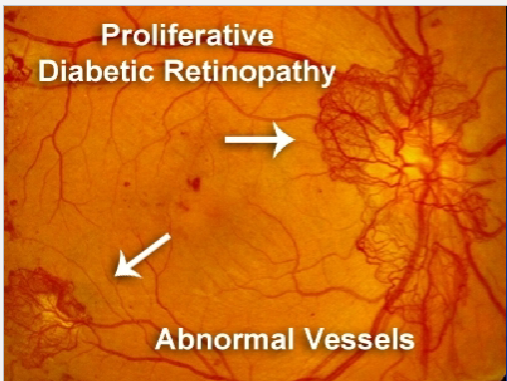
Neovascularization in response to ischemia
Disc, retina, iris
If untreated
→ vitreous hemorrhage, tractional retinal detachment
Macular Edema
Most common cause of mild-mod VA loss
Management
Frequency of exams
Type 1 – initial exam when post-pubertal and within 5 yrs of Dx
Type 2 – exam @ time of Dx
All:
generally examine q1yr unless poor glycemic control, HTN, anemia, proteinuria,
mod- severe NPDR or PDR which require more freq F/U
Pregnant + type I – first trimester + q3months
Treatment
Focal laser
Panretinal photocoagulation
Vitrectomy with laser
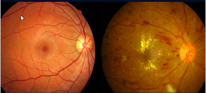


Dot Hemorrhages
Microaneuysms
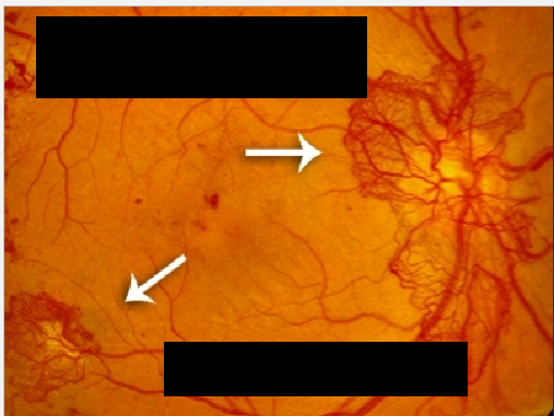
NVE = Neovascularization Elsewhere
NVD = Neovascularization of the Disc
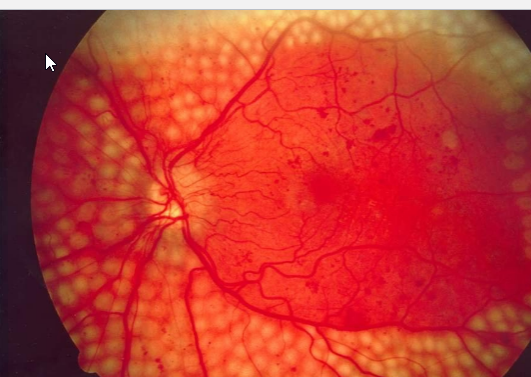
Panretinal photocoagulation
Describe Arteriolar Sclerosis
Severity of Eye disease?\
Eye symptoms?
Associated w/ Thickening and sclerosis of arterioles
Associated w/ BP Elevation
Classifications?
Management?
Arteriolar Sclerosis
Severity of Eye disease
Extent relates to duration + severity of HTN
Eye Symptoms:
Thickening and sclerosis of arterioles
↑ light reflex width (copper → silver wire)
Changes in the color of blood vessels
A-V nicking
***hardened, thickened artery (arteriole) crosses over and compresses a vein (venule), making the vein look pinched or bulging on both sides ***
May predispose to BRVO if severe
Acute BP elevation
Fibrinoid necrosis →
exudates,
CWS (cotton wool)
flame hemorrhages,
optic disc swelling
Classification
Grade 0 – no changes
Grade 1 – barely detectable arterial narrowing
Grade 2 – obvious arterial narrowing with focal irregularities
Grade 3 – gr 2 + retinal hemorrhages or exudate
Grade 4 – gr 3 + disc swelling
Management
Control BP
Avoid nocturnal hypotension
ischemic optic neuropathy, glaucomatous field loss
Describe Pregnancy and the eye
List the Physiologic Δs
List the Pathologic Δs
Physiologic Δs
↓ IOP,
↓ corneal sensitivity,
↓ accommodation,
dry eye,
Δ in refraction
***Avoid changing glasses, contacts, refractive surgery***
Pathologic Δs
↑ risk of CSR, uveal melanoma
Pre-eclampsia/eclampsia
Scotoma, diplopia, dimness
Vascular Δs
Hemorrhages, exudates, retinal edema, disc swelling
Serous exudative RD in 10% of eclampsia
Diabetes – exacerbated retinopathy
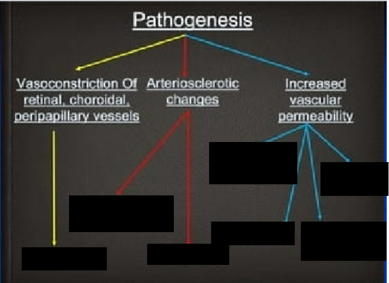


Cooper wiring + Nicking
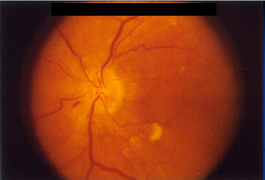
Hypertensive Retinopathy with cotton-wool spots
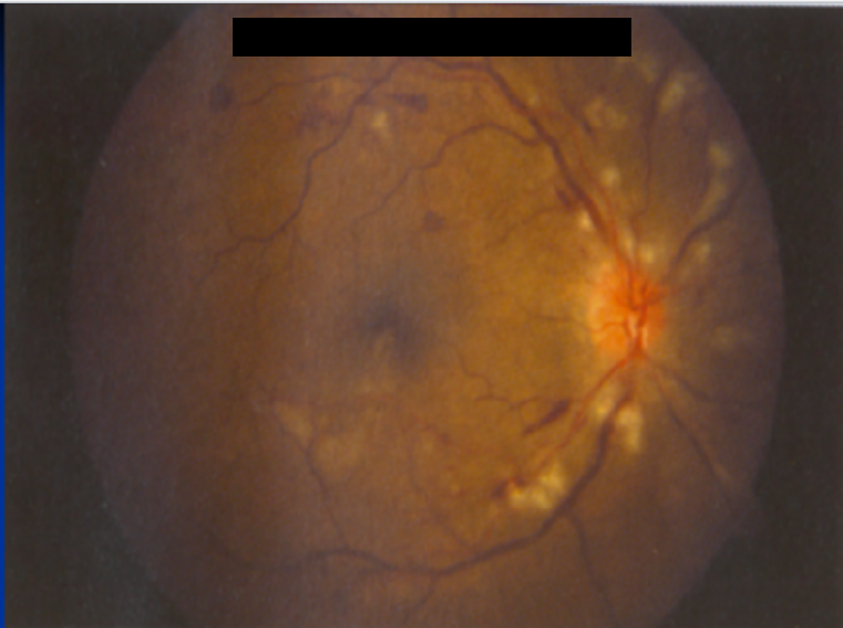
Severe Hypertensive Retinopathy
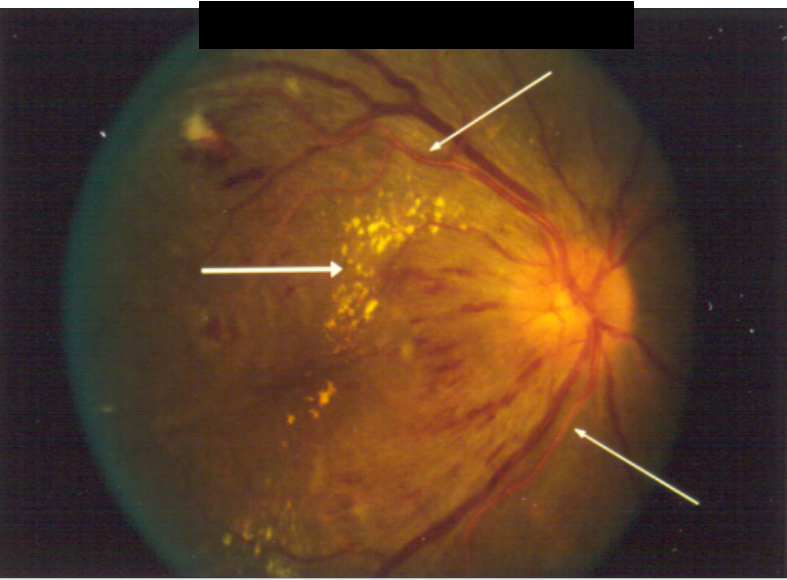
Malignant Hypertension Retinopathy
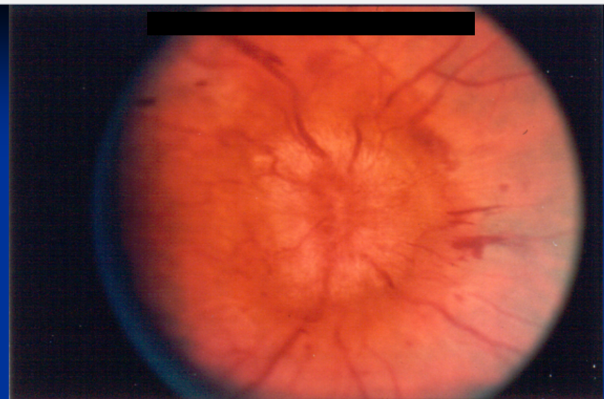
Malignant Hypertension with Papilledema
Describe Sickle Cell Anemia
Patho
Treatment
Sickle Cell Anemia
Patho:
Arteriolar occlusion
intravasc sickling → hemolysis → hemostasis → thrombosis → capillary non-perfusion
-> poor perfusion = retinal ischemia → neovascularization
Similar to Diabetes
Treatment:
Laser Tx – can prevent vision loss
NOTE:
SC and S Thal more likely to have eye involved
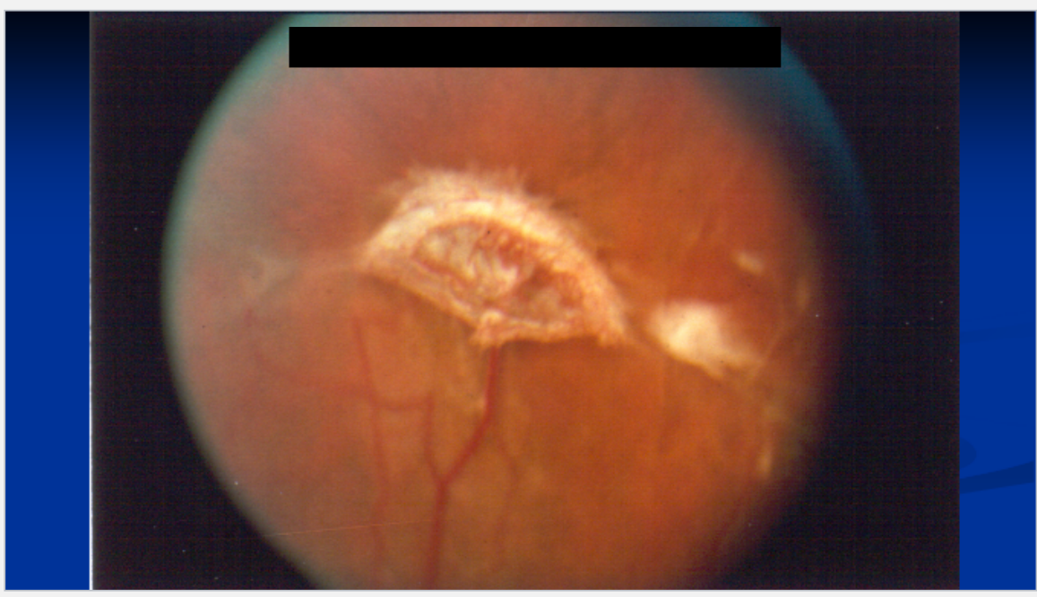
Seafan in SS Sickle cell retinopathy
Describe autoimmune influences on the eye:
Dry eyes
Causes
Symptoms
Tx
Anterior uveitis
Causes
Treatment
List RA Ocular Manifestations
Dry eyes:
Causes: Sarcoidosis, SLE, Rheumatoid arthritis, Thyroid
Healthy pts > 40yrs
Symptoms
Burning, grittiness esp in PM
crusting in AM
Tearing
Treatment:
lubrication
Anterior uveitis
Causes:
Ankylosing spondylitis, Reiter, Behcet
Juvenile RA – esp pauciarticular (asymptomatic)
Treatment:
Close F/U
RHEUMATOID ARTHRITIS: OCULAR MANIFESTATIONS
Dry eyes
Episcleritis
Scleritis
Corneal ulcers
Uveitis

Episcleritis

Episcleritis w/ ulcers forming

sclera so thin from inflammation→ see choroid underneath

Corneal Ulcer
Describe Grave’s effect on the eyes
Signs
Treatment
Graves
Signs
Retraction of upper + lower lids
Upper lid lag in ↓ gaze
Eyelid swelling,
conj vascular congestion
Treatment
Surgery for
severe proptosis,
diplopia 2° EOM involvement,
optic nerve decompression
Radiation/Steroids/Tepezza (Teprotumumab) for inflammatory TED

Thyroid Eye Disease Eyelid Retraction

Thyroid Eye Disease Fibrosis of Inferior Rectus
Describe Sarcoidosis
What is it
Epidemiology
Abnormal Labs
Ocular Involvement
Tx
Sarcoidosis
What is it?
Focal non-caseating granulomas
Epidemiology:
MC African-American females 20 – 40 yrs
Abnormal Labs
↑ Ca++, ↑ACE, abnormal CXR
Ocular involvement
Conj, lacrimal gland → dry eye
Ant/post uveitis
Retinal perivasculitis,
Hemorrhages,
neovascularization
CNS involvement (if retina affected)
Treatment:
Early topical or systemic steroids may prevent complications
Cataract, glaucoma, iris to lens adhesion

Mutton-Fat Keratic Precipitates in Sarcoidosis
Describe AIDS
List the common complications and describe them
Less common complications?
AIDS
Common Complications
AIDS retinopathy
Cotton wool spots
CMV retinitis
Leading cause of visual loss in AIDS
Hemorrhagic necrosis of retina
More common if CD4<100
Kaposi’s sarcoma
Less common Complications
Herpes zoster, simplex, toxoplasmosis
Oculomotor dysfcn 2° CNS involvement

HIV Retinopathy with Roth Spots and Cotton-Wool Spots
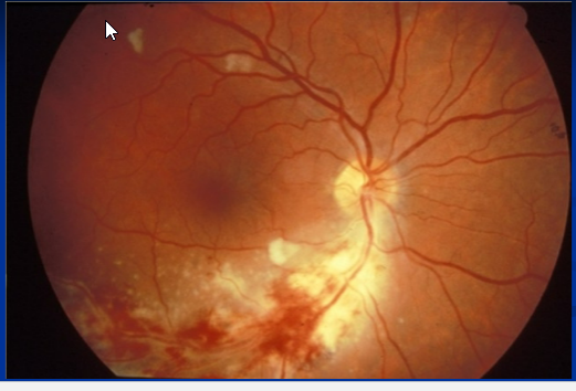
CMV Retinitis in AIDS
Describe Syphilis
Clinical Imp
Primary Complication?
Epi
Manifestations
Secondary Symptoms
Tertiarty?
Syphilis
Clinical Imp:
permanent visual loss if dx and tx are delayed
Primary Complication : Acute interstitial keratitis
Epi:
Age 5 – 25 yrs
Manifestations: Bilateral vs unilateral
Pain + photophobia
Diffusely opaque cornea with ↓ VA
Late – ghost vessels + opacities
Secondary Symptoms:
Pain, redness, photophobia, blurred vision, floaters
Iritis, choroiditis, and/or exudates around disc + vessels
Tertiary Symptoms:
Chorioretinitis
diffuse neuroretinitis and vascular sheathing
Describe Malignancy and the eye:
Primary?
Describe metastasis
MC?
Localization?
Other?
List the Radiation complications
Cornea, Lens, Optic nerve, Retina
List the chemo complication
Malignancy
Primary ocular malignancy
rare
Metastasis
MC: Breast; lung
Usually Localize to choroid
but EOMs, optic nerve can be affected
Lymphoma, leukemia
Radiation complications
Cornea
keratitis / dryness
Lens
cataract
Optic nerve
neuropathy
Retina
vasculopathy
Chemo
Carmustine
In which disease is an early diagnosis crucial in?
What symptoms warrent an immidiantly referral to an eye doc? for him to do what?
What should be considered in any ocular dif. diagnosis
Early diagnosis of diabetic retinopathy is crucial to the ultimate success of treatment, which is effective for both nonproliferative and proliferative retinopathy.
Any patient with an autoimmune disease or systemic infection who presents with a red eye, decreased vision, photophobia, or floaters should be referred to an ophthalmologist for a slit-lamp examination to look for subtle but vision-threatening intraocular inflammation.
TB, Scarcoid and Syphilis should be considered in any ocular differential diagnosis
What can fracture near optic canal cause?
What is a blowout fracture?
What can an ethmoid fracture cause?
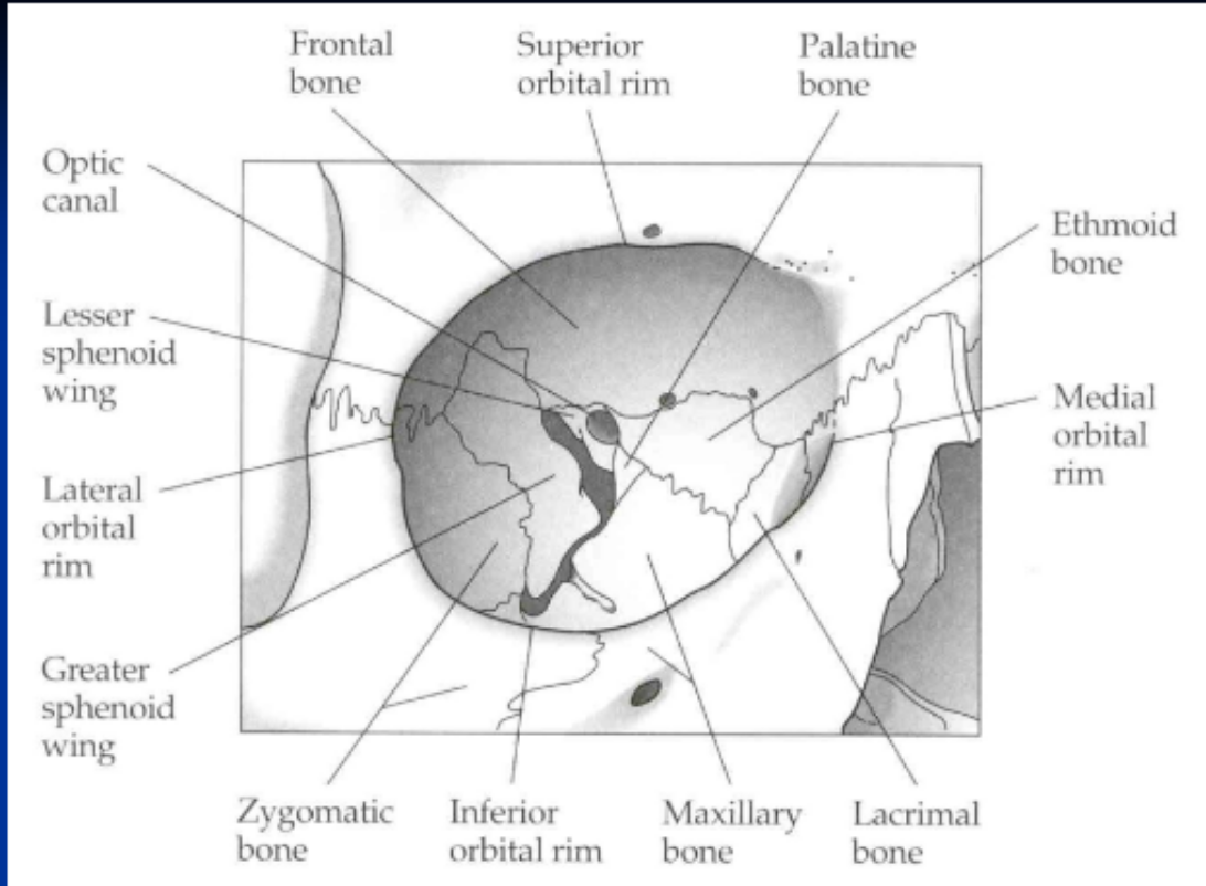
Fracture near optic canal → traumatic optic neuropathy
4% to 11% of pts → globe injury
Blowout fracture:
Orbital floor fracture into maxillary sinus
Ethmoid Fracture:
Very Thin
→ subcutaneous emphysema of the eyelids



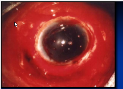
Ruptured Globe
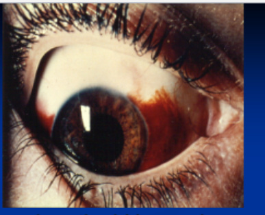
Subconjunctival hemorrhage

Dislocated lens
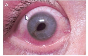
Peaked Pupil (ruptured globe until proven otherwise)
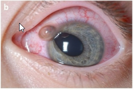
Iris coming out of eyeball
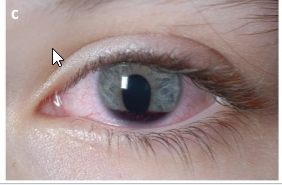
Blood coming from iris; hyphema; ant. chamber
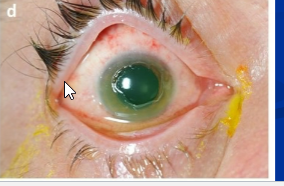
Corneal Abrasion

Retinal sclopataria

Object in eye
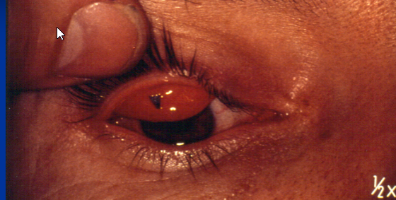
Foreign body under the tarsus

Full thickness eyelid laceration

Morgan Lens

Pressure Patch
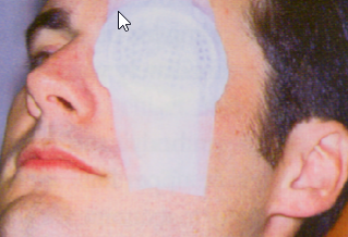
eye shield

subperiosteal abcess

Compartment Syndrome
How treat compartment syndrome in orbit
lateral canthotomy canthelis,

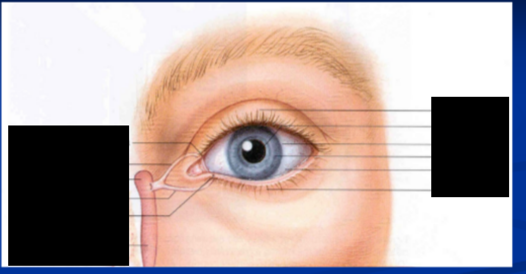


Dog bite eyelid lacerations

Eyelid laceration involving canaliculi

Post-traumatic inability to supraduct or elevate the eye

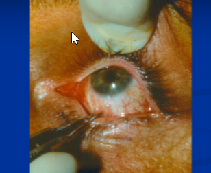
Forced duction tes
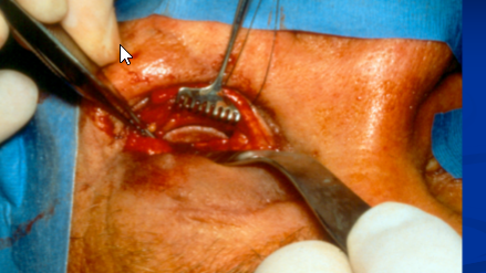
Repair of orbital floor with implant
Which is worst? Acid or Akali Chemical burn; why?
What should you do?
Chemical burn
Alkali worsen than Acid
more rapid penetration
OPHTHALMIC EMERGENCY
ALL chemical burns require immediate and perfuse irrigation to achieve pH of 7,
Then, ophtho referral
List the Urgen and Semi-urgent optho situations
Urgent Situation
Penetrating injuries of the globe
Conjunctival or corneal foreign bodies
Hyphema
Lid laceration
(sutured if not deep and neither the lid margin nor the canaliculi are involved)
Traumatic optic neuropathy
Radiant energy burns
(snow blindness or welder’s burn)
Corneal Abrasion
Semi-urgent Situation
Orbital fracture
Subconjuctival hemorrhage in blunt trauma
Refer patient within 1-2 days


What does a teardrop-shaped pupil and a flat anterior chamber signify?
What do you avoid in a pt w/ suspected perforating injury?
What do you do immediately in a chem injury
A teardrop-shaped pupil and a flat anterior chamber
= perforating ocular injury;
look for uveal tissue where the teardrop pupil is pointing.
Avoid any pressure on the globe in a patient with a suspected perforating injury.
In a chemical injury, irrigate immediately with water