hematology exam 1 - hematopoiesis and erythrocyte formation
 Call Kai
 Learn
Learn Practice Test
 Spaced Repetition
 Match
Match Flashcards
 Knowt Play
1/13

Earn XP
Description and Tags
ucsd extension
Name | Mastery | Learn | Test | Matching | Spaced | Call with Kai | Chat |
|---|
No analytics yet
Send a link to your students to track their progress
14 Terms
describe process of hematopoiesis
process of blood cell production
occurs continuously and regulated
cell renewal, proliferation, differentiation, and maturation
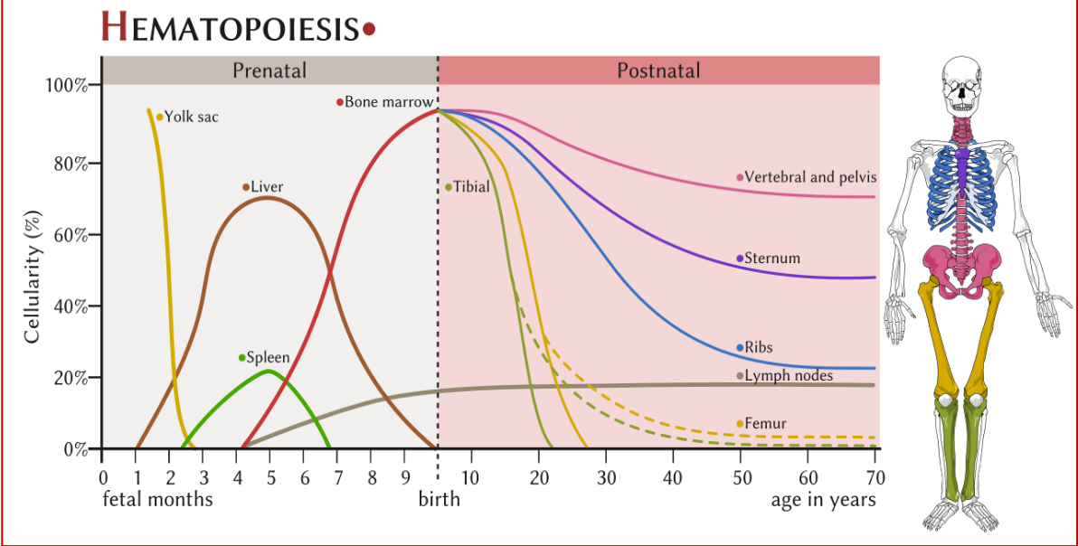
main centers of cell production (hematopoiesis) from fetal development until adulthood
mesoblastic phase
hepatic/liver phase
medullary phase
adult phase
mesoblastic phase
starts around 19 days and occurs b/w 2-10 wks
occurs in the vessels or intravascularly
embryonic hemoglobin is produced
primitive nucleated erythroblasts originate in the embryonic yolk sac lining, or mesoderm from mesenchymal stem cells
hepatic/liver phase
starts at 5-7 wks, occurs between 1-9 months, peaking at 5 months, in between medullary and yolk sac phase until 1-2 wks after birth
nucleated RBCs will migrate from yolk sac to the liver until the fetus reaches 7 months → changes to bone marrow
+ spleen (3-6 months), kidney, thymus, and lymph nodes
recognizable cell clusters or blast being to arise
megakaryocytes begin to be produced
fetal hemoglobin (HbF) present, start of adult hemoglobin (HbA)
medullary phase:
occurs in the bone marrow
begins at 5 months, and by 6 months, bone marrow becomes primary site of hematopoiesis in the fetus
liver and spleen cease hematopoiesis at birth
active sites switch to bone marrow
bone marrow produces all cell types
as well as lymph nodes, spleen, liver, and thymus
provides stable, resourceful environment for proliferation and maturation of blood cells
can find diff stages of maturation of all cell lines during this phase
adult phase (hematopoiesis)
contains cells that are developing: erythroid, myeloid, megakaryocytes, and lymphoid
lymphoid cells will develop in the marrow, spleen, thymus, and lymph nodes
occurs in the red bone marrow or extramedullary (outside of bone marrow)
after age 18, hematopoiesis is primarily occurring in the sternum, ribs, pelvis, and vertebrae
hematopoiesis phases from fetal development to postnatal graph
what are each stages of erythrocyte formation, development, and differentiation
proerythroblast
basophilic erythroblast
polychromatic erythroblast
orthochromatic erythroblast
reticulocyte
mature red cell
proerythroblast
first step in erythrocyte formation (earliest morphologically distinguishable erythroid precursor)
diameter: 20-25 micrometers
nucleus takes up 80% of the cell (high nuclear to cytoplasm ratio); round
stains bluish-purple (basophilic due to RNA)
lacy chromatin
only 1 to 2 nucleoli present
golgi complex may be visible
chromatin is distinct and spread out
ready to divide at 12 hrs
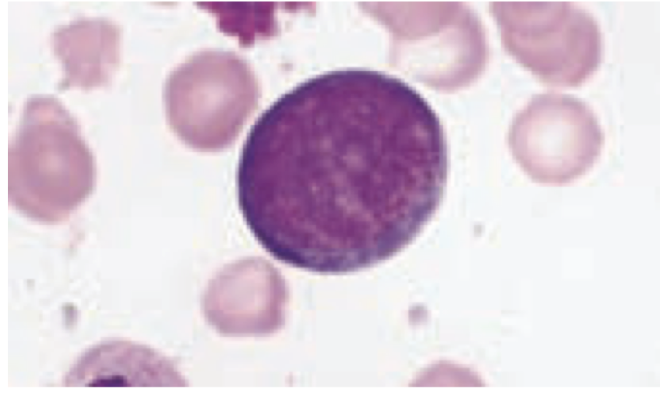
basophilic erythroblast (normoblast)
2nd stage of erythrocyte formation
diameter: 16-18 micrometers (similar to pronormoblast but smaller)
nucleus: 75% of cell
chromatin clumps and becomes coarse
heterochromatin is dark violet, euchromatin is lighter → forms a wheel with spokes appearance
cytoplasm:
blue with pink, basophilic (higher number of ribosomes/RNA)
nucleus is deep purple
require 20 hrs to develop
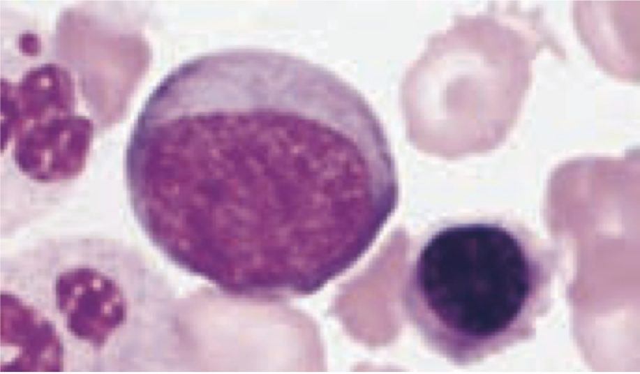
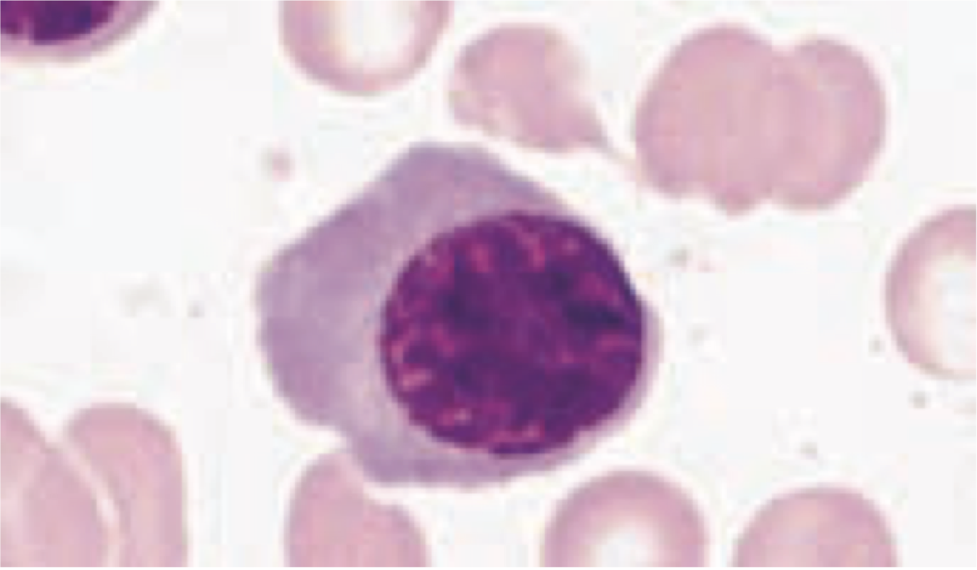
polychromatic erythroblast (normoblast)
3rd stage of erythrocyte formation
diameter: 12-15 micrometers
½ size of mother cell
nucleus:
smaller nucleus and more cytoplasm (chromatin is being condensed)
irregular chromatin and coarsely clumped due to chromatin aggregation
nucleoli are NO LONGER visible
cytoplasm:
abundant gray-blue/pink mixture from Hb and RNA
Hb is apparent in blue cytoplasm
30 hrs to mature
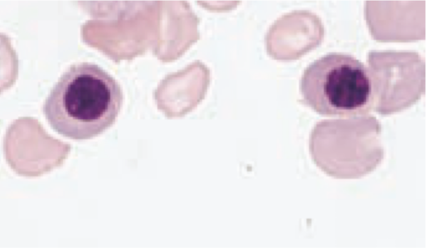
orthochromatic erythroblast (normoblast)
4th stage of erythrocyte formation
diameter: 10-15 micrometers
nucleus:
low nucleus:cell ratio
solid, blue-black nucleus that begins to degenerate that contains nonlinear clumped chromatin
nuclear chromatin is heavily condensed
at the end of this stage, nucleus is fragmented/structureless
phase contrast microscopy: can observe cytoplasmic projections protracting and retracting
preparing for nucleus ejection
cytoplasm:
after final mitotic division, concentration of hemoglobin increases within → pink/salmon color, but slight tinge of blue
not able to divide at this stage or moving forward bc the cell can’t perform DNA synthesis
maturation time: 48 hrs
reticulocytes
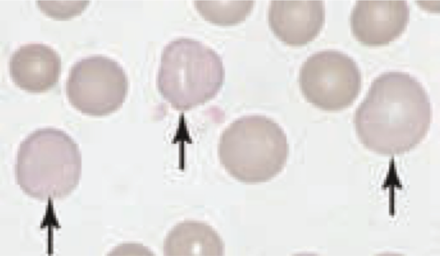
5th stage of erythrocyte formation
diameter: 7-10 micrometers
cytoplasm:
blueish tint and slightly larger than RBCs
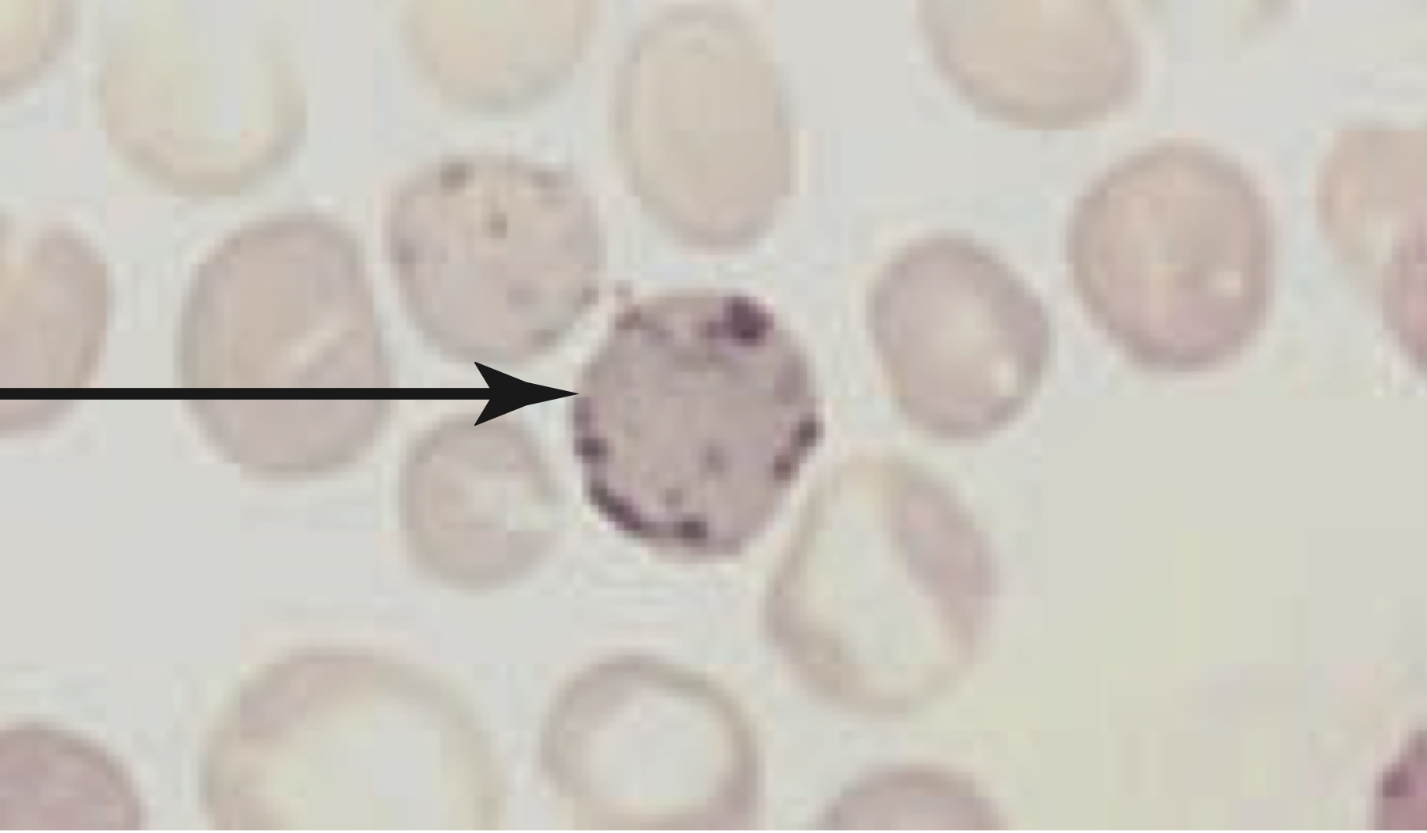
contain residual RNA but no nucleus → bluish tinge w/ Romanowsky stains (supravital stains - new methylene blue-N)
when nucleus is first ejected, the cell has irregular lobe shape
when remaining 20% of cell’s Hb is made
react w/ these stains to produce precipitation of RNA + mitochondria
supravital stains = purplish-blue inclusions spread out
new methylene blue N
brilliant cresyl blue
released in 2-3 days and circulates for 1-2 before maturing
ribosomes disappear as cell matures to carry more Hb
granular filamentous ribosomes
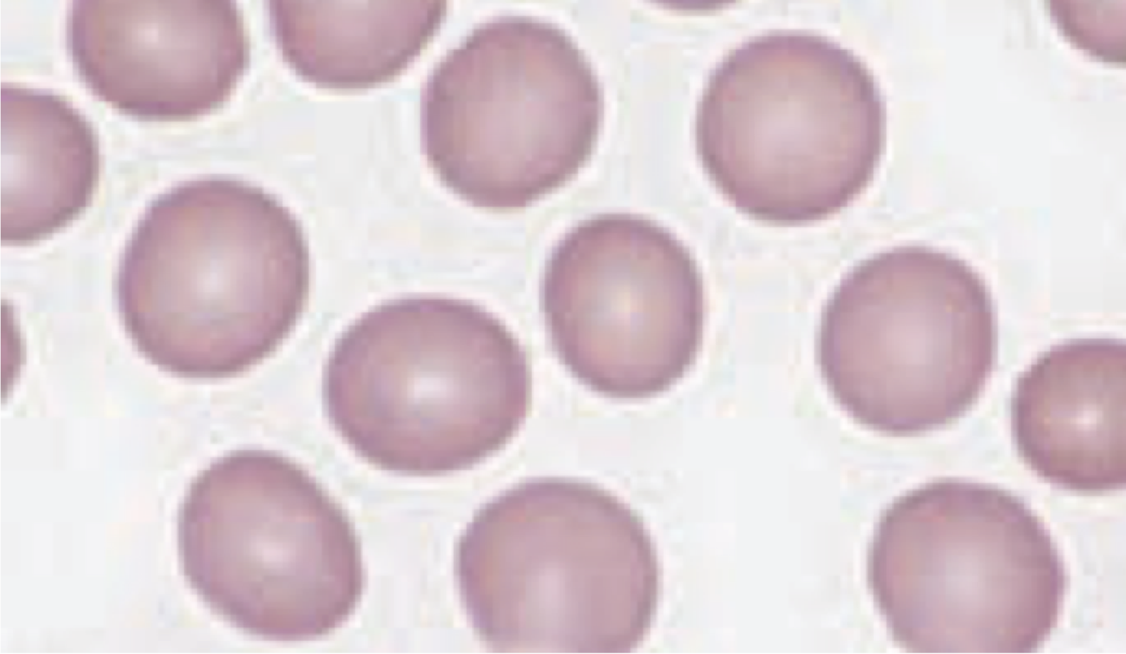
mature red cell
6th stage of erythrocyte formation
diameter: 7-8 micrometers
volume: 80-100 fL
bioconcave disc
stains pink to orange bc of Hb (acidophilic)
do NOT have cellular organelles and enzymes to synthesized new lipids/proteins
can’t be repaired
can’t synthesize Hb
carries O2 from lungs and CO2 back
flexible to allow for passage throughout the body