Lecture 16 - DNA Replication
 Call Kai
 Learn
Learn Practice Test
 Spaced Repetition
 Match
Match Flashcards
 Knowt Play
1/48

There's no tags or description
Looks like no tags are added yet.
Name | Mastery | Learn | Test | Matching | Spaced | Call with Kai | Chat |
|---|
No analytics yet
Send a link to your students to track their progress
49 Terms
Griffith experiment set up
Griffith's experiment with Streptococcus pneumoniae in 1928 was one of the first experiments to identify a "heritable factor". There were two strains of the bacteria: a virulent "S" (smooth) strain that causes disease and a non-virulent "R" (rough) strain. Griffith analyzed what accounted for bacterial pathogenesis. He conducted experiments where he injected mice with different forms of the bacteria. He found that:
•
Living R strain did not cause disease.
•
Living S strain did cause disease.
•
Heat-killed S strain did not cause disease.
•
However, a mixture of heat-killed S strain and living R strain did cause disease and the ability to kill was inherited by the descendants of the "transformed" bacteria.
Griffith experiment answer
Griffith concluded that the R bacteria had been "transformed" into virulent S bacteria by some heritable substance. He termed this heritable component the "transforming principle". This experiment showed that there was a heritable component that could change the properties of a cell, but it did not identify the chemical nature of this component.
Avery and mcleod
Avery, McCarty, and MacLeod sought to identify the chemical nature of Griffith's "transforming principle". They prepared soluble extracts from heat-killed S bacteria and treated these extracts with enzymes that destroyed protein, DNA, lipid, or polysaccharide. They then asked which treatment would destroy the transformation of R bacteria into S bacteria. Their results showed that bacterial transformation was lost when DNA was destroyed by nucleases. Their conclusion was that the DNA of the S cell, and not protein, was responsible for the heritable changes during bacterial transformation. This experiment provided strong evidence that DNA was the genetic material.
Nucleotides
Nucleotides are the building blocks of nucleic acids. A nucleotide consists of a sugar (deoxyribose in DNA, ribose in RNA), a phosphate group, and a nitrogenous base (adenine (A), guanine (G), cytosine (C), and thymine (T) in DNA; uracil (U) replaces thymine in RNA).
•
In both deoxyribose and ribose sugars, the carbons are numbered 1' through 5'.
1’ carbon
◦
The 1' carbon is where the nitrogenous base is attached. The specific base determines whether the nucleotide contains adenine, guanine, cytosine, thymine, or uracil. This base is crucial for storing genetic information through its sequence and for complementary base pairing between DNA strands (A with T, and C with G).
2’ carbon
The 2' carbon distinguishes between DNA and RNA. Deoxyribose, found in DNA, has a hydrogen atom (-H)attached to the 2' carbon. Ribose, found in RNA, has a hydroxyl group (-OH) attached to the 2' carbon. This difference affects the stability of the nucleic acid. DNA is more stable than RNA due to the absence of the hydroxyl group on the 2' carbon.
3’
◦
The 3' carbon has a hydroxyl group (-OH) that is involved in forming the phosphodiester bond with the phosphate group of the next nucleotide in the chain. This bond creates the sugar-phosphate backbone of the DNA and RNA strands. The 3' OH end is essential for DNA polymerase to add new nucleotides during DNA replication. DNA polymerase can only add nucleotides to the 3' end of a pre-existing strand.
5’ Carbon
The 5' carbon is where the phosphate group is attached. The phosphate group of one nucleotide forms a phosphodiester bond with the 3' hydroxyl group of the preceding nucleotide. A DNA or RNA strand has a 5' end with a free phosphate group or a nucleotide linked to it, and a 3' end with a free hydroxyl group. This directionality, or polarity, is crucial for processes like DNA replication, which proceeds in the 5' to 3' direction.
DNA helix formation
DNA is a double-stranded helix formed by complementary base pairing of nucleotides. The two strands run in opposite orientations, meaning they are antiparallel. The sugar-phosphate backbone is on the outside of the helix, and the nitrogenous bases are on the inside, held together by hydrogen bonds (two between A and T, and three between G and C). The uniform diameter of the helix is maintained by purine pairing with pyrimidine.
dna replication - initiation
Initiation: Replication begins at specific sites called origins of replication, which are often A/T rich. The number of origins varies among species. In eukaryotes, there are multiple origins to allow for faster replication of long, linear chromosomes.
unwinding
Unwinding: At the origin, DNA helicase binds to each strand of the double helix and breaks the hydrogen bonds between the two strands, separating them and opening up a replication "bubble". This creates two replication forks where the DNA strands are actively unwinding.
stabilization
Stabilization: Single-strand DNA binding proteins bind to the separated DNA strands to prevent them from rejoining.
relieving strain
4.
Relieving Strain: Topoisomerase binds to the double helix ahead of the replication fork and relieves the torsional strain that is placed on the double helix as it unravels.
priming
Priming: DNA polymerase cannot initiate DNA synthesis on its own; it requires a primer. Primase, a DNA-directed RNA polymerase, synthesizes a short, complementary RNA primer (about 10 nucleotides) using the parental DNA as a template. The primer has a free 3' OH end, which is necessary for DNA polymerase to begin synthesis.
elongation
6.
Elongation: DNA polymerase catalyzes the addition of deoxyribonucleotides (dNTPs) to the 3' OH end of the primer, extending the new DNA strand in the 5' to 3' direction. The incoming dNTP base pairs with the template strand, and DNA polymerase catalyzes the formation of a phosphodiester bond. The energy for this reaction comes from the hydrolysis of two phosphate groups from the dNTP.
leading strand synthesis
7.
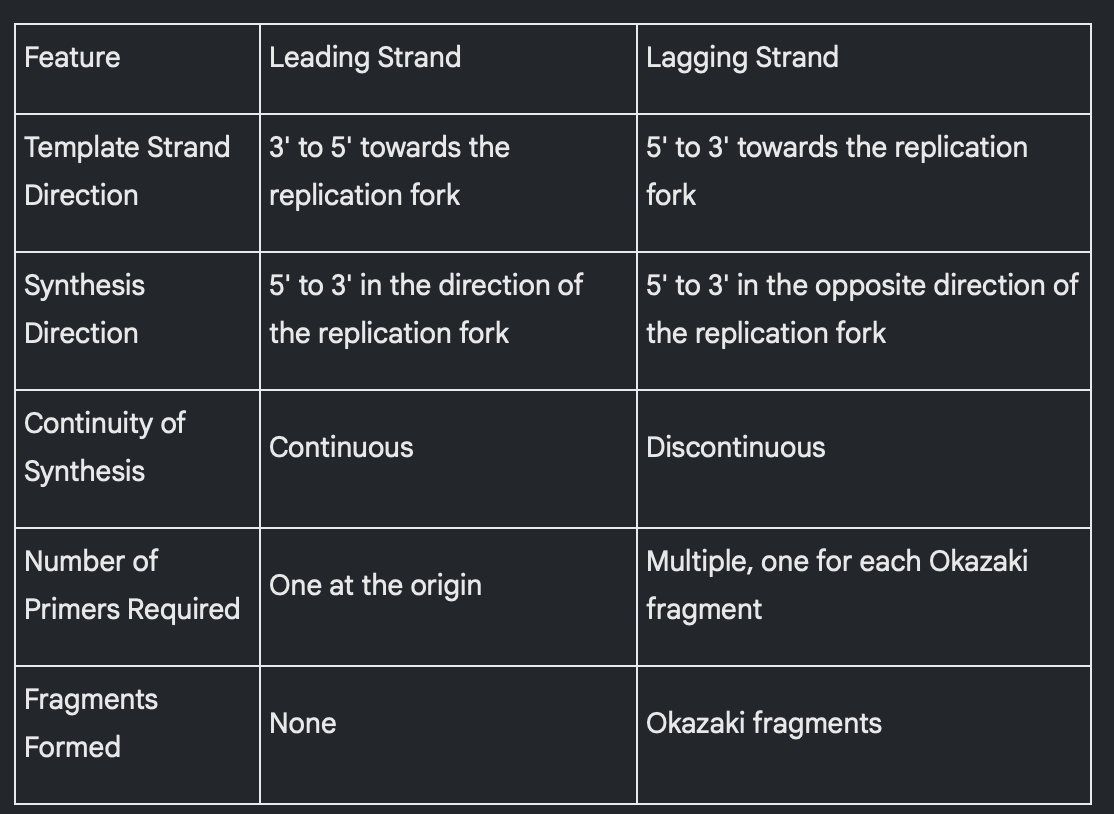
Leading Strand Synthesis: One of the parental strands runs in the 3' to 5' direction towards the replication fork. The new strand synthesized along this template is called the leading strand. DNA polymerase synthesizes the leading strand continuously in the direction of the moving replication fork, requiring only one initial primer at the origin of replication.
lagging strand synthesis
8.
Lagging Strand Synthesis: The other parental strand runs in the 5' to 3' direction towards the replication fork. The new strand synthesized along this template is called the lagging strand. Because DNA polymerase can only synthesize in the 5' to 3' direction, and this direction is away from the replication fork, the lagging strand is synthesized discontinuously as a series of short segments called Okazaki fragments. Each Okazaki fragment requires a new RNA primer.
rna primer removal and gap filling
RNA Primer Removal and Gap Filling: Once the Okazaki fragments are synthesized, the RNA primers are removed, typically by an exonuclease. The resulting gaps are then filled with DNA by DNA polymerase.
joining of fragments
0.
Joining of Fragments: Finally, DNA ligase catalyzes the formation of phosphodiester bonds to join the Okazaki fragments together, creating a continuous DNA strand.
dna replication illustration
The origin of replication with separated DNA strands forming replication forks.
•
The leading strand being synthesized continuously in the 5' to 3' direction towards the fork, starting from a single primer.
•
The lagging strand being synthesized discontinuously in the 5' to 3' direction away from the fork, showing multiple RNA primers and short Okazaki fragments.
•
The roles of helicase in unwinding, single-stranded binding proteins in stabilizing, primase in synthesizing RNA primers, and DNA polymerase in elongating the new strands.
•
The eventual removal of RNA primers and the joining of Okazaki fragments by ligase.
comparison of leading and lagging strands
dna polymerase
DNA Polymerase: This enzyme catalyzes the addition of dNTPs to the 3' OH end of a pre-existing strand (either DNA or RNA primer), synthesizing new DNA in the 5' to 3' direction. It also has a 3' to 5' exonuclease "proofreading" activity to detect and remove mismatched nucleotides.
primase
Primase: This is a DNA-directed RNA polymerase that synthesizes short RNA primers using the parental DNA as a template. These primers provide the necessary 3' OH end for DNA polymerase to start DNA synthesis. Primase does not need a primer to initiate RNA synthesis.
Okazaki fragments
Okazaki Fragments: These are the short segments of DNA synthesized on the lagging strand during DNA replication. They are formed because DNA polymerase can only synthesize in the 5' to 3' direction, which is away from the overall direction of lagging strand synthesis at the replication fork. These fragments are later joined together by DNA ligase.
helicase
Helicase: This enzyme binds to each strand of the double helix at the replication fork and breaks the hydrogen bonds between the two strands, unwinding the DNA and separating them.
single stranded binding proteins
Single-stranded binding proteins: These proteins bind to the separated single-stranded DNA and prevent them from rejoining or forming secondary structures that would impede DNA replication.
ligase
Ligase: This enzyme catalyzes the formation of phosphodiester bonds to join the sugar-phosphate backbones of DNA fragments. Specifically, it joins the Okazaki fragments on the lagging strand and also joins the newly synthesized DNA to any replaced segments where RNA primers were removed.
structure of telomeres
•
Structure: Telomeres are specialized DNA sequences located at the ends of eukaryotic chromosomes. In humans, these ends consist of many repeats of the sequence 5’ –GGGTTA-3’. The length of the telomere sequence can vary among species but can be up to 10,000 nucleotides long. There are no genes located at the ends of chromosomes within the telomeric regions.
function of telomeres
•
Function: Telomeres protect the ends of linear chromosomes from being degraded and prevent them from fusing with other chromosomes. They also solve the "end" problem faced by linear DNA during replication. During lagging strand synthesis, after the last RNA primer is removed from the 5' end, DNA polymerase cannot fill in the resulting gap because there is no upstream 3' OH group to add to. This leads to a shortened 5' end after each round of replication, which could potentially result in gene loss. Telomeres, being repetitive non-coding sequences, act as buffers to prevent the shortening from affecting essential genes.
enzyme telomerase
The enzyme telomerase "repairs" the ends of DNA in some cells. Telomerase is typically active in embryonic cells and highly proliferative cells (like stem cells) in adults. It carries its own RNA template that it uses to extend the 3' end of the lagging strand template, allowing DNA polymerase to then complete the synthesis of the lagging strand. Interestingly, high levels of telomerase activity are observed in 90% of human cancers, leading to the hypothesis that telomerase inhibitors might be potential cancer therapeutics.
anti parallel
•
Anti-parallel: This describes the orientation of the two strands of a DNA molecule, which run in opposite directions. One strand runs from the 5' end to the 3' end, while the other runs from the 3' end to the 5' end. This anti-parallel arrangement is crucial for the double helix structure and the mechanism of DNA replication.
double stranded
•
Double stranded: A DNA molecule is typically composed of two strands of nucleotides that are held together by hydrogen bonds between their bases. This double-stranded structure, along with the complementary base pairing, provides a mechanism for DNA to be replicated.
single stranded
Single stranded: During DNA replication, the double helix unwinds, creating regions of single-stranded DNA that serve as templates for the synthesis of new DNA strands. Also, RNA primers, which initiate DNA synthesis, are single-stranded. Single-strand DNA binding proteins bind to these separated strands to prevent them from rejoining.
5’ end of dna or rna
5’ end of DNA or RNA: This refers to one end of a nucleic acid strand where the 5' carbon of the sugar molecule is attached to a phosphate group.
3’ end of dna or na
3’ end of DNA or RNA: This refers to the other end of a nucleic acid strand where the 3' carbon of the sugar molecule has a free hydroxyl (OH) group. DNA polymerase can only add new nucleotides to this 3' OH end of a pre-existing strand or primer.
complementary
Complementary: The two strands of DNA are complementary because of specific base pairing rules. Adenine (A) always pairs with thymine (T) via two hydrogen bonds, and guanine (G) always pairs with cytosine (C) via three hydrogen bonds. This complementary base pairing is essential for accurate DNA replication, as each strand serves as a template to create a new, complementary daughter strand.
base pairing
•
Base pairing: This refers to the specific hydrogen bonding between the nitrogenous bases in DNA: adenine (A) with thymine (T), and guanine (G) with cytosine (C). Chargaff's rule also highlights this principle, stating that the amount of A equals the amount of T, and the amount of G equals the amount of C in a DNA molecule.
template strand
•
Template strand, Daughter strand: During DNA replication, each of the original DNA strands acts as a template strand for the synthesis of a new, daughter strand. The sequence of the daughter strand is determined by the complementary base pairing with the template strand, resulting in two DNA molecules, each consisting of one old (template) and one new (daughter) strand (semi-conservative replication).
polarity
Polarity: This refers to the inherent directionality of a DNA or RNA strand due to the orientation of the sugar-phosphate backbone, defined by the 5' end and the 3' end. The anti-parallel arrangement of the two DNA strands reflects their opposite polarity.
origin of replication
Origin of replication: These are specific sites on a DNA molecule where DNA replication begins. At these origins, the two DNA strands separate, forming a replication "bubble" with two replication forks moving in opposite directions (bidirectional replication). Eukaryotic chromosomes have multiple origins of replication to speed up the replication of their long, linear DNA.
leading strand lagging strand
Leading strand, Lagging strand: Because DNA polymerase can only synthesize DNA in the 5' to 3' direction, the two new strands are synthesized differently at a replication fork. The leading strand is synthesized continuously in the direction of the replication fork movement, requiring only one primer. The lagging strand, on the other hand, is synthesized discontinuously in the opposite direction of the fork movement, as a series of short segments called Okazaki fragments, each requiring a new primer.
primer
•
Primer: DNA polymerase cannot initiate DNA synthesis on its own; it requires a short, pre-existing strand with a free 3' OH end to add nucleotides to. This short strand is called a primer, and in DNA replication, it is a short RNA sequence synthesized by the enzyme primase. The RNA primers are later removed and replaced with DNA.
dna polymerase
•
DNA polymerase: This is a key enzyme that catalyzes the synthesis of new DNA strands by adding nucleotides to the 3' OH end of a pre-existing strand or primer. It adds each deoxyribonucleotide triphosphate (dNTP) that base pairs with the template strand, forming a phosphodiester bond. DNA polymerase also has a 3' to 5' exonuclease “proofreading” activity to correct mismatches. Different types of DNA polymerases exist with specialized roles.
primase
•
Primase: This is an RNA polymerase that synthesizes short RNA primers using the parental DNA as a template. It does not require a primer itself. These RNA primers provide the 3' OH end necessary for DNA polymerase to start synthesizing a new DNA strand.
ligase
•
Ligase: This enzyme joins the Okazaki fragments of the lagging strand together by catalyzing the formation of phosphodiester bonds between them. It also seals any remaining gaps in the DNA after RNA primers are replaced with DNA.
helicase
•
Helicase: This enzyme unwinds the double helix at the origin of replication by breaking the hydrogen bonds between the two DNA strands, forming a replication fork.
single strand binding protein
•
Single strand binding protein: These proteins bind to the separated single strands of DNA near the replication fork, preventing them from re-annealing or forming secondary structures.
telomere
•
Telomere: These are specialized DNA sequences located at the ends of eukaryotic chromosomes that protect them from degradation and prevent the loss of genes during DNA replication. In humans, telomeres consist of many repeats of the sequence 5’–GGGTTA-3’.
telomerase
•
Telomerase: This is an enzyme that "repairs" the ends of DNA in some cells, particularly embryonic cells and highly proliferative cells, by adding telomeric repeats. It carries its own RNA template that is complementary to the telomere repeat sequence. High telomerase activity is also found in many cancer cells.