Pediatric Module on Oral Health: Key Terms and Definitions
 Call Kai
 Learn
Learn Practice Test
 Spaced Repetition
 Match
Match Flashcards
 Knowt Play
1/72

There's no tags or description
Looks like no tags are added yet.
Name | Mastery | Learn | Test | Matching | Spaced | Call with Kai | Chat |
|---|
No analytics yet
Send a link to your students to track their progress
73 Terms
Infancy through toddlerhood (0-3 years)
Emotions undiscerned
• Behavior changes - crying
• Separation anxiety
• Second year - language skills
• Third year - NO! Why?
An initial oral evaluation visit should occur within 6 months of eruption of the first primary tooth and no later than 12 months of age.
Preschool (3-6 years)
Developing:
• Process of self control
• Frustrations
• Fears
• Power of reasoning
• Attention Span:
• 3-year-old: 4-8 minutes
• 4-year-old: 8-12 minutes
• 5-year-old: 12-28 minutes
School Age (6-12 years)
Literate
Body image / establish self-identity
Ability of independence
Influence of peers in school
Why are primary teeth important?
Help children speak clearly.
Help children chew naturally and eat.
Space holder for permanent teeth (maintain arch length).
Guide path for permanent teeth eruption.
Severe early childhood caries
1) Any sign of smooth-surface caries in a child < 3 years of age.
OR
2) From ages 3-5 years, 1 or more cavitated, missing
(due to caries), or filled smooth surfaces in primary maxillary anterior teeth.
OR
3) A decayed, missing, or filled score ≥ 4 (age three), ≥ 5 (age four), or ≥ 6 (age five).
Comprehensive clinical examination
Components of a comprehensive clinical examination include:
• general health/growth assessment (e.g., height, weight, BMI calculation, vital signs);
pain assessment;
extraoral soft tissues examination;
temporomandibular joint assessment;
intraoral soft tissues examination;
oral hygiene and periodontal-risk assessment;
intraoral hard tissue examination;
assessment of the developing occlusion;
radiographic assessment, if indicated;
caries-risk assessment; and
assessment of cooperative potential/behavior of child.
facial symmetry
facial profile
positions of maxilla/mandible
vertical facial relationships
airway assessment
arch form
symmetry
spacing
crowding
presence/absence of teeth
midline discrepancies or posterior crossbones
overbite/overjet
Facial profile
Class I: slightly convex
Class II: truly convex
Class III: straight or concave
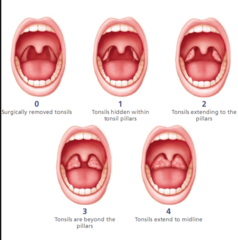
Mallampati score

Brodsky classification system
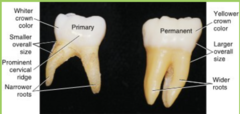
Deciduous teeth

Primary teeth morphology
Crowns are smaller and more bulbous
Greater cervical constriction
Narrow occlusal table with anatomy not well defined
Thinner enamel & dentin
Larger pulp chamber and higher pulp horns
Enamel rods in cervical area directed occlusally
Broad and flat proximal contacts
Whiter in color
Primary teeth eruption sequence
calcification starts in utero
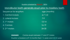
Permanent teeth eruption sequence
Maxilla: 61245378 Mandible: 61234578
6 months - First primary tooth
• 3 years - Complete primary teeth eruption
• 6 years - Permanent lower incisors and permanent first molars • 6-12 years - Mixed dentition
• 6-9 years - Early Mixed Dentition
• 10-12 years - Late Mixed Dentition
• 12 years - Permanent second molars

Radiograph by caries risk (0-5 years old)
Primary dentition: 2 BW, anterior occlusal, PAs as needed

Radiograph by caries risk (> 6 y/o)
Mixed dentition:
2-4 BW
PAs as needed
Panoramic

Anterior PA
caries/pulpal pathology
evaluate previous treatment
trauma, follow-up
evaluate resorption of primary teeth and development of permanent teeth
detect dental anomalies
use a size 2 sensor
lay the sensor flat and have patient gently bite down on the sensor between their teeth (horizontal sensor)
Early childhood caries
≥1 dmft (noncavitated or cavitated) in
children ≤71 months (
Main caries causing bacteria
Mutans streptococci
use sucrose to make extracellular polysaccharides stick the plaque together
Lactobacilli
produce predominantly lactic acid from fermentable carbohydrates
Bacteria transmission
Children can acquire cariogenic bacteria from anyone who is in close contact with them.
From birth to 3 year old - the critical time for caries causing bacterial transmission but can last life-long
Children with early cariogenic bacteria colonization before 4 years are four times more likely to get caries than those don't or get later
The transmission of the bacteria - saliva-sharing activity
Special Health Care Needs oral health conditions
Dental caries
o Orally defensive/behavior problems, diet, brushing - autism
o Sweetened medication and dry mouth
Enamel hypoplasia - young cancer patients, infancy infections
Calculus build-up/periodontal disease - G-tube fed
Dental crowding/malocclusion/dental anomalies
Bruxism and wear facets
Fracture of teeth or trauma - seizure
Poor cooperation
High risk to dental caries and periodontal diseases
Limited access to dental careAdvanced behavior management tools such as general anesthesia is
need to treat if advanced diseases developed (dental caries and
excessive dental calculus build up).
High risk for undergoing general anesthesia.
Anti-caries therapy for children
1. Current antimicrobial treatment: Broad spectrum antibiotics: PVPI, SDF(in-office)
2.Natural sugar substitute: xylitol, erythritol etc. Inhibits MS transmission and colonization
Xylitol 2g 3-4 times/day
¡ Wipes or syrup for infants
¡ Mints/candy/lolipops for children >3 yrs old
¡ Gums for children >6 yrs old
3. Modify oral and plaque pH environment:
Sodium bicarbonate (baking soda)
¡ Add to toothpaste
Wipeteeth (1/4TSP in 1OZ of water/rinse)
High and extreme risk
Pediatric patients with one (or more) white spot and/or cavitated lesion(s) are high- risk patients.
POE every 6 month
CRA every 3-4 months or at each operative treatment
Fluoride varnish every 3 month
Active caries found à Caries arrest/control: Silver
diamine fluoride/ GIC for cavitated caries lesions
Sealants for pits/fissure at risk
Pediatric patients with one (or more) cavitated lesion(s) and hyposalivary or special needs are extreme-risk patients.
Sealants
Up to 90% of caries in school-aged children occurs in pits and fissures
The teeth at highest risk are permanent first and second molars
Fluoride has least preventive effect on the pits and fissures
Fuji Triage sealants
Fuji Triage Sealants
Not as strict isolation requirement
Especially useful for:
ü Patient with behavior
management problems
ü Patient with special care needs
ü Partially erupted molars with high caries risk (enamel hypoplasia)
Piaget's 4 stages of cognitive development for children
Sensorimotor Stage (0-3yrs)
§Separation anxiety §Egocentric
Preoperational Stage (3-6yrs)
§Limited ability to
reasoning
§Love pretend play
Concrete Operational Stage (7-11yrs) Understand some logic and reasoning
Formal Operational Stage (12+yrs)
Abstract reasoning, hypotheticals
Sensorimotor stage
Emotional/Cognitive Development
§Build basic trust with parents and separation anxiety §Start to build simple concepts but limited language ability
§Fear of unfamiliar objects/noise
üKnee-to-knee exam or laying on parent in chair
üBest to have parents present
üBe calm, playful, positive and effective üPre-cooperative (
Preoperational stage (3-7 years)
Emotional/cognitive development
Curious to learn new things and start to express themselves
Understand/loves fantasy world & pretend play
Teachable, improved but limited ability to reasoning
React dramatically to real or perceived fears
✓ Require firm but kind handling
✓ Make fun and pretend play can be effective
✓ Allow child to have a sense of accomplishment from
successful appointment - positive reinforcement
Concrete operational stage (7-11 years)
Emotional/cognitive development
Expose to education and social development in schoo
l Decreased parental influence and increasing influence from peers
Develop abstract concepts and logical thinking/reasoning
✓ Motivate patient with reasoning
✓ Educate with fantasy, facts and respects
✓ Motivate patient using competition as reward
AAPD Behavior guidance techniques
non-verbal comunication
tell-show do
positive/negative reinforcement
positive and smart commands
effective communication
distraction
involve parents in the process
voice control/modulation
desensitization
Protective stabilization
Indications:
- Quick procedure: exam, extraction
- Autism/Developmental delay
- Sedated
- NOT just because parents want it
• Mandatory documentations
- Informed consent
- Indication and type of restraint
- Duration of restraint
- Behavior evaluation/rating during stabilization
¡ A patient cannot be restrained safely because of underlying medical or systemic problems
¡ Check tightness and duration of stabilization at regular intervals
¡ Extremities or the chest must not actively restrict circulation or respiration
¡ For patient's safety, terminated ASAP when a patient is experiencing severe stress
Why do we treat primary teeth?
1. Prevent odotogenic pain
2. Prevent infection
3. Preserve the space for the adult tooth (space loss)
Space maintenance of primary anterior teeth
The canines serve as an anchor
There is non-significant space loss after loss of primary anterior teeth
Space maintenance: One primary molar, between two teeth (in a quadrant)
can happen in primary or mixed dentition
Unilateral band/loop space maintainer
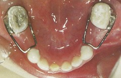
Space maintenance: One primary molar, the most distal tooth in the quadrant
Primary dentition only
Unilateral distal shoe space maintainer
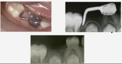
Space maintenance: Two primary molars, adjacent to each other in the quadrant
Primary and mixed dentition
mandibular: lower lingual holding arch
maxillary: nance, transpalatal arch

Odontogenic Infection in Primary Teeth
Localized:
Pain
Swelling
Redness
Parulis
Sinus tract
Percussion sensitivity
Systemic
Fever
Lymphadenopathy
Malaise
Elevated white blood cell count
Treating odontogenic infection
Extraction is often the best treatment, but primary tooth pulpectomy or
antibiotic therapy is indicated in certain clinical situations.
Antibiotic treatment is not indicated:
- If tooth is extracted
- If dental infection is contained within the pulpal tissue or the immediate
surrounding tissue
Antibiotic Therapy for odontogenic infection
Antibiotic therapy is indicated:
- patient has a concurrent systemic medical problem
- patient has cellulitis, spreading of infection from localized area
- Systemic manifestations (e.g. fever)
Rapidly increasing signs of infection
- Fever and chills, malaise, fatigue, weakness
Involvement of anatomical danger zones
Large or rapidly progressing abscess/cellulitis
Factors to consider :
- Severity of infection
- Medical status of child
- Ability to obtain adequate anesthesia
- Feasibility for treatment
Antibiotics
Bactericidal (killing potential)
Penicillin
Amoxicillin
Augmentin
C lindamycin @ high dose
Bacteriostatic (growth inhibiting)
Clindamycin
Erythromycin
Tetracycline
Cellulitis
Definition: "A painful swelling of the soft tissue of the mouth and face resulting from a diffuse spreading of
purulent exudate along the fascial planes that separate
the muscle bundles." (Sapp)
Patient may be febrile with red and/or swollen face
Child appears acutely ill
Treatment
- extraction of the infected tooth
- antibiotic therapy
- Hospitalization if signs and symptoms indicate
serious systemic infection
- IV antibiotics may be indicated!
Indications for extractions
Extensive caries & unrestorable tooth
- Apical periodontitis
- Abscess
- Fractured root/ crowns
- Over-retained primary teeth
- Supernumerary teeth
Assessing trauma
medical history
¡Allergies to medications
Bleeding disorders e.g. Hemophilia, Von Willebrand ¡Cardiac disease (requires SBE prophylaxis?) ¡Current medications
Neurological disorders e.g. seizures
Status of tetanus prophylaxis
do no harm
Can the patient tell you...
What is your name?
What place is this?
Is it morning, afternoon, or night time?
Always ask questions sensitive to the child's age and cognitive development
Neurological assessment of a child with head trauma
signs of neurological issue:
Nausea
Vomiting
Drowsiness
Blurred vision
Loss of consciousness
Headache or neck ache
Blood or clear fluid from the nose (rhinorrhea) or ears (otorrhea)
Numbness
Amnesia
Was there LOC?¡Head exam: laceration, hematoma, skull fracture ¡Temperature, pulse, RR, BP
¡CN III: are pupils equal, round, responsive to light and accommodate (PERRLA)
¡CN III, IV, VI: extraocular movements intact (EOMI)
¡CN VII (Facial): close eyes, smile, frown, look for symmetry
Tetanus
Typically part of the DTAP (diphtheria-tetanus- pertussis) immunization series completed at age 5
¡Booster of tetanus toxoid at 11-12 years and every 10 years after.
¡If a child sustains a "dirty wound", the booster should be administered if it has been >5 years since his/her last booster.
EO exam for trauma
Neurologic concern
Blood or clear fluid from the nose (rhinorrhea) or ears (otorrhea) §Facial asymmetry
Hemorrhage
Wounds
Bruising (ie Battle sign) ¡Deviated/limited opening ¡Palpation of facial skeleton ¡ROM of mandible
¡ Soft tissue injury § Contusion
Abrasion
§ Laceration
IO exam for trauma
Crown fracture
¡ Pulp exposure
¡ Displacement
¡ Disturbance in
occlusion
¡ Tooth mobility
¡ Percussion
¡ Pulp vitality
Type of fractures
Uncomplicated crown fracture ¡Enamel
¡Enamel + Dentin
¡Treatment: Restore
Complicated crown fracture
¡Enamel, Dentin Fracture with Pulp Exposure
¡Treatment: Pulp treatment or extraction
¡ Root fracture ¡Monitor or Extraction
¡Alveolar fracture
Classification of dental trauma
Concussion (jolt, but no mobility, tender but appear normal)
monitor
Subluxation (trauma to teeth with class I, II mobility, but no displacement from the socket, sulcular bleeding)
monitor
Extrusion (extrude out of tooth socket, elongated crown and mobile)
Primary: extraction
Permanent: reposition/splint
Intrusion (intrude inward, into the tooth socket, shorten crown)
primary: monitor for future spontaneous re-eruption
permanent: re-eruption, surgical reposition/splint, ortho later
Luxation (lateral, facial, and lingual displacement)
primary: ext if severe
permanent: reposition/splint
Avulsion (completely out of socket)
primary: discard tooth
permanent: replant and splint
Tooth fracture:
-Uncomplicated fracture (only enamel or dentin exposure)
-Complicated fracture (pulpal tissue exposure).
Traumatized primary tooth consequences
¡Discoloration
§Yellow (calcific metamorphosis)
§Grey (hemosiderin deposit)
¡PCO (pulp canal obliteration) ¡Pulpal necrosis with abscess ¡Early exfoliation
¡Internal resorption ¡Ankylosis
Trauma permanent developing teeth sequelae
Determining factor - age at the time of trauma
¡Possible sequelae: ¡Discoloration of the enamel
blood pigment in dentinal tubules
¡Enamel hypoplasia (Turner's tooth)
¡Disturbances in eruption
¡Root dilaceration
¡Complications/arresting of tooth development
3 weeks post trauma= abscess and fistula develops --> RCT
Pathological sequelae of dental trauma
Pulpitis
} PDL (periodontal ligament cells) infection
} Pulp necrosis & infection
} Coronal discoloration
} Inflammatory resorption
◦ Internal resorption
◦ External resorption: root or surface resorption
} Replacement resorption (ankylosis)
} Pulp canal obliteration
} Damage of the developing permanent tooth buds
(primarily due to intrusion of primary tooth trauma)
Crown fractures with pulp exposure
Pulp exposure
¡Objectives: Maintain pulp vitality, restore normal esthetics and function
¡ Options:
§Direct pulp capping
§CaOH (DyCal) §Pulpotomy or Cvek
§MTA (preferred), or CaOH
§Pulpectomy (if pulpal necrosis development later on)
Lateral luxation
Tooth displaced laterally
¡Crown palatal/lingual
¡Tooth may be locked firmly into place
¡Usually not mobile nor tender to touch
¡Radiographic Findings:
¡Increased PDL space
¡Apex displacement toward or through the labial bony plate
Reposition as soon as possible and stabilize with flexible
wire splint for 2-3 weeks
¡Prognosis:
¡Dependent on stage of root development and amount of displacement
¡Damage to the PDL can lead to inflammatory and replacement resorption (ankylosis).
Intrusion
Apical displacement ¡Shortened/missing crown ¡Discontinuous PDL
Apex open:
Avulsion
The sooner the replantation, the better the prognosis
§A high percentage of teeth replanted within 5 minutes will have the PDL restored within a few weeks.
¡RCT is an important component of long term survival of the tooth
storage media:
} Milk
} Saliva
} Physiologic saline
} Hank's buffer solution (HBSS)
Not desirable media: water, causes rapid lysis of cells and increases inflammation on replantation
Every effort should be made to replant 15-20 min - acceptable, PDL healing is expected.
} Less than 60 min of extra oral dry time or in proper storage media, the chance for periodontal healing exits, and the chance of less severe inflammatory response.
} Greater than 60 min, PDL cells are not expected to survive
Extraoral dry time >60 minutes: PDL non viable
§Clean PDL cells off in 1% doxycycline solution
§Soak 15-20 min 2% NaF gel to slows osseous replacement §Flexible splint for 1-2 weeks
§Rx Antibiotic (Penicillin or Amoxicillin for 10 days)
§Tetanus update
¡Initiate RCT 7-10 days post injury (endo procedure such as apexification, revascularization, or obturation with gutta percha, depending on open or close apex)
Indications for N2O use
•Reduce anxiety and stress
•Mildly apprehensive, fearful child
•Control gagging (due to breathing through the nose)
•Mild to moderate asthma (not while having active wheezing attack)
•Reduce aware of time and fatigue
•When specifically requested by the patient
•Safe with several types of medically compromised children:
•Epilepsy and seizure disorders
•High function autistic and Down's syndrome pts
•Sickle Cell Anemia (Need to keep oxygen high)
Contraindications to N2O
•Psychiatric Disorders
•Developmental delayed
•Claustrophobic patients
•Severe behavioral problems, very apprehensive patients
•Pulmonary disease (COPD)
•Respiratory infections, otitis media, active coughing
•Any nasal obstruction
•Chronic mouth breathers
•First trimester pregnancy (toxicity to cellular mitosis of fetus)
•Drugs users (may experience traumatic, frightening and unpredictable pt's reaction)
•Methylenetetrahydrofolate reductase (MTHFR) deficiency (increase plasma homocysteine , thus increase venous thrombosis, stroke), and treatment with bleomycin sulfate (high concentration of oxygen may cause lung damage in patients with this therapy)
Disadvantage to N2O
•High Cost of equipment and refill of gases
•Potentiation (synergistic effects)- Combination with other sedative meds into deep sedation
•Staff training (required CPR or BLS training)
•Weak agent (lack of potency), not good for moderate to severe anxious patients
•Inconvenience-when working on maxillary anterior teeth with nasal hood
•Patient must breathe through the nose
•Potential toxicity due to chronic exposure (spontaneous abortion, congenital malformation, liver and kidney damage, neurological diseases, thus important to check for scavenger system)
•Pregnancy contraindicated
•Bioenvironmental pollution (contribution to greenhouse effect)
Mechanisms of N2O
•Anxiolytic effect:
- involves activation of GABAA receptor through benzodiazepine binding site.
•Analgesic effect:
-Initiated by neuronal release of endogenous opioid peptides and activation of opioid receptors
•Rapid uptake and absorbed quickly from alveoli, passing down a gradient into other tissues/cells (CNS), and excreted quickly from the lung.
Stages of inhalation sedation
•10-20% concentration, nitrous oxide will begin to produce some sedation
•Described as warm tingly feeling
•20-30% concentration
•Numbness of extremities
35-50% concentration
•Enhanced sedation and some analgesic effects
•Feeling heavy
•Trance-like eyes
•Room noise distinct but distant
•Numbness in hand, feet, and thighs
Symptoms of N2O sedation
BEarly to ideal sedation:
ØLight dizziness
ØIncrease in heart rate and peripheral vasodilation
ØTingling and numbness of hands and feet
ØWave of warmth
ØFeeling of euphoria
ØFeeling of lightness or heaviness of extremities
Diffusion Hypoxia
•If oxygen is turned off and the patient re-breathes room air or air in mask, Diffusion Hypoxia can occur
•
•Rapid release of N2O from blood stream into alveoli, thus diluting the concentration of oxygen
•Symptoms
•Headache
•Disorientation
•Nausea
•Lethargy
•5 minutes of 100% oxygen flow should be administered at the end
LA mechanism
•Diffuse (uncharged) through cell plasma membrane
•Bind intracellular receptor
•Close Na+ channel
•Inhibit depolarization/action potential to propagate, thus no nerve impulse
•Why in acute infection would inhibit the effectiveness of L.A.?
Types of LA
•Ester (benzocaine, novocaine, tetracaine): tendency for allergic reaction
•Amide (lidocaine, mepivacaine, prilocaine, bupivacaine, articaine): less allergic reaction greater potency
Properties of LA
1.Potency
2.Toxicity
3.Onset time (time required for complete conduction blockage)
4.Duration (increase protein binding capacity, vasoconstrictor epià increase duration)
CNS reactions to LA toxicity
•CNS excitation
•Signs of numbness or tingling
•Dizziness, disoriented
•Drowsiness, transient loss of consciousness, seizure
•Muscle twitching, tremors,
•Slurred speech, shivering
•Respiratory depression
Cardiovascular reaction to LA toxicity
•Increase HR and BP initially, then followed by
•Vasodilation, fall in BP
•Bradycardia, cardiovascular collapse, cardiac arrest.
WHY PEDIATRIC PTS ARE VULNERABLE TO L.A. TOXICITY?
•Agents absorbed more rapidly from tissues into blood stream (due to higher cardiac output and higher tissue perfusion)
•Liver enzyme system less mature, thus detoxify at slower rate
•Immature CNS and cardiovascular systems are more susceptible to toxicity
Very important to know maximum safe dosage
Consideration for special needs patients and LA
•Cardiac patients (ie. VT, hypoplastic left ventricle, arrhythmia, uncontrolled hypertension, PAH):
1.Epinephrine increases heart rates (tachycardia), increase BP
2.Lidocaine overdose suppresses cardiac function and CNS activity, seizure, and pulmonary depression.
3.Avoid block injections for patients on multiple anti-coagulants (direct infiltrations instead to avoid potential bleeding. This also applies to patients with bleeding disorders such as Hemophilia A and B).
•Patients with liver diseases:
1.Metabolized by liver (via cytochrome P450 3A4 dealkylation)
2.Prolonged half-life of lidocaine by 3-4 times (normal time is 1.5 to 2 hrs)
3.Increase potential drug toxicity and drug interactions.
•Patients undergo oral sedation with the use of diazepam (Valium):
1. Diazepam inhibits cytochrome P450, thus increases plasma lidocaine levels; predisposes pts to lido toxicity and respiratory depression.
Mepivacaine
•Mild vasodilation = long duration
•Good for post-op pain control in pediatric dentistry
Recommended for pulpal revascularization procedure
Articaine
•Better penetration, more effective numbness = controversial
•Contraindication for pt < 4 years old
Bupivacaine
•Very long duration (4-5 hrs)
•Good for post-op pain control for G.A. patients
HOW MANY MG OF ANESTHETIC IN 1 CARPULE OF 1.8 ML OF 2% LIDOCAINE?
•First step of calculation, 2% = 2/100 (g/mL). Then 2nd step is to multiple by the volume:
•2/100 (g/mL) x 1.7 (mL) = 0.034 g = 34 mg {multiply by 1000 to convert g to mg)
•100% of epi = 100/100 (g/mL) = 1 (g/mL)
•You then dilute 100% of epi by 100,000 times:
1 (g/mL) x 1/100,000
•The volume is 1.7 mL in one carpule:
1 (g/mL) x 1/100,000 x 1.7 (mL) = 0.000017 g = 0.017 mg of epi