PATH 201 - LECTURE 4
 Call Kai
 Learn
Learn Practice Test
 Spaced Repetition
 Match
Match Flashcards
 Knowt Play
1/107

There's no tags or description
Looks like no tags are added yet.
Name | Mastery | Learn | Test | Matching | Spaced | Call with Kai | Chat |
|---|
No analytics yet
Send a link to your students to track their progress
108 Terms
What is the process of blood cell production called
haemotopoeisis
what is a hematopoietic cell
immature stem cell that produces all the different types of blood cells
how many cells can arise from a haematopoietic cell
millions
what are teh two main lineages of blood cells
myeloid
lymphoid
which lineage are blood cells/erythrocytes a part of
myeloid
what blood cells are part of the myeloid lineage
erythrocytes
platelets
basophil
eosinophil
monocyte
neutrophil
what blood cells are part of the lymphoid lineage
B cells
T cells
NK cells
what is the site of haematopoiesis in 0-2 month fetus
yolk sac
what is the site of haematopoiesis in 2-7 month fetus
liver
spleen
what is the site of haematopoiesis in 3-9 month fetus
bone marrow
thymus
what is the site of haematopoiesis in infants
bone marrow of all bones
what is the site of haematopoiesis in adults
bone marrow of central skeleton. proximal humerus and femur
is all of our bone marrow active
no part of it is active (red) and part of it is inactive (yellow)
what is yellow bone marrow mostly made up of
adipocytes
compare active bone marrow in infants and adults
infants have mostly all red bone marrow
as we age it regresses into the central part of the skeleton
adults have 50/50
what are the two types of lymphoid tissue
primary lymphoid and secondary lymphoid tissue
where is primary lymphoid tissue
bone marrow
thymus
where are B and T cells produced
primary lymphoid tissue
(bone marrow and thymus)
where is secondary lymphoid tissue
spleen
lymph nodes
tonsils and adenoids
gut associated lymphoid tissue
what is the function of secondary lymphoid tissues
lymphocyte activation
immune cells are set up in follicles in sites where we have interactions with pathogens
these follicles allow antigen presentation and the development of immune responses in local sites
what is the function of the spleen
filters circulating blood
removes old/damaged cells
stores platelets
checks for pathogens that have capsules around them
what are people without a spleen more prone to
pathogenic organisms that have polysaccharide capsules around them that hide them from the immune ssytem
what is the kidneys role in red blood cell production
20% of our blood passes through kidneys
it detects oxygenation of the blood
if it detects low oxygenation it releases erythropoietin which is growth factor for RBCs
what is the liver's role in haematopoeisis
site for fetal haematopoiesis
makes proteins for when we have infections
breaks down Hb
provides growth factors for platelets
produces coagulation factors
what are the two main ways that blood can be examined
qualitative
quantitative
describe the quantitative method of blood examination
blood that is drawn is run through a haemotology analyser
it uses laser light to determine cell size and granularity
electrical impedance measures cell number and volume
light absorbance measures haemoglobin
what is done if quantitative exam is not conclusive
qualitative exam
describe qualitative exam
blood smear
view under microscope
allows us to see morphology
if qualitative and quantitive were not conclusive, what is done
bone marrow examination
what are the two forms of bone marrow exam
aspirate and biopsy
describe bone marrow aspirate
just taking the fluid portion
describe bone marrow biopsy
chunk of bone is also taken
what conditions may a bone marrow exam be used for
unexplained anaemia
blood conditions with too many or too few of types of blood cells
cancers that have spread into marrow
hemochromatosis (excess iron absorption)
fevers of unknown origin
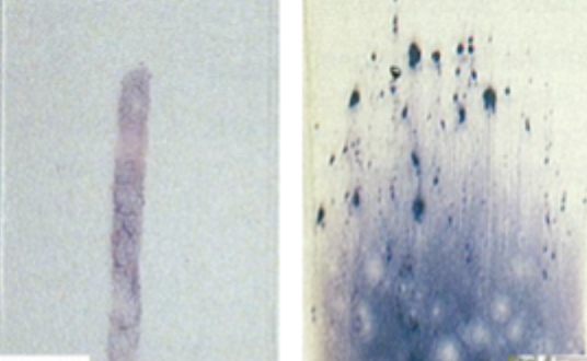
what form of an exam is being displayed
left is bone marrow biopsy
right is bone marrow aspirate
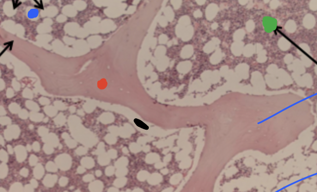
decribe what each of the coloured dots is referring to
blue: fat cells get washed out so we are left with these large spaces
green: millions of haematopoeitic stem cells and all of the cells that have divided from them
black: bone walls that are full of osteoblasts
red: bone tissue, if we look super closesly, can see blue dots which is osteocytes sitting within lacunae
describe the shape of platelets
round/oval
describe the size of platelets
2-4 micrometres diameter
describe the nucleus of a platelet
anucleate
what are platelets derived from and where
fragmentation of megakaryocytes in the bone marrow
describe the granularity of platelets
slightly granular
describe the shape of erythrocytes
flexible biconcave disk
describe the size of RBCs
6-8 micrometers
what is the central pallor
the centre of an RBC is thinner than the rest and so under a light microscope it shows up as a lighter circle
what does it mean if the central pallor doesnt exist in RBC
it means the cell is shaped differently and something might be going on
like a disease
what proportion of the RBC is central pallor
1/3
what does the cytoplasm of RBC contain
haemoglobin
enzymes
why is it important for haemoglobin to be within RBCs
it would thicken the blood too much if it was free floating
how does an RBC carry out energy production
glycolysis
what is the function of haemoglobin
delivers oxygen to tissues and returns carbon dioxide to the lungs
describe the structure of Hb
tetramer with 2 globin chains
describe the lifecycle of an RBC
erythropoetin released by kidney
RBC produced in bone marrow
circulate in blood for 120 days
old cells are killled and phagocytosed
what determines when RBCs are too old and need to be killed
as theyre circulating, they undergo changes to the surface proteins as a sign of aging
macrophages in the spleen recognise these proteins and phagocytose the RBC
another way is cells must squeeze through sinusoids of MPS, if their ability to squeeze and rebound to original shape is diminished, then thats another sign of their age, and macrophages in the MPS will phagocytose them
what happens to RBCs that have been phagocytosed
the haem gets split into iron and bilirubin
iron goes back into the blood
bilirubin goes into the liver into biles (give it the yellow colour)
what are immature erythrocytes
reticulocytes/polychromatic cells
when are reticulocytes released from the bone marrow
2 dyas
what does reticulocytes contain
residual RNA and organelles
how long does it take for a reticulocyte to mature
1-2 days in the blood
what happens to a reticulocyte as it matures
looses its RNA and organelles
what is the difference between polychromatic cells and reticulocytes
essentially the same thing
reticulocytes arent visible with normal stains - need special staining
polychromatic cells show up as purple on giema stained blood film (basically routine staining)
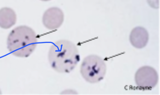
what type of cell is this
reticulocyte
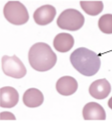
what type of cell is this
polychromatic cell
describe the membrane of RBC
super strong and super elastic
describe the components of the membrane of RBC
phosphlipid bilayer with cholestrol
glycophorin proteins
spectrin and ankyrin
what do glycophorin proteins do in the RBC membrane
protect against injury and damage
what do spectrin and ankyrin do in the RBC membrane
part of cytoskeleton
anchor everything in place
what allows blood cells to be able to deform
biconcave shape
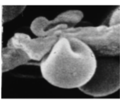
whats this image showing
an RBC squeezing through a sinusoid in the MPS
what is anaemia
low haemoglobin
what does low hameoglobin mean
less oxygen carrying capacity
less ability for cells to make ATP
what are some symptoms of anaemia
weakness
fatigue
angina
shortness of breath (trying to take more o2 in)
what are some signs of anaemia
pallor
tachycardia
jaundiced eyes
what causes jaundiced eyes due to anaemia
too much RBC breakdown>too much billirubin>yellow
what causes tachycardia due to anaemia
ischaemic pain due to build up of lactic acid
how are anaemic classified based on cell size (MCV)
small cell is microcytic
big cell is macrocytic
normal cell is normacytic
how are anaemia classified based on cell colour (MCH)
hypochromic: pale
normochromic: normal
relates to Hb btw
what causes macrocytic cells
problem with maturation
missing things like b12 and folate that are essential to cell division and so cells arent dividing at the same rate and we end up with bigger cells
what is normochromic anaemia
mean cell haemoglobin is within the normal range
MCH
what is aetiology
referring to the cause of the disease
what are the 3 classes of anaemia based on aetiology
reduced RBC or Hb production
haemolysis
blood loss
describe causes for anaemia due to reduced RBC or Hb production
nutritional deficiencies
globin gene defects
kidney disease (low erythropoetin)
bone marrow disease
what nutritional deficiencies can lead to anaemia
iron
b12
folate
what disease is an example of a globin gene defect
thalassaemia
what disease is an example of bone marrow disease
leukemia
what is haemolysis
increased destruction of RBC
how does someone get haemolytic anamia
can be inherited or accquired
how can haemolytic anaemia be inherited
sickle cell disease
heriditary spherocytosis
G6PD
how can haemolytic anaemia be acquired
malaria
autoimmune disease>antibodies attack RBCs
describe blood loss anaemia
pretty self explanatory
u loose blood from acute trauma, surgery, menorrhagia
describe what happens to a cell during sickle cell disease
when the cell is deoxygenated, an area that is phobic to the blood is exposed, it tries to avoid it and the cell collapses (sickles)
why is sickle cell shape a problem
gets stuck in the vessels and occludes them
they also have decreased oxygen carrying capacity
describe the genetic mutation that causes sickle cell
chromosome 11 point mutation resulting in altered B-blobin
glutamine to valine
why is sickle cell disease selected for in some places
selected for in areas where malaria is endemic
malaria cannot infect sickle cells
what is hereditary spherocytosis
it is a defect in the membrane proteins of RBCs that causes them to become spherical
affects proteins such as ankyrin
makes RBCs less deformable, so they get trapped in the sinusoids and get destroyed by macrophages
describe the structure of the membrane proteins in an RBC
2 spectrin polypeptide chains a and b form a chain like heterodimer
spectrin then vinds to actin and ankyrin
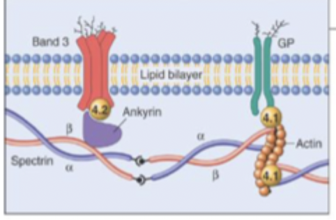
what does hereditary spherocytosis look like on a blood film
missing central pallor
polychromasia (presence of immature Reticulocytes)
describe the consequences of hereditary spherocytosis
anaemia
jaundice, gallstones (bilirubin)
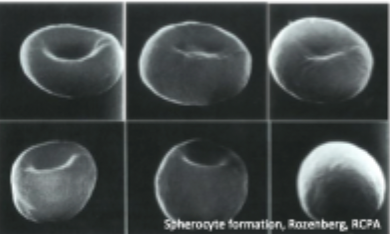
what is happening here
the cells are becoming more spherical due to hereditary spherocytosis
what is G6PD
glucose 6 phosphate dehydrogenase
enzyme that produces glutathione and NADH
oxidised glutathione converted into GSH which protects against oxidant radicals
what is G6PD deficiency
lack of protective HSG
damage from oxidant radicals
RBC membrane damage
creates heinz bodies: unfolded, corss linked Hb that looks like a perm