TCP Week 5 (Pediatric Ortho)
 Call Kai
 Learn
Learn Practice Test
 Spaced Repetition
 Match
Match Flashcards
 Knowt Play
1/121

There's no tags or description
Looks like no tags are added yet.
Name | Mastery | Learn | Test | Matching | Spaced | Call with Kai | Chat |
|---|
No analytics yet
Send a link to your students to track their progress
122 Terms
scoliosis
- Lateral deviation >10º, often w/ rotational component
- Usually convex to the R in thoracic spine
- Predominantly idiopathic (80%), classified by age (adolescent, juvenile, infantile)

scoliosis epidemiology
- 2-4% of US population has scoliosis
- M = F *but the F compression risk is higher
- 90% of cases will not require intervention
epiphysis
end of a long bone
scoliosis s/sx
CC: usually ASX
Frequently reported by caregiver, + screenings
- poor posture
- hips sticking out
- uneven shoulders
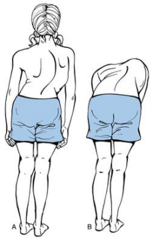
scoliosis PE
inspecting from behind patient for asymmetry at shoulders and hips
- Adams Forward Bend Test
- + Scoliometery

red flags of scoliosis
- L thoracic curve (spine is butting up against heart - not usually idiopathic)
- Leg length discrepancy (very progressed)
- Significant pain
scoliosis diagnostics
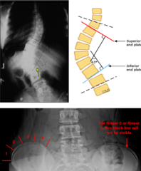
- X-ray: AP/Lat entire T/L spine
- Measure Cobb Angle (>10º = scoliosis)
- Riesser Sign (eval of bone age). More concerning if they're still growing.
MRI only if + neuro findings (or red flag sx)
scoliosis management
refer to ortho/spinal surgeon if >10º or progression on serial images, symptomatic
scoliosis management based on angle (
observation
*if skeletally mature, less concern it will worsen
scoliosis management based on angle (25-40º)
bracing (ONLY if skeletally immature, so younger pts)
brace worn up to 20 hrs/day
scoliosis management based on angle (40-50º)
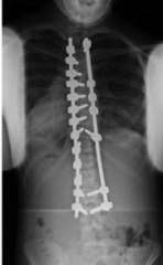
surgery
arrests the curve progression
spondylolysis/listhesis continuum of disease
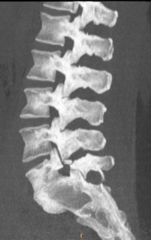
lysis: fracture of the pars interarticularis
lithesis: anterior subluxation
spondylolysis/listhesis pathophysiology of disease
lysis: repetitive stress -> stress/complete fx
listhesis: both pars fx -> anterior subluxation

Spondylolysis/lithesis epidemiology
6-7% of general population, 7-21% adolescent athletes
Risk factors: contact sports with repetitive hyperextension
L5 (80%), anterolithesis of L5 relative to S1
spondylolysis/listhesis history
CC: insidious lower back pain with activity
Generally, more pronounced with listhesis
Improves with rest
red flag of spondylolysis/listhesis
radicular pain/weakness
spondylolysis/listhesis PE
Limitations in lumbar flexion/extension
Potential step offs with listhesis
One-legged hyperextension/stork standing test:
- Patient stands on one leg and leans back
- No pain with flexion
spondylolysis diagnostics
No definitive gold standard (ombination of X-rays, CT scan, SPECT scan)
X-ray: AP/Lateral/Oblique
- Scottie Dog on oblique X-ray
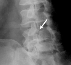
spondylolisthesis diagnostic imaging
X-ray and CT
Classification based on anterior translation

classification of spondylolisthesis
Grade 1 - 0-25%
Grade 2 - 25-50%
Grade 3 - 50-75%
Grade 4 - 75-100%
Grade 5 - > 100% (spondyloptosis - complete anterior subluxation)

spondylolysis management
generally, observation, managed by PCP
surgical if fracture has failed observation, activity modification
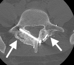
spondylolisthesis management
establish care with pediatric spine or physiatry
Observation: Asymptomatic patients
PT/activity modification: Symptomatic, < grade 3
Bracing: Symptomatic, < grade 3 & failed PT
Operative: Symptomatic, > grade 3 or neuro deficits
developmental dysplasia of the hip
Abnormal relationship between proximal femur and acetabulum
Results in:
- dysplasia (shallow acetabulum)
- subluxation (partially displaced)
- dislocation of the hip
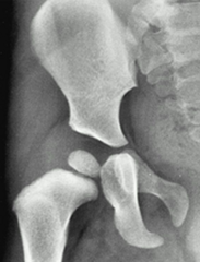
epidemiology of developmental dysplasia of the hip
most common orthopaedic disorder of newborns!
F > M, 6:1
More common in left hip (60%), bilateral (20%)
risk factors of developmental dysplasia of the hip
- first born
- female
- breech presentation
- family hx
developmental dysplasia of the hip history
More a PE finding than history finding
- Findings vary based on age & ambulatory status
- Several associated special tests and findings for dx
Picked up generally at well-infant check
Repeat testing until child is walking, especially in cases with risk factors
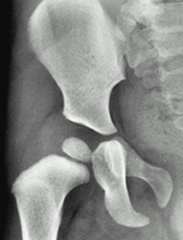
signs/maneuvers for developmental dysplasia of the hip (0-6 months)
Asymmetric Thigh Folds: From dislocated hip being displaced proximally
Galeazzi Sign: Differences in knee height level while supine
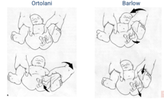
Ortolani Maneuver: Detects if hips are already dislocated, reduces hip
Barlow Maneuver: Detects if hips are unstable, dislocates hip

signs/maneuvers for developmental dysplasia of the hip (6 months-walking)
Asymmetric Thigh Folds: From dislocated hip being displaced proximally
Galeazzi Sign: Differences in knee height level while supine
Asymmetric Passive Hip Abduction: Limited abduction on dislocated side

signs/maneuvers for developmental dysplasia of the hip (walking)
- Leg length discrepancies
- Painless Limp
- Asymmetric Abduction
- Hyperlordosis
ortolani and barlow maneuvers
developmental dysplasia of the hip diagnostics
Limited screening usage here, again mostly on PE
Ultrasound: < 6 months with positive Barlow or Ortolani
X-ray: > 6 months
developmental dysplasia of the hip management
Refer if positive Barlow and/or Ortolani
Early treatment yields best results; treatment based on age
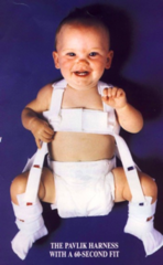
developmental dysplasia of the hip management (0-6 months)
- Pavlik Harness (90% successful)
- Flex 100°, Abduct to 50°
- Re-eval in three weeks by U/S
- Duration of 3 months
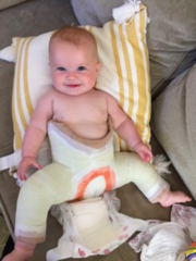
developmental dysplasia of the hip management (6-12 months)
- Closed Reduction under anesthesia (80% successful)
- Followed by a spica cast
- Duration of 2-4 months
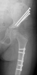
developmental dysplasia of the hip management (12-24 months)
- Failed conservative measures
- Require big, invasive surgeries
- But rarely needed
Legg-Calve-Perthes
Idiopathic avascular necrosis (causes bone to shrink) of the proximal femoral epiphysis
*blood supply to femoral head is lost*
Disruption -> Revascularization -> Resorption -> Collapse
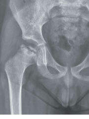
Legg-Calve-Perthes epidemiology
Rare: 1/10,000 children, 7 years old (+ 5 yrs)
Generally unilateral (90%)
M > F
possible connection to coagulopathies
Legg-Calve-Perthes history
CC: painless limp
- In children it can be an aching discomfort
- Refers to thigh or knee
Legg-Calve-Perthes PE
- Atrophy on affected side
- Limited range of motion, loss of INTERNAL ROTATION
- Flexion contracture 0-30º
- Leg Length Discrepancy is LATE FINDING
Legg-Calve-Perthes diagnostics
Multimodal, but X-ray first
On XRAY: Crescent Sign (subchondral fx) = avascular necrosis
If positive --> bone scan/MRI to confirm

Legg-Calve-Perthes management (
Refer to pediatric orthopedics
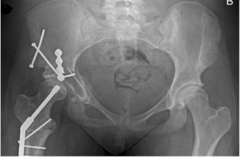
Slipped Capital Femoral Epiphysis (SCFE)
Common condition of the proximal femoral physis (growth plate)
Slippage of the metaphysis relative to the epiphysis, main cause of premature OA in young adults
During periods of rapid growth (10-16 yrs old)
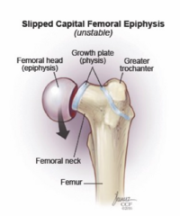
Slipped Capital Femoral Epiphysis (SCFE) epidemiology
Most common disorder affecting adolescent hip!
- 10/100,000 adolescents
- M > F, BMI 25-30, mostly unilateral (60-70%)
- More common in Pacific Island, Black, and Hispanic populations
Slipped Capital Femoral Epiphysis (SCFE) history
CC: Vague hip, groin, thigh, knee pain
- Can be atraumatic (more common)
- Can be traumatic after a fall or while playing sports
- Primary caregiver notices a limp
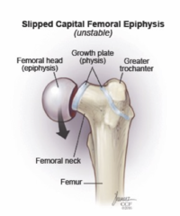
Slipped Capital Femoral Epiphysis (SCFE) PE
Antalgic gait, externally rotated foot at rest, leg length discrepancy (shorter)
Loss of hip IR, abduction, flexion,
Drehmann Sign (when flexing hip, it will ER)

Slipped Capital Femoral Epiphysis (SCFE) diagnostics
X-Ray: AP, Lateral, and frog-leg of BOTH hips
S-Sign: on frog-leg, sharp discontinuity of the normal S shape of femoral head and neck
MRI can pick up pre-slip conditions (edema)
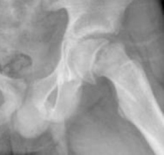
Slipped Capital Femoral Epiphysis (SCFE) management
- REFER, REFER, REFER
- Success dependent upon prompt referral
- Always operative, >90% successful
**Consider prophylactic contralateral treatment in

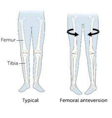
femoral torsion
Rotation deformity of femoral head in acetabulum (anteversion or retroversion)
Part of the process that causes in-toeing
Thought to be related to intrauterine position
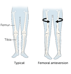
femoral torsion epidemiology
- Minimal prevalence data since it's a mostly benign and transient condition
- F > M , 2:1
- Associated with DDH, metatarsus adductus, congenital muscle torticollis
femoral torsion history
CC: no pain, often just a primary caregiver concern
Should not cause ambulatory issues, but may cause tripping during running activities
Often more pronounced later in the day
femoral torsion PE
Bilateral, asymmetry is a red flag!!
Check hip motion while prone:
- IR usually > 70º (nl 20º-60º)
- ER usually < 20º (nl 30º-60º)
Craig's test: assessing trochanteric prominence. >15º is anteversion.
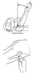
femoral torsion diagnostics
Usually not required
If unilateral or progressive bilateral + failed conservative treatment, and surgery being considered (rare) then CT or MRI

femoral torsion management
Largely SELF-LIMITING (grow out of it)
Managed by PCP
PT can help with hip and core strengthening
Rare: formal ortho work up if continues > 10 years old, causes ambulatory issues
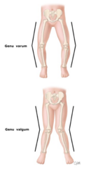
Genu Varum/Valgum
Varum: bowleg
Valgum: knock-knee

genu varum/valgum epidemiology
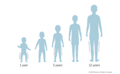
Anatomic variations over the first decade
Varus -> Neutral -> Valgum
genu varum/valgum Hx and PE
CC: Generally asymptomatic. If older anterior knee pain, worse with running
History: Rickets, trauma, fractures, neoplasms
Varum: medial malleoli touching, knees deviate from midline
Valgum: medial aspect of knee touching, malleoli deviate from midline
genu varum/valgum Dx
Generally, not needed
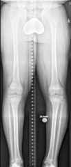
X-ray if >6 cm varum/valgum and not following typical progression
Image should be from hip to the ankle
If unilateral, then MRI
genu varum/valgum management
Reassurance, observation
Refer:
• Unilateral
• Varum beyond age 2
• Valgum beyond age 12
Surgery only if > 12, failed PT, gait disturbances (RARE)
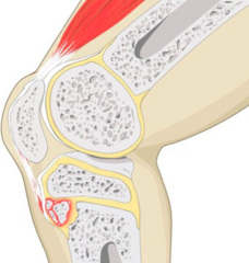
Osgood-Schlatter
Osteochondrosis (abnormal overgrowth) of tibial tubercle
Apophysitis (inflammation) of growth plate
Fragmentation of tibial tubercle
Osgood-Schlatter epidemiology
- During adolescence
- Common in athletes (jumping, running sports)
- M > F
Osgood-Schlatter Hx
CC: generalized anterior knee pain, worse with activity
- Swelling and pain at tibial tubercle
- During phases of rapid growth
Osgood-Schlatter PE
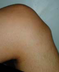
- Thick, indurated patellar tendon (palpable)
- Prominent tibial tubercle
- Pain with resisted knee extension
Osgood-Schlatter Dx
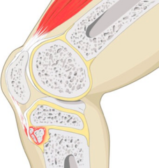
X-ray: AP/Lateral, looking for irregularity, fragmentation of the tubercle, ossicles within the tendon
MRI: pre-surgical planning

Osgood-Schlatter management
Self-limiting, usually resolves when patient reaches skeletal maturity
Mostly controlled by PCP
Activity modification, NSAIDs, PT
Rarely surgery
ACL
ACL prevents torsion and anterior translation of the knee
One of the primary stabilizers of the knee
Adjacent to the PCL in the intercondylar notch

ACL tear epidemiology
- 400,000 injuries a year
- F > M, 4:1
- Secondary to non-contact or contact
- High risk: soccer, basketball, skiing, football
ACL tear history
CC: pop after quick cut or traumatic injury
- Generally, discomfort, not necessarily pain
- Progressive swelling, instability
- After a few days can walk straight
ACL tear PE
General apprehension with movement
Limited ROM due to swelling
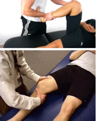
Tests:
• Anterior Drawer Test
• + Lachman (most sensitive) (how far can you pull tibia forward pushing femur down)
• + Pivot shift
ACL tear diagnostics
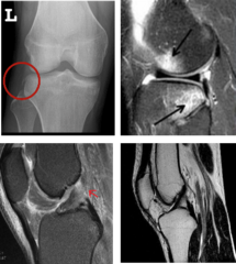
X-ray: AP/Lateral, Segond fracture (pathognomonic for ACL)
MRI: Necessary to confirm dx, kissing contusion
ACL tear management
- Refer to orthopaedics
- Non operative if > 40, low demand
Operative
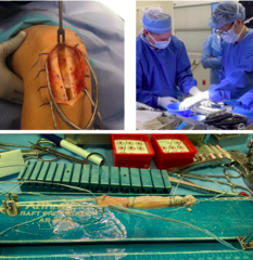
- Reconstruction: using grafts (allograft vs autograft)
- Repair: emerging approach
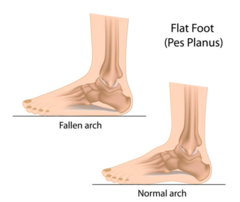
pes planus
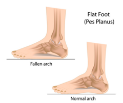
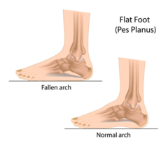
AKA flat feet
Loss of medial longitudinal arch of the foot, heel valgus deformity
Arch region of foot touches the ground
pes planus epidemiology
- Relatively common, 20-30%
- Normal developmental profile, arch develops around 10 yrs old
- Flexible... flat only when wt bearing (95%)
- Rigid... flat even if non wt bearing
pes planus history
CC: mostly asymptomatic
- Reported excessive valgus of the hindfoot
- Outersole wear pattern
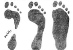
pes planus PE
Severe pronation, can see bunion
Gait analysis... shoes on and off
Achilles alignment, angulated rather than vertical
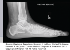
pes planus dx
X-ray: Must be weight-bearing
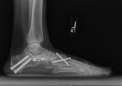
pes planus management
Managed by PCP, non-operative
Physical therapy
Orthotics (supportive, gradual introduction)
Rarely surgery (failed all conservative options or if rigid form w no arch at all)
metatarsus adductus
Generally, self-limiting that condition that causes the forefoot to turn inwards
Visible at birth
Flexible or rigid
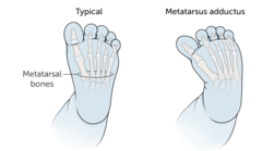
metatarsus adductus epidemiology
MC foot deformity!
Bilateral in about 50%
Associated with DDH (15-20%)
Risk: family history, breech presentation, sleeping position of baby
metatarsus adductus Hx
CC: mostly asymptomatic
Primary care-giver will report in-toeing
Usually reported within the first year of life

metatarsus adductus PE
Diagnosable on inspection
Flexible: can be corrected manually versus rigid which is non-correctable
Normal heel bisector line should fall between 2nd and 3rd toe

metatarsus adductus diagnostics
Not needed
X-ray: only if older or rigid form
metatarsus adductus management
Managed by PCP
90% will resolve by age 4
• If child can actively correct = no treatment
• If child can be passively corrected = serial stretching by parents
Rarely operative (if persists > 4 years old)
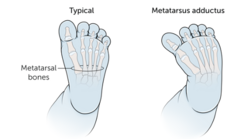
tarsal coalition
Congenital connection of two or more tarsal bones
Calcaneus/navicular or calcaneus/talus
Progresses, starts cartilaginous but will ossify with age

tarsal coalition epidemiology
Rare, only 2% of general population
Frequently bilateral
Autosomal dominant inheritance pattern
tarsal coalition Hx
CC: mostly asymptomatic
Brought in by primary caregiver
Foot pain and stiffness develops during adolescence, as lesion ossifies
tarsal coalition PE
Pes planus, forefoot abduction
Limited subtalar range of motion
Hindfoot remains valgus upon toe-standing

tarsal coalition Dx
X-ray
Calcaneovavicular coalition - "anteater sign" (elongated anterior process of calc)
Talocalcaneal coalition - "talar beaking" (superior projection of distal talus)

tarsal coalition management
Refer to foot and ankle specialist
Treatment based on severity of pain and deformity
Conservative - orthotics, casting, 30% success
Operative - if failed conservative
Congenital Talipes Equinovarus
(Club Foot)
Contracture of the medial tendons of the foot with subsequent deformity of Achilles, ankle, hindfoot, and midfoot
Characterized by fixed ankle plantar flexion, inversion of subtalar joint, medial subluxation of talonavicular joint
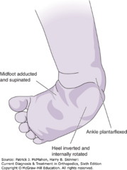
Congenital Talipes Equinovarus
(Club Foot) epidemiology
Rare, 1 in 1,000 births
M > F, 2:1
Bilateral in 50%
Associated with neural-tube defects
Congenital Talipes Equinovarus
(Club Foot) history
CC: pain and callus formation along lateral border of foot
Brought in by primary caregiver
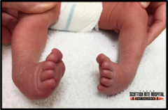
Congenital Talipes Equinovarus
(Club Foot) PE
Clinical diagnosis
Inverted, plantar flexed heel with supinated midfoot
*This is a rigid deformity, cannot be corrected passively
Congenital Talipes Equinovarus
(Club Foot) Dx
X-ray
Loss of the normal angle between talus and calcaneus, results in parallel relationship
U/S can detect in utero
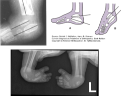
Congenital Talipes Equinovarus
(Club Foot) management
Refer immediately! Treatment should start at birth
*Passive manipulation: Ponseti method (90% success)
Operative if failed/relapse conservative
nursemaid elbow intro and epidemiology
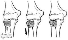
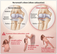
Subluxation of radial head, followed by entrapment of the annular ligament
Common injury in young children (birth - 6 years old)
Results from sudden pull on arm usually by a taller individual!
nursemaid elbow Hx and PE
CC: sudden pain and loss of function
Child will hold arm slightly flexed and pronated
No focal pain or tenderness to palpation
nursemaid elbow Dx
* Not needed if history of traction injury, child is 6 years or younger, consistent clinic exam
Normally straight to tx... X-rays or Ultrasound can be helpful if needed or to confirm diagnosis if needed
nursemaid elbow management
Closed reduction, but confirm no fracture first
- Supination-flexion method
- Hyperpronation method
Should be immediate resolution of pain and normal ROM

salter harris fractures
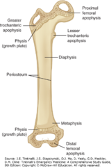
Series of fractures that impact the physis (growth plate)
Disruption through trauma can lead to chronic complications (e.g. growth arrest)
classification of salter-harris fractures
I: Separation through physis
II: Fracture through portion of physis that extended through metaphysis
III: Fracture through portion of physis that extended through epiphysis and into joint
IV: Fracture across metaphysis, physis, and epiphysis
V: Crush injury to physis
**SALTER to remember (see image)
