CBNS116 MT 2
1/136
There's no tags or description
Looks like no tags are added yet.
Name | Mastery | Learn | Test | Matching | Spaced |
|---|
No study sessions yet.
137 Terms
What are the three primary longitudinally arranged components of the brainstem
Tectum "roof"
dorsalposterior to cerebral aqueduct
SC and IC
Tegmentum "covering"
anterior to the ventricular space
complicated
contains CN nuclei and tracts and some long projecting fiber bundles as well as reticular formation
Larger structures
Medulla: inferior olivary nuclei and pyramids
Pons: pontain nuclei, pontocerebellar fibers, cerebral peduncle
what are the 3 major subdivisions of the brainstem
midbrain
pons
medulla
What artery supplies the medulla
vertebral arteries
What artery supplies the pons
Basilar artery
What artery supplies the midbrain
Posterior inferior Cerebral artery
What regions are rostral to caudally in the Posterior column- medial lemniscus system?
Posterior cuniculus (fasciculus gracillis and fasciculus cuneatus)
posterior column nuclei ( n. Gracillis and n. cuneatus) in the caudal medulla
second order neuron axons cross the midline in the anteromedial tegmentum called the medial lemniscus (ML)
ML synapes onto (Vental posterolateral n.) VPL for body and (ventral posteromedial neucleus) VPM
Inputs proceeds to primary somatosensory cortex
How does the somatosensory pathway travel down the spinal cord?
travels through the spinothalamic tract continues from the substantia gelatinous
What regions are rostral to caudally in the corticospinal tract system?
Motor movements
- primary motor cortex down through the large cerebral peduncles through the pons and then form the medulla
What does the pyramidal decussation mark?
The brainstem-spinal cord junction
Where does a small amount of proprioceptive information from the lower limbs travel up the anterior spinocerebellar tract
- travel up the ASCT and enters the cerebellum via the superior cerebellar peduncle
Where does proprioceptive information from the lower limbs travel up the anterior spinocerebellar tract
travels up the PSCT formed by axons of Clarkes column nucleus
proprioceptive info enters via dorsal root and ascends to T-L levels of spinal cord
PSCT then ascends in the dorsolateral tegmentum level of the caudal pons where they join to form the Inferior cerebellar peduncle
Where does proprioceptive information from the upper limbs travel up the anterior spinocerebellar tract
Ascends via the Cuneo cerebellar tract ( CCT) within the Fasciculus cuneatus to reach the lateral cuneate nucleus
CCT fibers then join PSCt fibers in the Inferior cerebellar peduncle
What is the reticular formation? What activating system apart of? What functions does this regulate?
found on all brain stem levels and is all does not form a clear well defined nucleus
Part of reticular activating system of the brainstem core that is the power supply
regulates levels of consciousness and attention
What is the trigeminal nerves function?
- Trigeminal nerve is the head version of the spinal nerve specifically the posterior column nuclei and substantia gelatinous
what is the spinal trigeminal nucleus and tract similar to? where does the STN project to?
brainstems version of substatia gelatinosa and lissauers tract of the spinal cord
followed rostrally thought the level of inferior pons starting at the mid pons level in the same location of the vagus nerve
What is the Medual longitudinal fasciculus? What is the function?
-MLF is small fiber bundle that lies in the midline tangentium.
coordinate eye movements
What is the PAG and where is it located? What is the function
Grey matter that surrounds the cerebral aqueduct- only located in the midbrain
- apart of descending pain system discussed as a associated with the reticular formation
What is the decussation of the superior cerebellar peduncles? Where is it located? Function?
cerebellar output fibers crossing on their way to contralateral VA/VL of the thalamus
at caudal midbrain
fibers have to cross to coordinate voluntary motor output controlled by motor cortex
What is the red nucleus? where is it located? function?
receives crossed cerebellar efferent fibers of the SCP
Project to inferior olivary n. of the rostral medulla--> project to CBL climbing fibers joining the spinocerebellar tract axons with the ICP
What is the substantia nigra? where is it located? function?
located just posterior to the cerebral peduncles
nucleus that provides dopaminergic input to the basal ganglia and therefore plays a critical role in facilitating voluntary movements
What is the VTA? where is it located?
midline nucleus that lies along the basal ganglia
part of an ascending pathway to the Nucleus accumbens involved in reward
What is the Raphe nucleus?
Raphe nucleus
- a small nuclei that "hugs" the midline seam of the brainstem
- produce and releases serotonin to the rest of the brain "juice machines"
What is the Medial zone
- most long ascending and descending projections within
What is the Lateral zone
- located in rostral medulla and caudal pons
- most autonomic CN (VII
What are the projections of the rostral zone of the Pontomesencephalic reticular formation? What is the function and pathway called?
- projects to the thalamus
What are the projections of the caudal zone the pontomedullary reticular formation? What is the function and pathway called?
- Caudal zone projects to the spinal cord as a reticulospinal tract
- Generates motor reflexes and produces basic movements
-Collection of upper motor neurons fibers end on lower motor neurons in the anterior horn of the spinal cord.
What are the projections of the reticular formation? What is its function?
- interacts with the motor cranial nerve nuclie inntervating musculature of the head and neck
- stereotyped repetitive motor activities/reflexes including stretching
What fibers make up the central tegmental tract?
afferent fibers of the RT
Where is the location of the central tegmental track? What is it?
developed at mid pons level
rather diffuse fiber bundle located in the center of the tegmentum on each side 'best seen at pons'
What type of the breathing does the medulla control?
automatic (subconsciously) respiratory rhythms
What areas of output control voluntary breathing control?
motor cortex areas
What instance showed the importance of the medulla for breathing?
Chicken that was cut above the pontomedullary junction and continued to breath
when chicken was cut below the pontomedullary junction
Where does the medullary pacemaker respiratory neuclei project to?
-Projects to collection of lower motor neurons in the anterior horn of C3 or C5 cerviclal spine segments that give rise to the phrenic nerve that innervates the diaphragm
- Also projects to expiratory and inspiratory lower motor neurons at thoracic levels
What does having the wind knocked out of you mean?
Temporary paralysis of the diaphram
What is the cardiovascular center made of?
Collection of cells in the reticular formation is relaly nucleus of CN -X and X
What is the nucleus solitarius
visceral sensory nucleus of the the brainstem
- cardiovascular center
What is the solitary tract? What does it do? What does it receive inputs from?
The tight fiber bundle of the small nucleus solitarius
Receives inputs from CN IX and X from baroreceptors and chemoreceptors located in the carotid body and aortic arch
controls heart rate and blood pressure is then mediated by direct projections from the nucleus solitarius to the parasympathetic and sympathetic preganglionic ganglia
What does the PAG function?
Participates in pain control pathway
amplify or suppressed
What neurotransmitters does pain suppression rely on?
enkephalins and endorphins
What happens to noxious stimuli that gets passed through the limbic system?
Leads to emotional responses tied to painful stimuli to form strong
What is the sequence of steps in the pain suppression pathway?
Spinomesencephalic axons ( axons from the STT to the PAG) activate projection neurons in the PAG via disinhibition.
PAG project to raphe nuclei along the midbrain of the brainstem
What is ultimately responsible for the feeling of pain supression
reduced output from s. gelatinosa neuron forming the spinothalamic tract
What region secretes seritonin?
Raphe nuclei
What region secretes norepinephrine?
locus ceruleus
What regions secretes dopamine?
substantia nigra
What region secretes ach?
Neurons of the rostral reticular formation
- these cells project to the thalamus
most ach input to the cortex comes from the basal forebrain
What region secretes histamine
midbrain reticular formation
- hypothalamus as well
What muscles does the CN III innervate?
Innervates medial rectus
What does SO4LR6
SO- superior oblique
4- CN 4
LR- lateral rectus
6- CN 6
What is one clinically important eye muscle innervated by CN3? What is it responsible for?
Medial rectus-
- responsible for moving the eye medially (when you cross your eyes
T or F: the location of the CN nuclei are typically close to where each nerve enters/ exits the brainstem
T
What muscle does the CN 4 innervate? what information does it carry?
Carries somatic motor output to the superior oblique muscle of the eye
Where is the trochlear nucleus seen in a brain?
Seen indentign the medial longitudinal fasciculus int he caudal midbrain section
Why is the trochlear nerve unique?
emerges from the dorsal surface of the brainstem just caudal to the inferior colliculus
Where is the abducens nucleus located and what is its function?
located in the caudal pons and primarily responsible for the facial colliculus
- abducts the eyes
- abducens nerve exits at the pontomedullary junction
What is the function of the hypoglossal nerve?
general motor innervation to the tongue
What are some symptoms of CN III damage?
- deviation of eye laterally ( lateral stabismus)
- medial rectus paralyzed so now lateral rectus muscle unopposed
- impaired vertical eye movements
- among the remaining eye muscles innervated by CNIII is the superior rectus that raises the eye
- pitosis
- drooping of eyelid due to paralysis of muscle that raises upper eyelid
- dialation of the pupil
- paralysis of pupilary sphincter muscles supplied by the PNS fibers
- lack of pupil constriction
What are some symptoms of CNVI damage?
- Abducens PaLsy
- damage to the left abducens nerve
- causes no abduction to the left from the left eye
- Lateral gaze paralysis
- damage to the left abducens nucleus
- paralysis of the gaze to the left
- Damage to the MLF
- causes paralysis of the left medial rectus during lateral gaze
- left eye cannot adduct
- "one-and-a- half"syndrome
- combo of lateral gaze paralysis and damage to MLF
- causes paralysis of gaze to left and only right eye moves
What is the function of the trigeminal nerve?
provides brachial motor innervation to the muscles of mastication and is the primary somatosensory nerve for the head
What information reaches the face and the top of the head via the trigeminal nerve?
fine touch and proprioception as well as pain and temperature
How is information carried in the head? What is this similar to?
relayed trough the trigeminal ganglion
The DRG of the head
What are the 3 divisions of CN 5
- ophthalamic nerve (V1)
- supplying an area around the eye and forehead
- maxillary nerve ( V2)
- which includes the Upper jaw or maxilla
- Mandibular nerve (V3)
- supplying area around the lower jaw or mandible
- all brachial motor fibers are carried through the mandibular nerve
What is the trigeminal main sensory nucleus function?
fine touch and proprioception
What is the trigeminal spinal nucleus function?
pain and temperature
What is the trigeminal motor nucleus function?
supply muscles of mastication
What is the main sensory nucleus pathway
cells in the main sensory nucleus innervated from the trigeminal ganglion
efferents from MSN cross midline and join medial lemniscus (which carries all body pain and temp info)
ML fibers terminate in VPM of the thalamus
What is the spinal trigeminal nucleus pathway?
light mylinated fibers (carry nociceptive info) enter the brainstem at the pons level and descend to reach spinal V n. ipsylateral
second order fibers cross the midline and join controlateral spinothalamic tract (pain and temp)
VPL and VPM
What are the special motor fiber cranial nerves
V
What are the muscles of mastication the CN V innervate
Temporalis muscle
Masseter muscle
The medial and lateral pterygoids
the anterior belly of the digastric muscle
What are the muscles of mastication the CN VII innervate
muscles of facial expression
small muscle in middle ear (strapedius)
What are the muscles of mastication the CN IX innervate
supplies one of the pharyngeal muscles that contracts
What are the muscles of mastication the CN X innervate?
supplies all of the pharyngeal muscles except one
supplies laryngeal muscles for voice box (larynx)
What are the muscles of mastication the CN XI innervate
Supplies the sternocleidomastoid and trapezius muscles
What is the jaw jerk reflex? what muscle does it use
muscle stretch reflex similar to knee jerk reflex
- uses masseter muscle when it is stretched by a downward tap on chin
What does the jaw jerk reflex assess the integrity of?
both sensory and motor components of CN V3 ( mandibular division)
What is hyperflexia
enhancement of jerk jaw reflex after upper motor neuron damage
What is the gag reflex?
this is the reflex that occurs when you stick your fingers down ur throat
How does the gag reflex work?
you rub the mucosal membrane of the pharynx which activates visceral sensory fibers carried in the CN IX that travel back to reach the nucleus solitarius. Then interneurons project to namb which innervates the pharygeal musculature via CN X
What does the gag reflex test the integrity of?
tests the integrity of IX and X and some of their connections in the brainstem
What is the corneal blink reflex? how is this reflex tested?
we naturally blink when we sense something foreign in our eye
tested using a qtip to gently rub the cornea of one eye and the reflex is to blink both eye
What is the circuit of corneal blink reflex?
sensory information carried by V1 of CN V to rach the spinal trigeminal and main sensory nuclei
Interneurons in the spinal trigeminal nucleus project bilaterally via the reticular formation to the motor neurons in the facial motor nucleus to forcefully blink both eyes
What does the corneal blink reflex assess the integrity of?
tests the integrity of both trigeminal nerves
What causes the medial medullary syndrome? what structures does this affect?
often caused by blocked anterior spinal artery
This affects the hypoglossal nucleus
What are the symptoms of the medial medullary syndrome?
patient will have paralysis to the tongue muscles ipsilateral to the lesion
- when tongue is stuck out
What causes the lateral medullary syndrome? What structures are damaged?
cause: Damage to the PICA or vertebral artery
damages
the inferior cerebellar peduncle,
Spinal trigeminal nucleus and tract,
vagus motor output from the namb supplying the larynx and pharynx,
descending sympathetic fibers from the hypothalamus
spinothalamic tract
What are the symptoms of lateral medullary syndrome?
loss of pain and temperature from the contralateral body and ipsylateral face (while sparing of tactile sensation)
hoarsness and difficulty swallowing (impaired larynx
vertigo
ataxia (loss of balance and equilibrium
a few other symptoms due to damage of the sympathetic
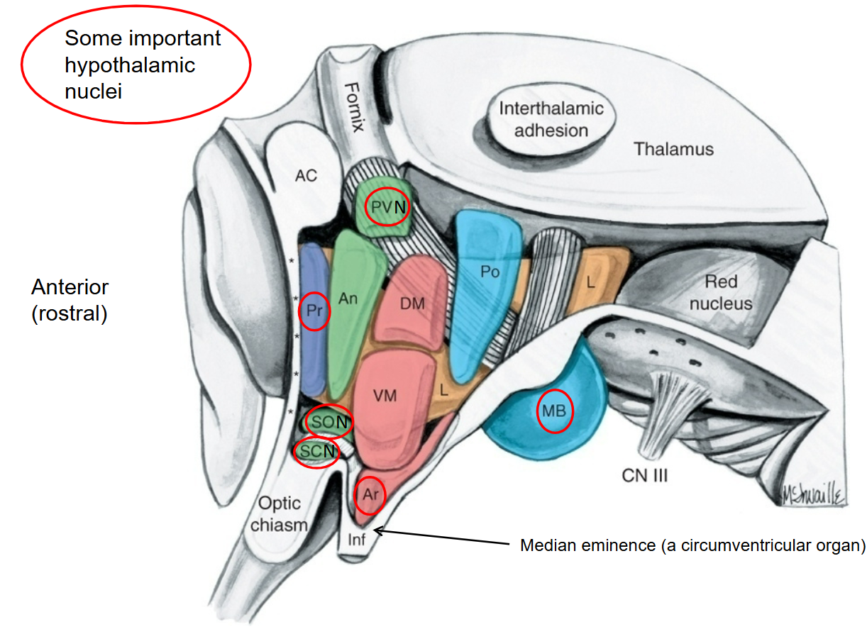
What is the lateral preoptic nucleus function? What area is it located in?
Important sleep promoting area ( how often we “fought” ourself for giving into strong urges to take a nap or keep studying)
preoptic area

What is the function of medial preoptic nucleus? What area is it located in?
Important in temperature regulation
when we feel a chill or are too warm, this increases our drive to add or remove clothing accordingly
preoptic area

What is the function of superchiasmatic nucleus?What area is it located in?
SCN is important in setting circadian rhythms, receiving direct input from the RGC of the retina.
Above the chiasm

What is the function of supraoptic nucleus? What area is it located in?
together with PVN secrete hormones antidiuretic hormones (ADH; vasopressin) and oxytocin into the posterior lobe of the pituitary gland (for water retention)
located immediately above the SCN

What is the function of Paraventricular nucleus? What area is it located in?
together with SON secrete hormones antidiuretic hormones (ADH; vasopressin) and oxytocin into the posterior lobe of the pituitary gland (for water retention)
most superior part of the anterior region

What is the function of Arcuate nucleus? What area is it located in? Why is the location important? What is it a satalite nucleus for
Controls feeding behavior
Receiving directly from NTS about stomach distension
Receptors that bind to peptide hormones, whose levels fluctuate based on nutrient glucose levels in blood
Median eminence is oneof the circumventricular organs, allowing the arcuate nucleus to freely monitor circulating hormone levels in the bloodstream w/o passing the BBB
nucleus, which the hypothalamus control, iss the anterior lobe of the pituitary
Located dorsal to the median eminence (middle region)

What does vasopression (ADH) do?
What is the function of mamillary bodies? What area is it located in? What circuit is it part of?
Site of termination of the fornix, primary anatomical output of the hippocampus that runs along the inferior edge of the septum pellucidum.
neurons then project to the anterior nucleus of the thalamus in the mammillothalamic tract and then to the cingulate cortex
located in the posterior region
part of the Papez circuit, which is involved in some aspects of memory and drive-related behaviors
What does the anterior lobe of the pituitary gland do?
Secretes the bulk of the endocrine hormones released by the pituitary gland
What are the 2 different types of the secretory cells in the anterior lobe of the pituitary? what are the functions of these
Named by preference for binding to acidic vs basic dyes
acidic- acid loving and stained with eosin
Basophils- stain well by basic dyes with hematoxylin
they secrete growth hormones and hormones that trigger secretion by other endocrine glands of the body
What does the posterior lobe of the pituitary gland do?
develops downward growth of the diencephalon and is therefore apart of the NS.
neurohypophysis is comprised of mainly axons and vascular tissues
What is the pituitary gland connected by?
The medial eminence ( the base of the tuberal part of the hypothalamus) by the infundibulum (infundibular stalk)

How does the hypothalamus control the pituitary for the anterior lobe? what nuclei are involved?
Neurons in the arcuate nucleus secrete hypothalamic releasing hor. or inhibiting hor. into a capillary bed in the median eminence.
the capillary bed gives rise to the portal vein though which the releasing or inhibiting hormones reach the acidophiles and basophils in the anterior lobe

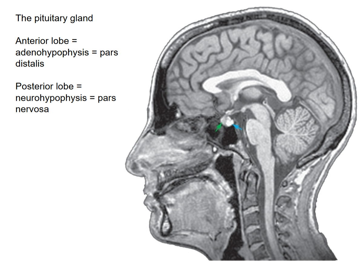
How does the hypothalamus control the pituitary for the posterior lobe? what nuclei are involved?
Neurons in the supraoptic and paraventricular nuclei of the anterior hypothalamus send long axons down the infundibulum to the neurohypophysis when the release ADH(vasopressin) and oxytocin directly into the capillary beds located there.
ADH signals the kidney to retain more water to prevent dehydration, while oxytocin is important in parturition( childbirth), its target organs being the uterus( cause contraction) and the mamillary glands ( milk secretion)
