1.4 - DNA & protein synthesis
 Call Kai
 Learn
Learn Practice Test
 Spaced Repetition
 Match
Match Flashcards
 Knowt Play
1/34

There's no tags or description
Looks like no tags are added yet.
Name | Mastery | Learn | Test | Matching | Spaced | Call with Kai | Chat |
|---|
No analytics yet
Send a link to your students to track their progress
35 Terms
What 3 things does a nucleotide contain?
Phosphate group
Pentose sugar
Nitrogenous base
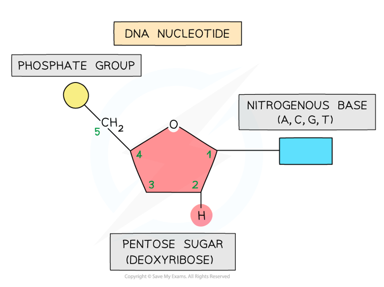
What are the pentose sugars in DNA & RNA?
DNA: deoxyribose
RNA: ribose
How do polynucleotide strands form?
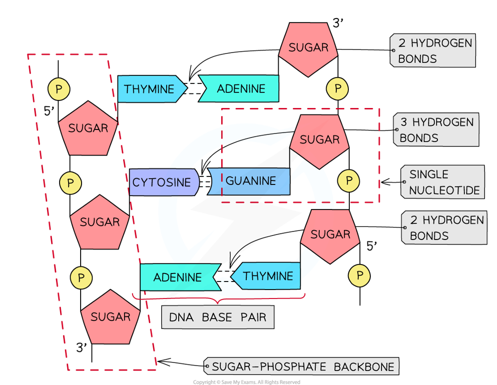
Condensation reactions between nucleotides to form strong phosphodiester bonds (sugar-phosphate backbone)
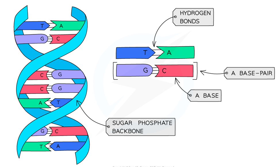
Describe the structure of DNA
Double helix of 2 deoxyribose polynucleotide strands → 2 sugar-phosphate backbones
Hydrogen bonds between complementary base pairs on opposite strands
adenine & thymine → 2 hydrogen bonds
guanine & cytosine → 3 hydrogen bonds
What are the purine bases & describe their structure?
Adenine & Guanine
Two-ring molecules
How to remember → pure As Gold (purines are large & gold is valuable)
What are the pyrimidine bases & describe their structure>
Thymine, Cytosine & Uracil
One-ring molecules
How to remember → CUT the Pie (smaller bases; easier to CUT)
What are the complementary base pairs in DNA?
Adenine (A) + Thymine (T)
2 hydrogen bonds between
Guanine (G) + Cytosine (C)
3 hydrogen bonds between
Name the complementary base pairs in RNA
Adenine (A) + Uracil (U)
2 hydrogen bonds between
Guanine (G) + Cytosine (C)
3 hydrogen bonds between
Why is DNA replication described as semi-conservative?
Strands from original DNA molecule act as templates
New DNA molecule contains 1 old strand & 1 new strand
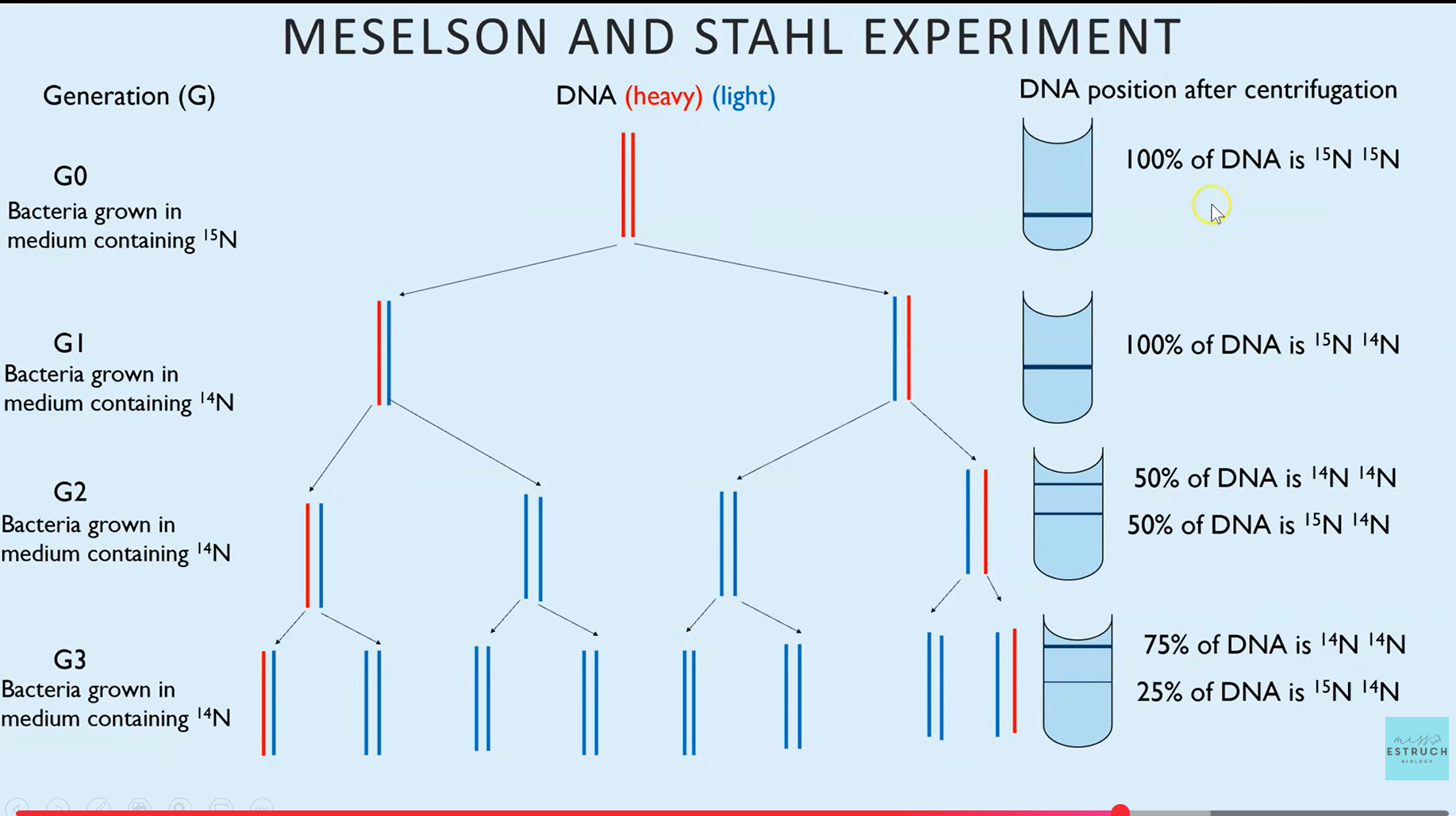
Describe Meselson & Stahl’s experiment
Generation 0
bacterium E.coli grown in heavy nitrogen medium (15N) & centrifuged
band is at the bottom of test tub → 100% DNA is 15N 15N
First generation
bacterium E.coli grown in light nitrogen medium (14N) & centrifuged
band is in middle of test tube → 100% is 15N 14N
Second generation
bacterium E.coli grown in light nitrogen medium (14N) & centrifuged
one band is in the middle of test tube, another band is at the top → 50% of DNA is 14N 14N & 50% of DNA 15N 14N
Third generation
bacterium E.coli grown in light nitrogen medium (14N) & centrifuged
one thicker band at the top of test tube & one thinner band in the middle → 75% of DNA is 14N 14N & 25% of DNA is 15N 14N
Explain the role of DNA ligase in DNA replication
Joins Okazaki fragments on the lagging strand by forming phosphodiester bonds between adjacent nucleotides, using ATP, to produce a continuous DNA molecule
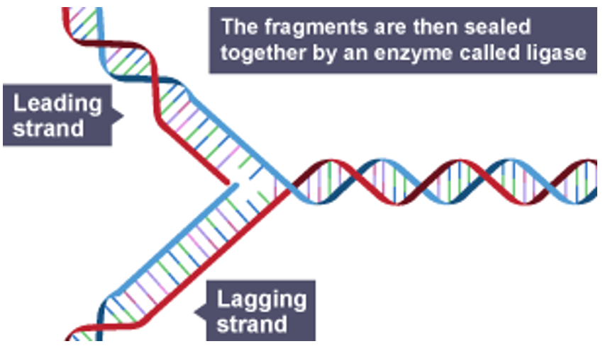
What is the role of DNA helicase in semi-conservative replication?
Breaks hydrogen bonds between complementary base pairs to form 2 single strands, each of which can act as a template
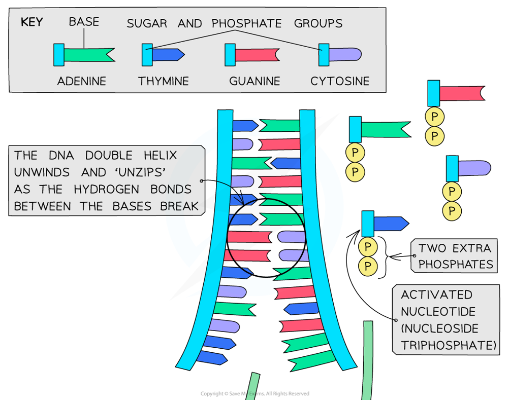
Outline the process of semi-conservative replication
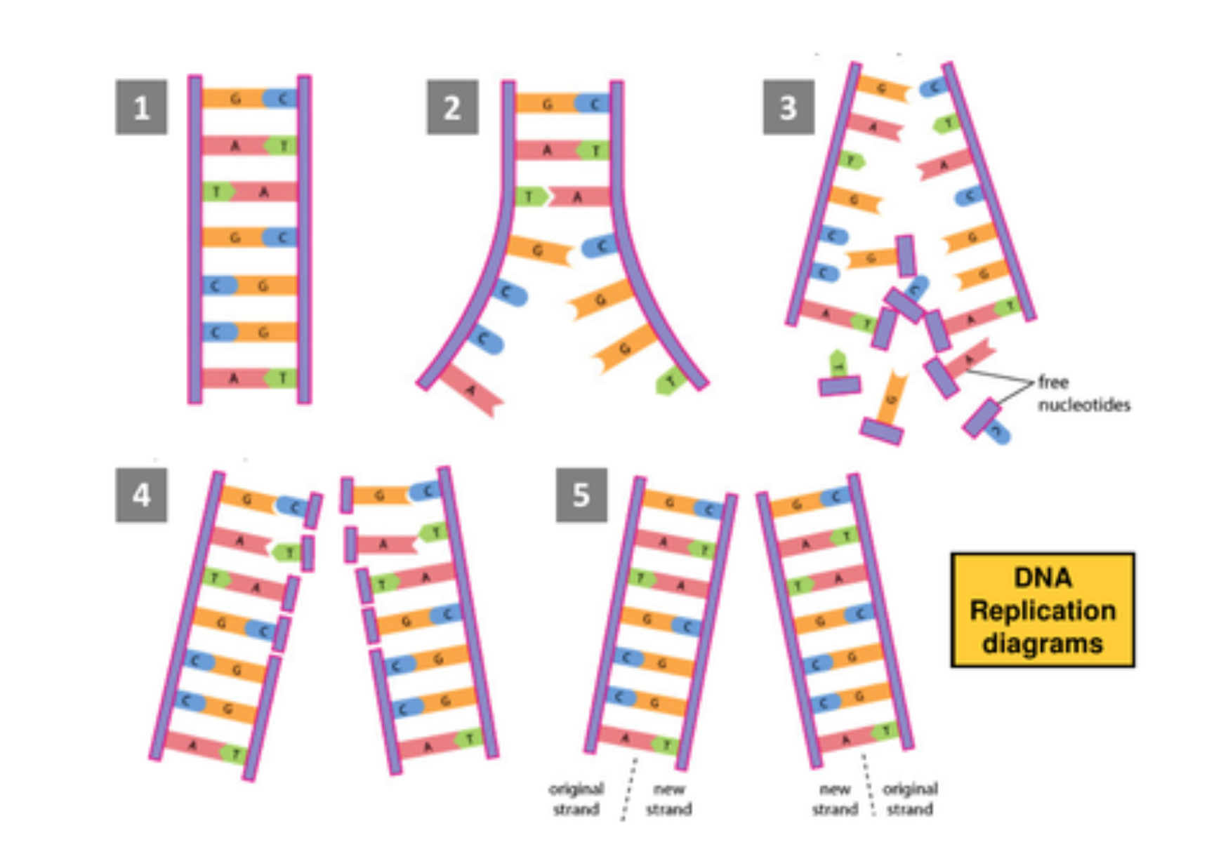
DNA double helix is ‘nicked’ & hydrogen bonds between complementary base pairs are broken
This occurs due to DNA helicase & results in the unwinding of the two polynucleotide strands
Bases are now exposed on each strand. Separated polynucleotide strands both act as templates for the addition of free, activated nucleotides
These pair with complementary bases that have been exposed by the unwound DNA (A+T & G+C)
DNA polymerase catalyses the linking together of activated nucleotides through covalent phosphodiester bonds. Two new nucleotide strands are formed
There are now two daughter DNA molecules, each of which contains one of the original strands of DNA & one new one
What is the definition of a gene?
A sequence of DNA bases that code for a polypeptide or a functional RNA
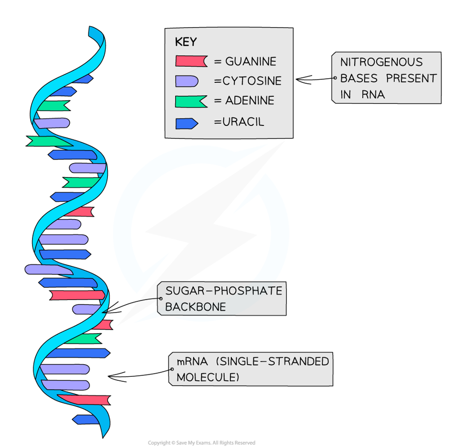
Describe the structure of mRNA
Single-stranded, linear molecule made of ribonucleotides
Each ribonucleotide consists of ribose sugar, phosphate group & one nitrogenous base: A, U, G or C
Nucleotides are joined by phosphodiester bonds, forming a sugar-phosphate backbone
The sequence of bases forms triplets called codons → each codon codes for one specific amino acid
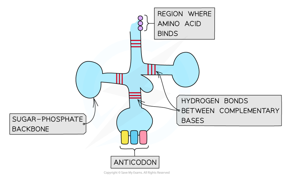
Describe the structure of tRNA
Single-stranded RNA molecule made of ribonucleotides, containing: ribose sugar, phosphate group & one nitrogenous base (A, U, C, or G)
Folded into clover-leaf shape due to hydrogen bonding between complementary bases
One end has an anticodon (triplet of bases complementary to an mRNA codon)
The opposite end has an amino acid binding site (where a specific amino acid binds)
What does transcription produce & where does it occur?
Produces mRNA (more specifically pre-mRNA)
Occurs in the nucleus
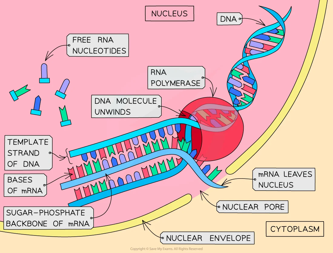
Outline the process of transcription
DNA helicase unwinds the DNA double helix
Free RNA nucleotides pair complementarily with exposed bases on the antisense strand (A+U & G+C)
As nucleotides are added, the DNA double helix rewinds behind it
RNA polymerase joins RNA nucleotides together, catalysing the formation of phosphodiester bonds
RNA polymerase detaches once stop codon is reached
What happens after a strand of mRNA is transcribed?
Splicing removes introns (leaving only exons) from pre-mRNA in eukaryotic cells
mRNA moves out of nucleus via nuclear pore & attaches to ribosome in cytoplasm
What is the function of mRNA?
Transfers the genetic code from DNA in the nucleus to ribosomes for translation into a specific polypeptide
What is the anti-sense strand of DNA?
The template strand of DNA which is transcribed
What does translation produce & where does it occur?
Produces proteins
Occurs in the cytoplasm on ribosomes
Outline the process of translation
mRNA attaches to a ribosome in the cytoplasm
A tRNA molecule with an anticodon complementary to the codon on the mRNA attaches (occurs at the start codon)
Another tRNA molecule with a complementary anticodon attaches to the next codon on the mRNA → a peptide bond forms between the amino acids
The ribosome moves along the mRNA to the next codon & the process repeats
This continues until a stop codon is reached
What is the role of ATP during translation?
ATP hydrolysis provides energy to form peptide bonds
What are 3 features of the genetic code?
Non-overlapping: each triplet is only read once
Degenerate: more than one triplet codes for the same amino acid (64 possible triplets for 20 amino acids)
Universal: same bases & sequences used by all species
What are DNA triplets?
Sequence of 3 bases that code for a particular amino acid
What is a start codon?
Initiates the translation of a polypeptide
What is a stop codon?
Terminates translation of a polypeptide & does not code for an amino acid
What are introns & exons?
Introns: non-coding sections of DNA (removed during splicing)
Exons: regions of DNA that code for amino acid sequences
What is a mutation?
Any change in the base sequence of DNA (often arise spontaneously during DNA replication)
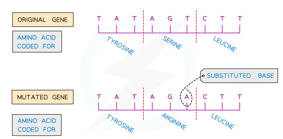
What is a substitution mutation?
One nucleotide in the DNA sequence is replaced by another → (likely to be a silent mutation which does not change amino acid sequence)
What is a deletion mutation & what is its effect?
A nucleotide in the DNA sequence is removed, leading to a frameshift
Significant since entire amino acid sequence downstream of mutation will be different
What is an insertion mutation & what is its effect?
Addition of one or more nucleotides to the DNA sequence, which causes a frameshift
Significant since entire amino acid sequence downstream of mutation will be different

What is sickle cell anaemia?
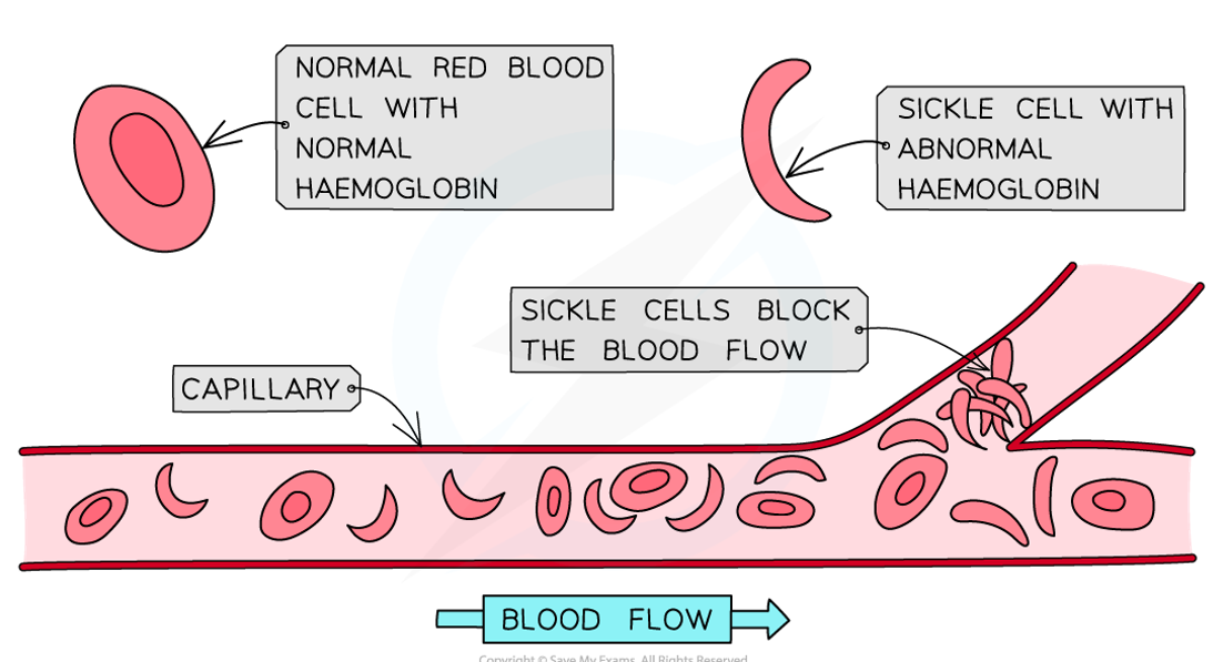
Genetic condition that is common among people originating from malaria-stricken areas that results in abnormal haemoglobin
Impaired ability to transport oxygen (blocks small capillaries) causing rapid heart rate, fatigue & dizziness
What causes sickle cell anaemia in humans?
Single point mutation in gene that affects protein chains making up haemoglobin:
in DNA: GTG (Glutamic acid) → GAG (Valine)
change in primary structure, meaning different tertiary structure → results in abnormal haemoglobin molecules that clump together & form long fibres that are sickle-shaped red blood cells
