A+P Lab 1: SKULL BONES
 Call Kai
 Learn
Learn Practice Test
 Spaced Repetition
 Match
Match Flashcards
 Knowt Play
1/71

There's no tags or description
Looks like no tags are added yet.
Name | Mastery | Learn | Test | Matching | Spaced | Call with Kai | Chat |
|---|
No analytics yet
Send a link to your students to track their progress
72 Terms
How much total cranial volume (in liters) are present in the skull?
1.35 Liters
Cranial Fossa
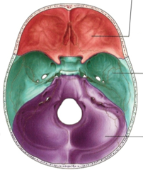
Anterior Cranial Fossa
formed by the frontal bone and part of the sphenoid bone

Medial Cranial Fossa
sphenoid, temporal and occipital bones

Posterior Cranial Fossa
supports the cerebellum of the brain

Frontal Bone
bone that forms the forehead

Supraorbital Margin
The superior rim of the eye socket located on the frontal bone.
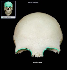
Supraorbital Foramen
opening above each orbit allowing blood vessels and nerves to pass

Parietal Bone
a bone forming the central side and upper back part of each side of the skull.

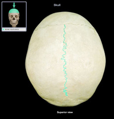
Sagittal Suture
between parietal bones
Occipital Bone
Bone that protrudes at the base of the skull

Foramen Magnum
A large opening at the base of the skull through which the brain connects to the spinal cord.

Occipital Condyle
ridges on left and right of foramen magnum

Temporal Bone
bone that forms parts of the side of the skull and floor of the cranial activity. There is a right and left temporal bone.

External Auditory (acoustic) Meatus
ear canal

Styloid Process
pole-like process extending downward from the temporal bone on each side of the skull

Jugular Foramen

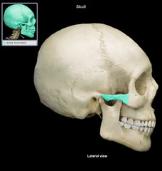
Zygomatic Process
a projection of the temporal bone that forms part of the zygoma
Malleus
"hammer"

Incus
"anvil"

Stapes
"stirrup"

Ethmoid Bone
Light spongy bone between the eye sockets; forms part of the nasal cavities.

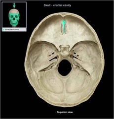
Crista Galli
ethmoid bone
Cribiform Plate
superior surface of the ethmoid; perforated by a foramina which allows passage of the olfactory nerves, which provide sense of smell

Perpendicular Plate
forms superior part of nasal septum

Fragile Nasal Turbinates (Concha)
long, narrow, curled shelves of bone that protrude into the breathing passage of the nose

Sphenoid Bone
forms part of the base of the skull and parts of the floor and sides of the orbit

Body of Sphenoid Bone
central section of sphenoid

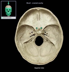
Sella Turcica
depression in the sphenoid bone where the pituitary gland is located

Superior Orbital Fissure

Optic Foramen
On either side ochiasmatic groove -transmits optic nerve (CN II) along with its meningeal coverings, and the ophthalmic artery
Maxilla
origin for teeth of upper jaw

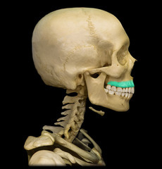
Alveolar Margin (of the Maxilla)
inferior margin of maxilla containing sockets for teeth
Palatine Process
forms the anterior portion of the hard palate (roof) of the mouth also forms parts of the nasal cavity and eye orbits

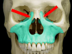
Frontal Process
Process of the zygomatic bone that extends upward to articulate with the frontal bone at the outer edge of the orbit
Mandible
origin for teeth of lower jaw

Ramus
An extension of a bone making an angle with the rest of the structure

Condyle (of the Mandible)
A ball like structure that forms the hinge between the maxilla and the mandible and is a part of the temporomandibular joint

Coronoid Process (of the Mandible)
flattened upward projection from the anterior margin of the mandibular ramus

Alveolar Margin (of the Mandible)
superior margin of the mandible; contains sockets in which the teeth lie

Zygomatic Bone
the arch of bone beneath the eye that forms the prominence of the cheek

Zygomatic Arch
cheek bone
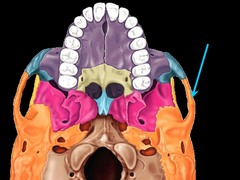
Nasal Bone
bridge of nose

What does the extension of cartilage at the end of the Nasal Bone provide?
distal flexion
Lacrimal Bone
small fragile bone making up part of the front inner walls of each eye socket and providing room for the passage of the lacrimal ducts

Lacrimal Fossa
canal that houses lacrimal sac; formed from the maxilla and lacrimal bone
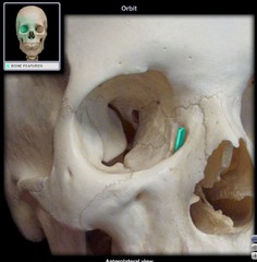
Palatine Bone
either of two irregularly shaped bones that form the back of the hard palate and helps to form the nasal cavity and the floor of the orbits
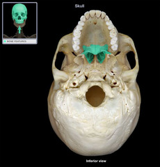
Hard Palate
roof of the mouth
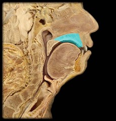
Soft Palate
muscular posterior (back) portion of the palate
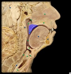
Vomer
nasal septum

Orbit
holds the eye and its nerves/blood vessels

Inferior Orbital Fissure
fissure in the orbit floor between maxilla and greater wing of sphenoid
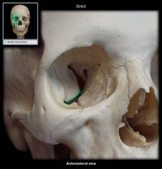
Optic Canal (Foramen)
the hole in the orbit. connects optic nerve to brain

How many bones form the edges of the Orbit?
7
How many Cranial Sutures are there?
5
Coronal Suture
the suture between the parietal and frontal bones of the skull

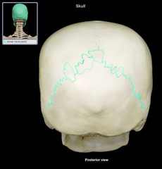
Lambdoid Suture
between parietal bones and occipital bone
Squamous Suture
Between parietal and temporal bones

Occipitomastoid Suture
connects occipital and temporal bones

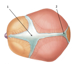
Anterior and Posterior Cranial Fontanel
How many Cranial Fossa are there?
3
If the Fossa are too small during early childhood, does the brain have space to grow?
Nasal Cavity
hollow space behind the nose

Nasal Septum
a wall of cartilage that divides the nose into two equal sections

Superior Nasal Concha
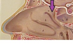
Middle Nasal Concha

Inferior Nasal Concha

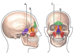
Paranasal Sinuses
air pockets that provide a shock-absorber like function and make the skull lighter in weight
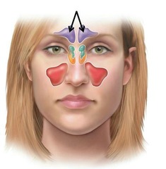
Frontal Sinus
cavity within the frontal bone
Ethmoid Sinus
Mucosa lined air spaces located above the Sphenoid Sinus and below the frontal sinus.

Maxillary Sinus
largest paranasl sinus; pyramidal; on cheek bone lateral to nasal bone

Sphenoid Sinus
sinus above and behind the nose
