4. action potentials and conductance
1/29
There's no tags or description
Looks like no tags are added yet.
Name | Mastery | Learn | Test | Matching | Spaced | Call with Kai |
|---|
No analytics yet
Send a link to your students to track their progress
30 Terms
leak channels
specific; always open for K and Na in resting cell so responsibe for resting potential
gated channels
open or closed
two types:
voltage gated
ligand gated: open when ligand/chem substance binds
the action potential
stimulating of a resting neuron creates disruption in the membrane that results n the opening of voltage-gated channels for Na
depolarization
any change that moves the membrane closer to zero
what threshold voltage generates an action potential?
-50-55 mV

stimuli
make Na channels open; at peak/channels K channels open
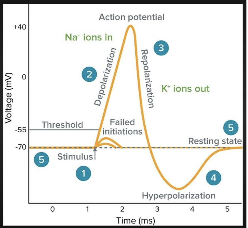
what causes action potential?
rapid inward influx of Na through voltage gated channels
depolarizes membrane to 0
first Na neutralize the K on the resting membrane and then established a (+) charge on interior membrane called the overshoot (peaks at +30-50 mV)
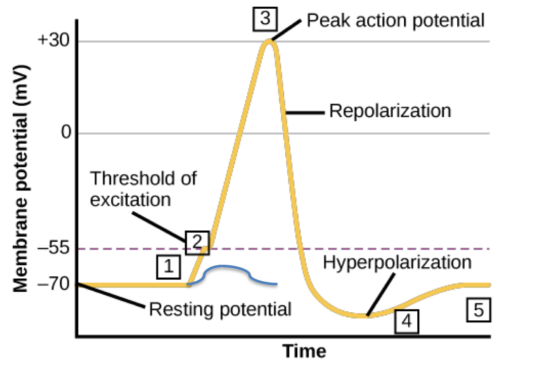
hyperpolarized
inward Na influx stops as Na vg channels close
outward influx of K starts as K vg open
but bc not all the K voltage channels close in time, the potential becomes hyper-polarized briefly
the dip is physically farther away from the threshold value
kinetics of Na or K is slower?
K
are Na and K vg channels stimulated by the same stimuli?
yes
three characteristics of action potentials:
all or none: reach the threshold its gunna happen
absolute refractory period: impossible to generate second action potential
relative refractory potential: possible to generate second action potential but require stronger stimulus to reach threshold
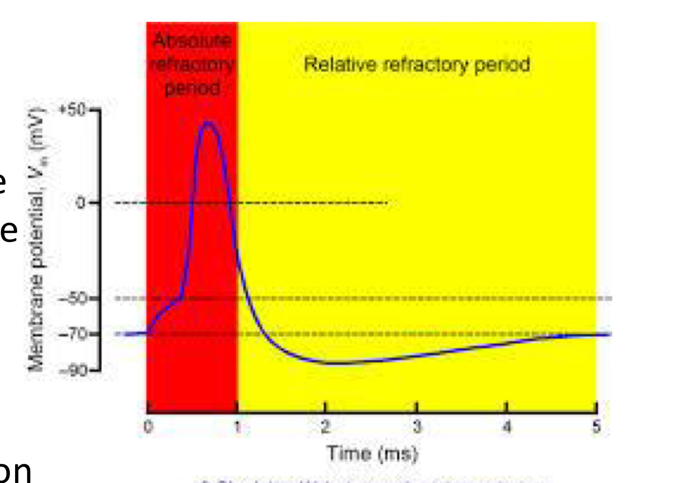
mechanism of absolute refractory period
sodium channel inactivation
*closed state can be reopened by stimulus but inactivated cannot; ball = inactivated
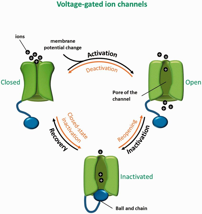
IFM
string of aas that block the channel
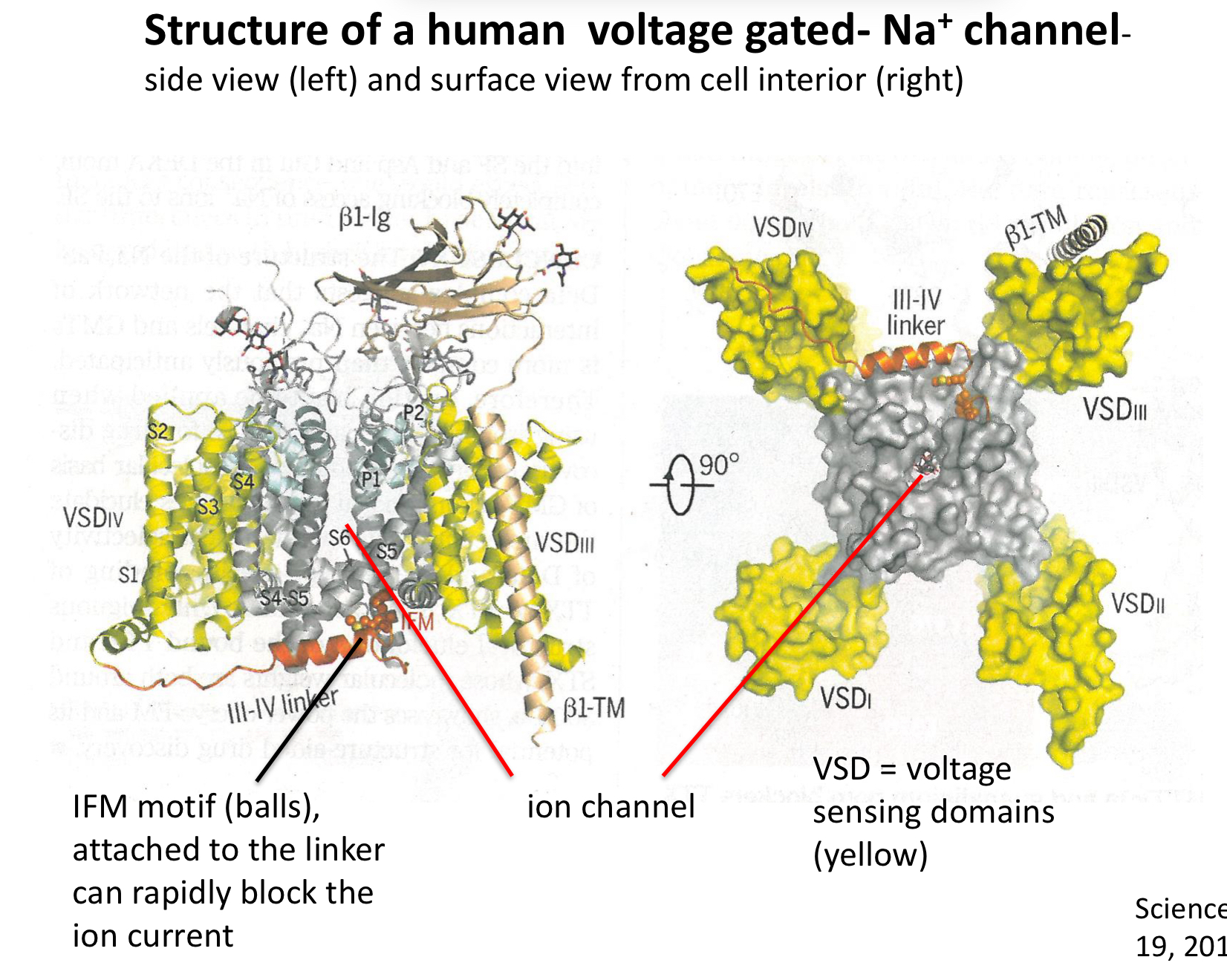
explain blockage of action potentials with local anesthetics
hydrophobic mines like lidocaine enter cell and move in and binds ball and chain to inhibit its release; keeps it closed
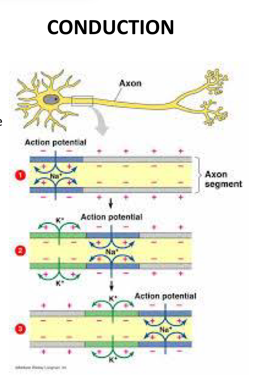
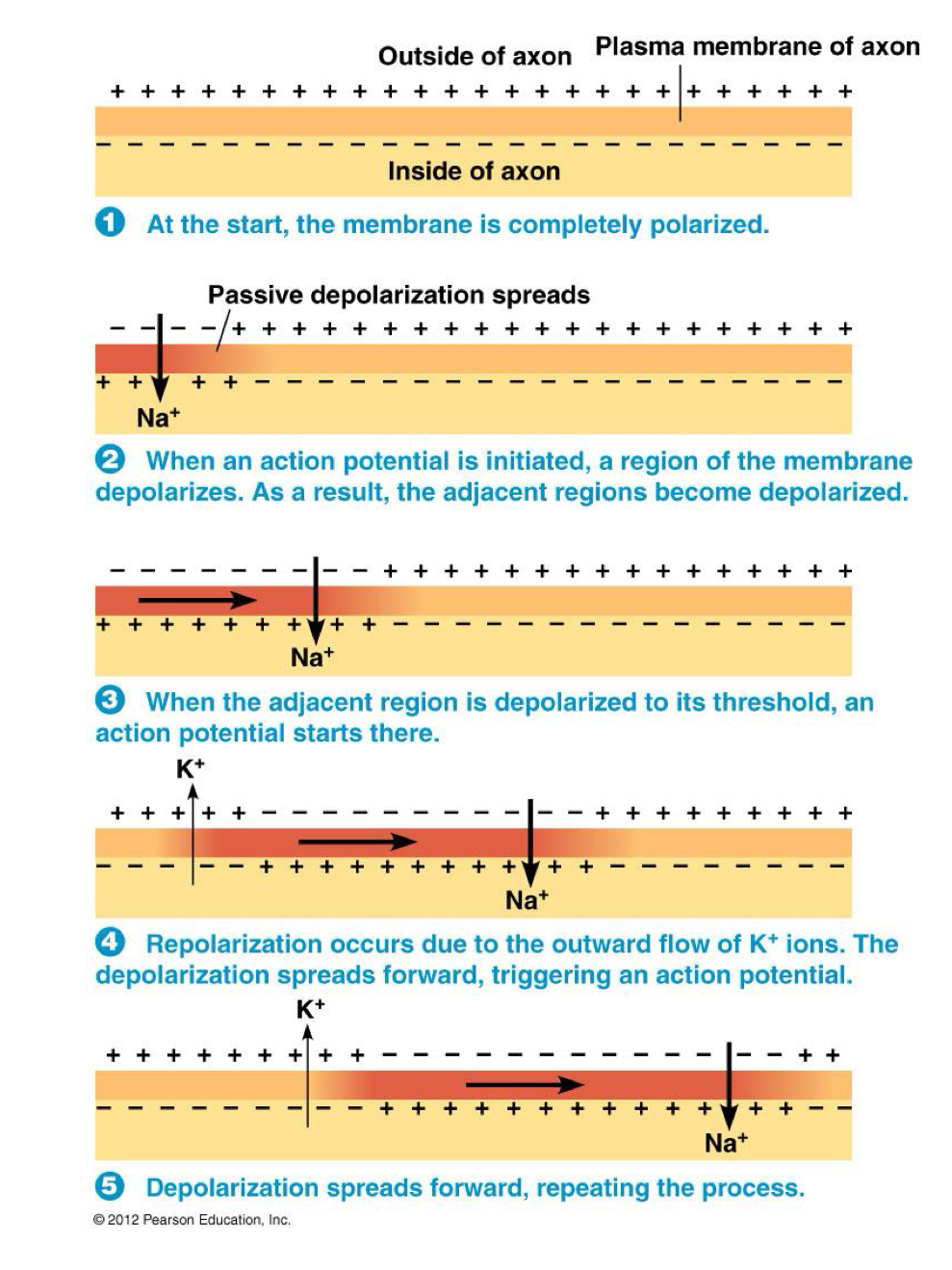
describe conduction
cell depolarized in segments; regenerate action potential as it continues each axon segment
green is refractory but action potentials cannot go back!
action potential Na (in or out) v refractory period K (in or out) to go back to (blank)
in
out
resting potential (first hyper polarization)
regenerative current
new action potential generated each segment of the axon
cable conduction
passive depolarization; not segments but a flow
electrical can conduct through myelin sheath
false; at nodes of ranvier you can conduct current (only nodes depolarize)
saltatory conduction
a rapid mode of signal transmission along myelinated neurons, where the electrical impulse jumps from one node of Ranvier to the next; depolarize axon in segments not entire axon like other conductions
faster 120m/s VS 0.5m/s
the leakier the cel, the spread of potential becomes
limited
Na can leak out membrane
length constant λ
distance that a graded electrical potential can travel passively down an axon before it decays to ~37% of its original amplitude
=√rm/ri
rm
resistance to ion current through membrane
high means membrane not leaky to Na; more chance for potential as charge (Na) stays inside
ri
resistance to ion flow in axoplasm (movement of things down axon); viz axial resistance = increase in conduction velocity
the lower the resistance, the easier it is for ions to move across the cell membrane
T or F: low ri and high rm means greater λ
true! means axon will depolarize to threshold further down from where injection occurs
length constant is a function of
axon diameter
myelination
T or F: larger axons have higher axial resistance
false they have lower axial resistance (ri)
T or F: myelin sheaths raise the rm
true
fastest neurons vs slowest diameter and myelination
fast: large diameter myelinated
slow: small diameter, non-myelinated
multiple sclerosis
myelin sheaths sleuth off
Na come at node but no normal channels so leaks!
exposes axon membrane which lacks Na channels but has K channels; nodes have Na channels
inward Na current dissipated in demyelinated region by outward K current which prevents depolarization from reaching Na channel in subsequent node
demyelination promotes conduction failure
sodium channels become abnormally distributed along demyelinated axons, moving away from the nodes of Ranvier where they are normally concentrated, which disrupts proper signal transmission. excessive sodium influx into axons, which in turn triggers a reverse sodium-calcium exchanger, causing a harmful buildup of calcium within the axon and contributing to axonal degeneration