Sheep Brain Structures and Landmarks
1/88
Earn XP
Description and Tags
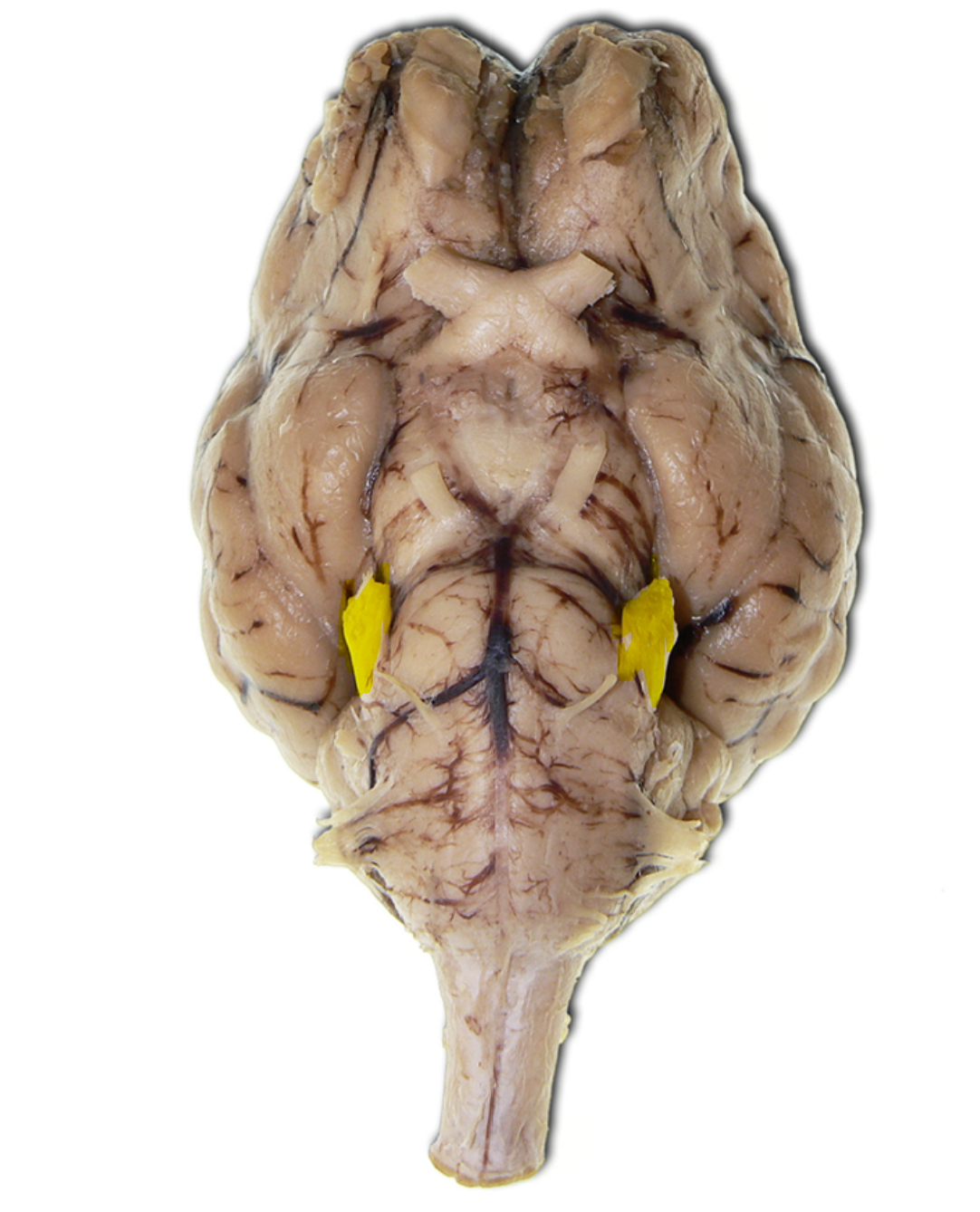
Overview of the cranial nerves and other key structures of the sheep brain.
Name | Mastery | Learn | Test | Matching | Spaced |
|---|
No study sessions yet.
89 Terms
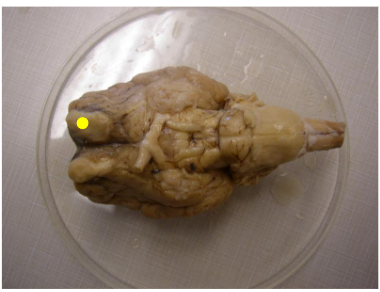
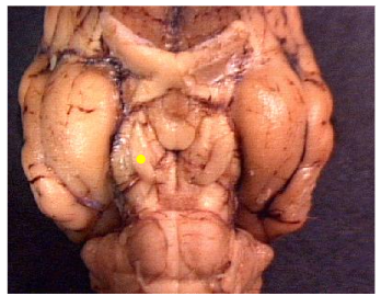
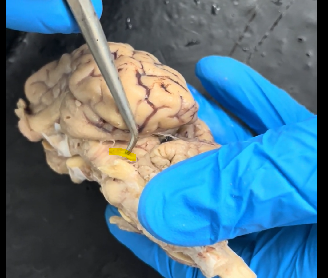
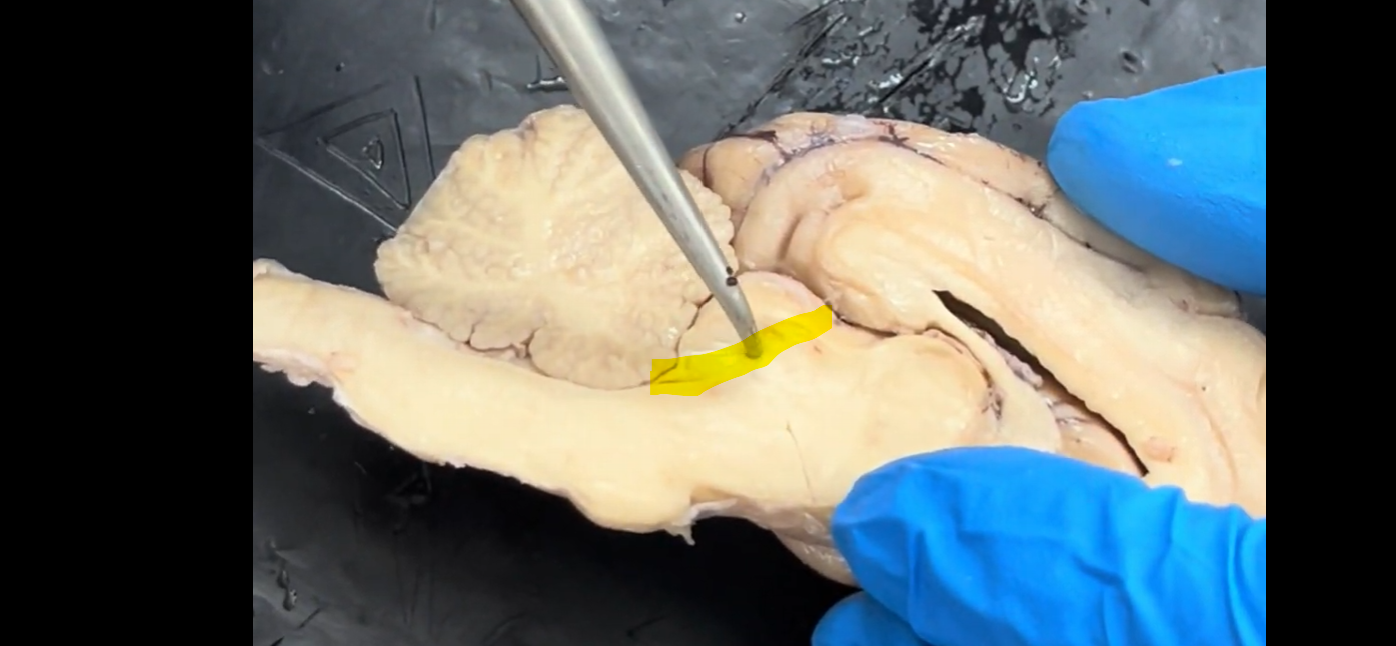
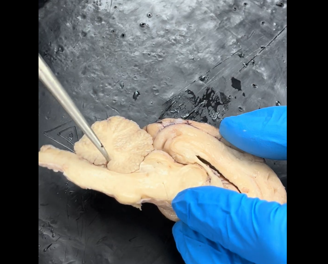
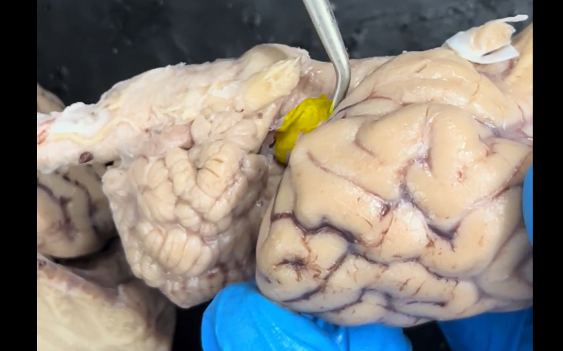
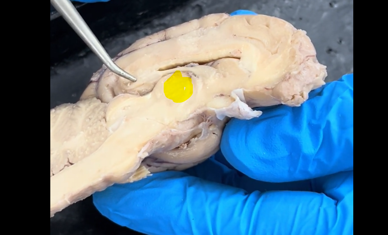
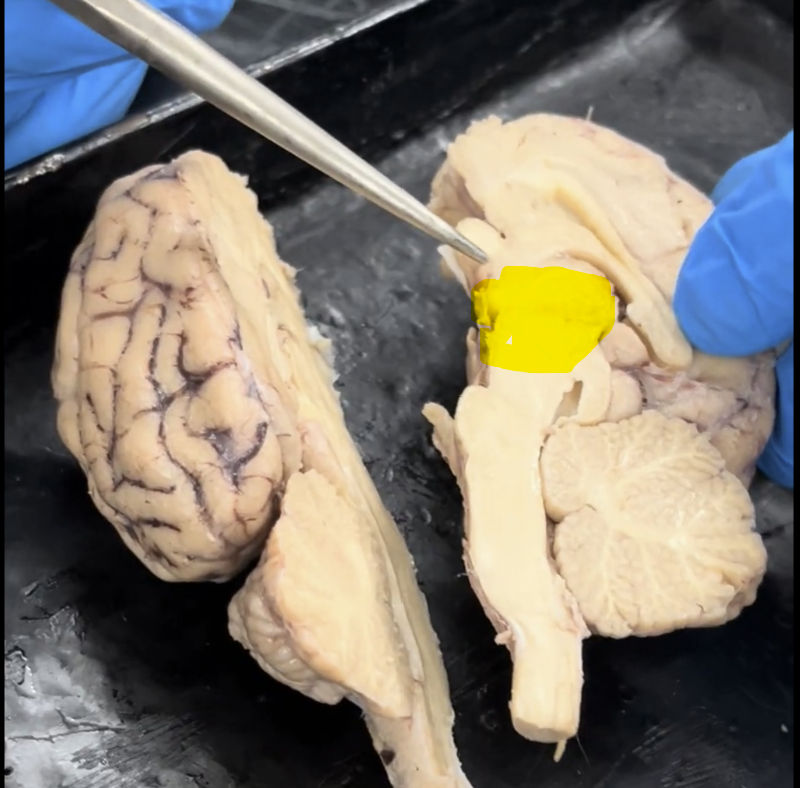
Name the part of the brain marked by the yellow dot.
Olfactory bulb (cranial nerve I).
I = 1
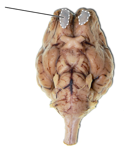
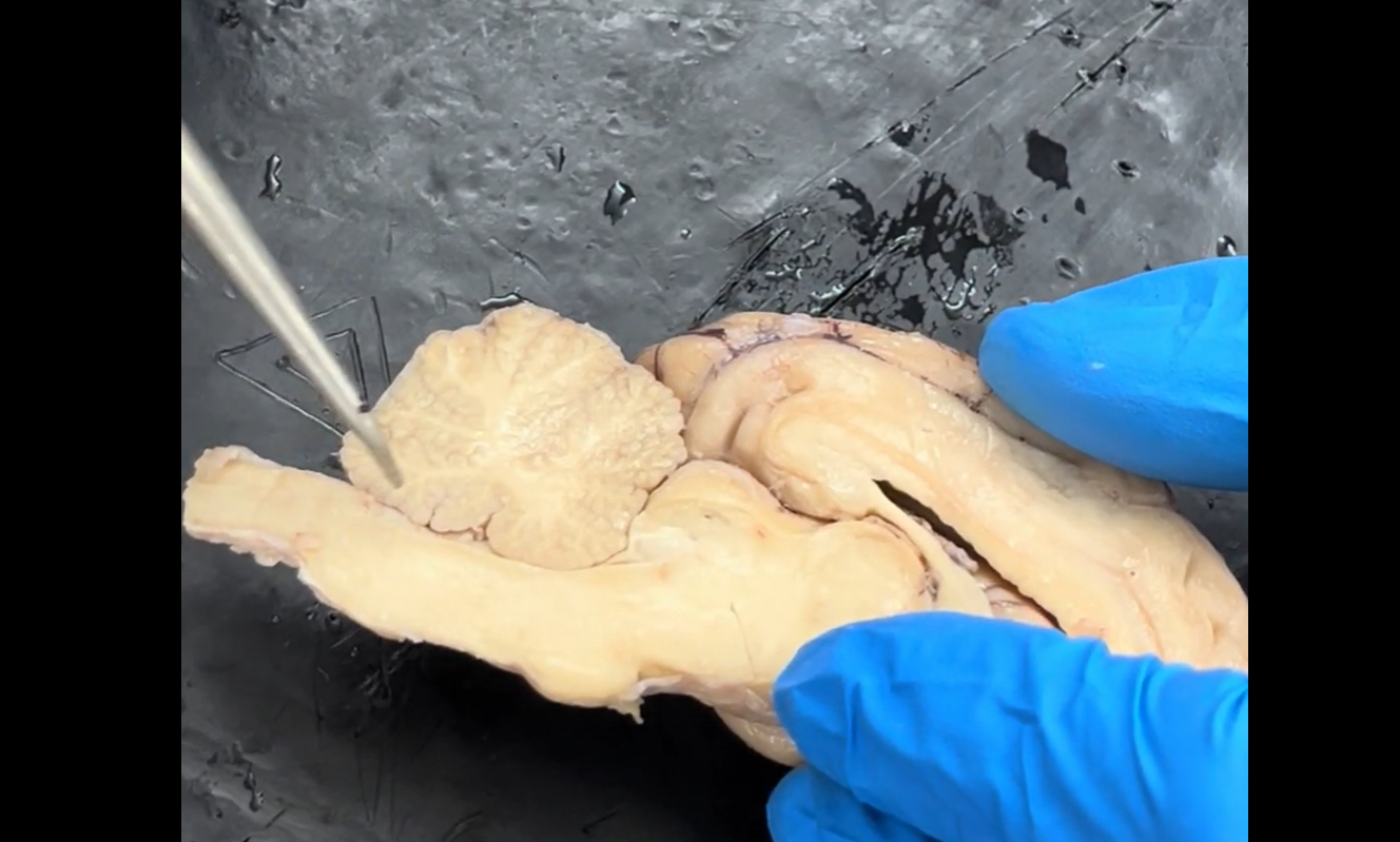
Name the part of the brain that is highlighted.
Olfactory bulb (cranial nerve I).
I = 1
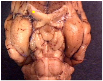
Name the part of the brain marked by the yellow dot.
Optic nerve (cranial nerve II).
II = 2

Name the part of the brain that is highlighted.
Optic nerve (cranial nerve II).
II = 2
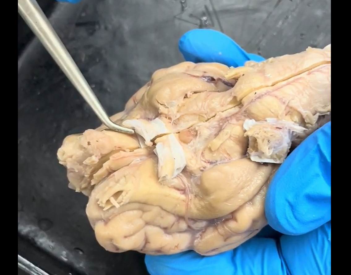
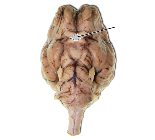
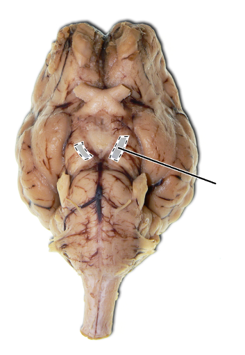
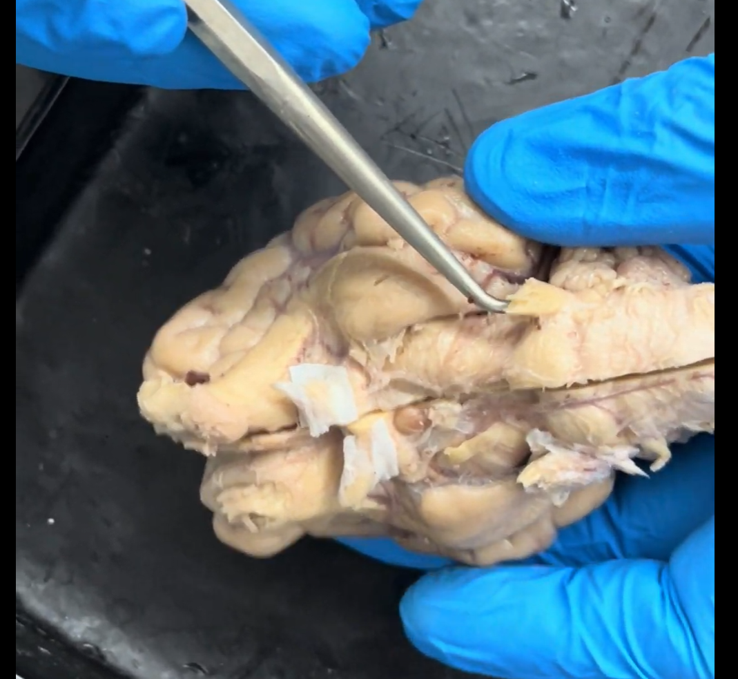
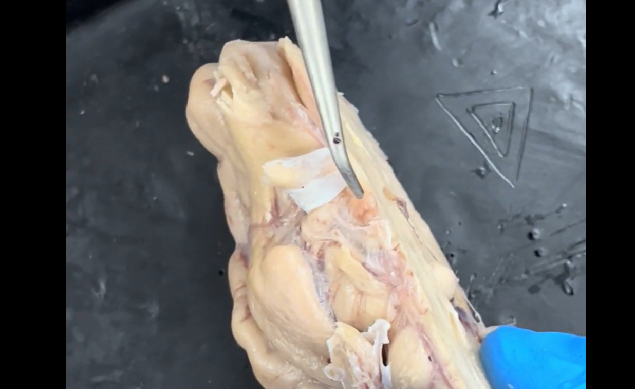
Name the structure the probe is pointing to.
Optic nerve (cranial nerve II).
II = 2
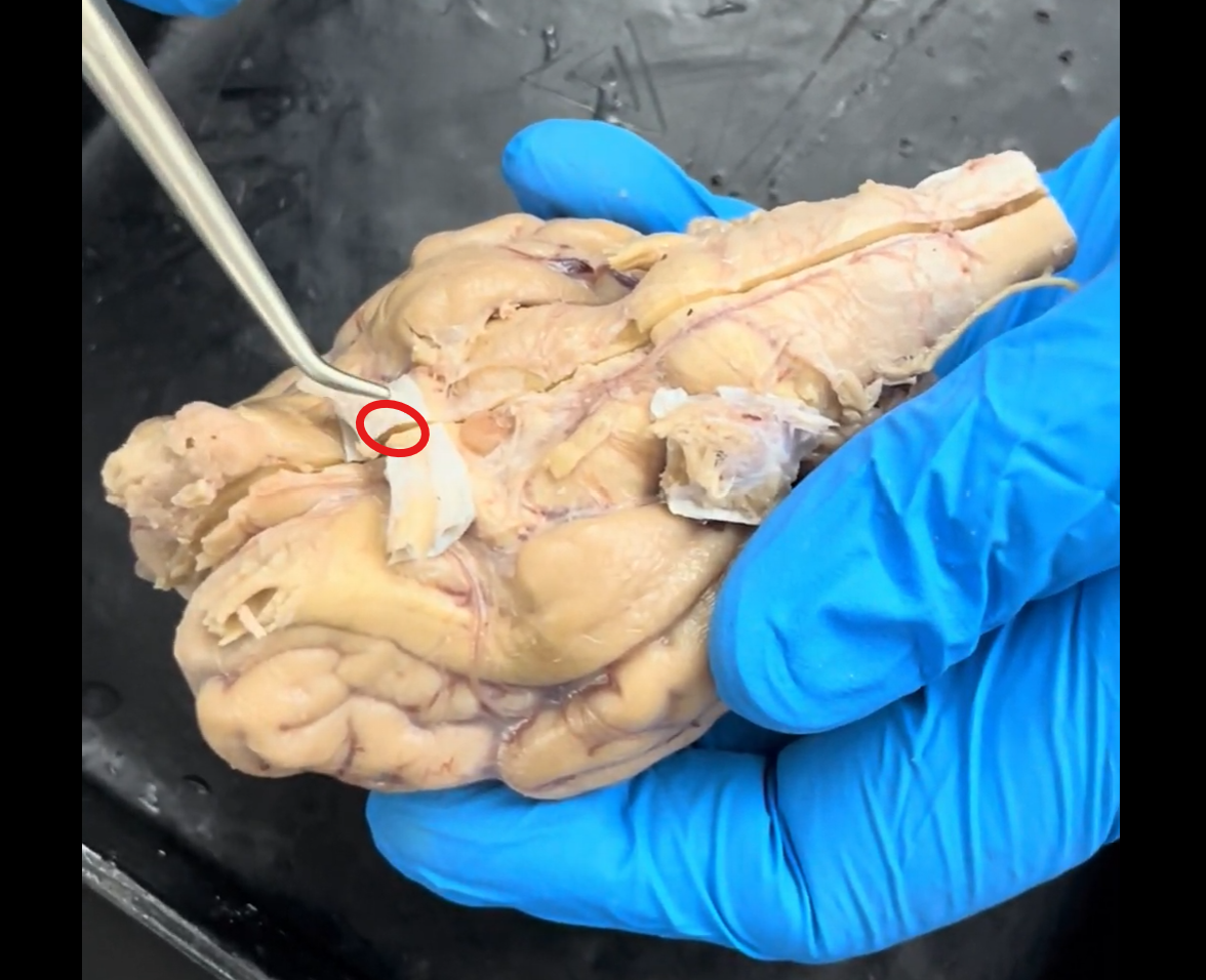
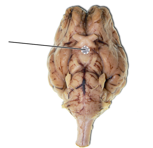
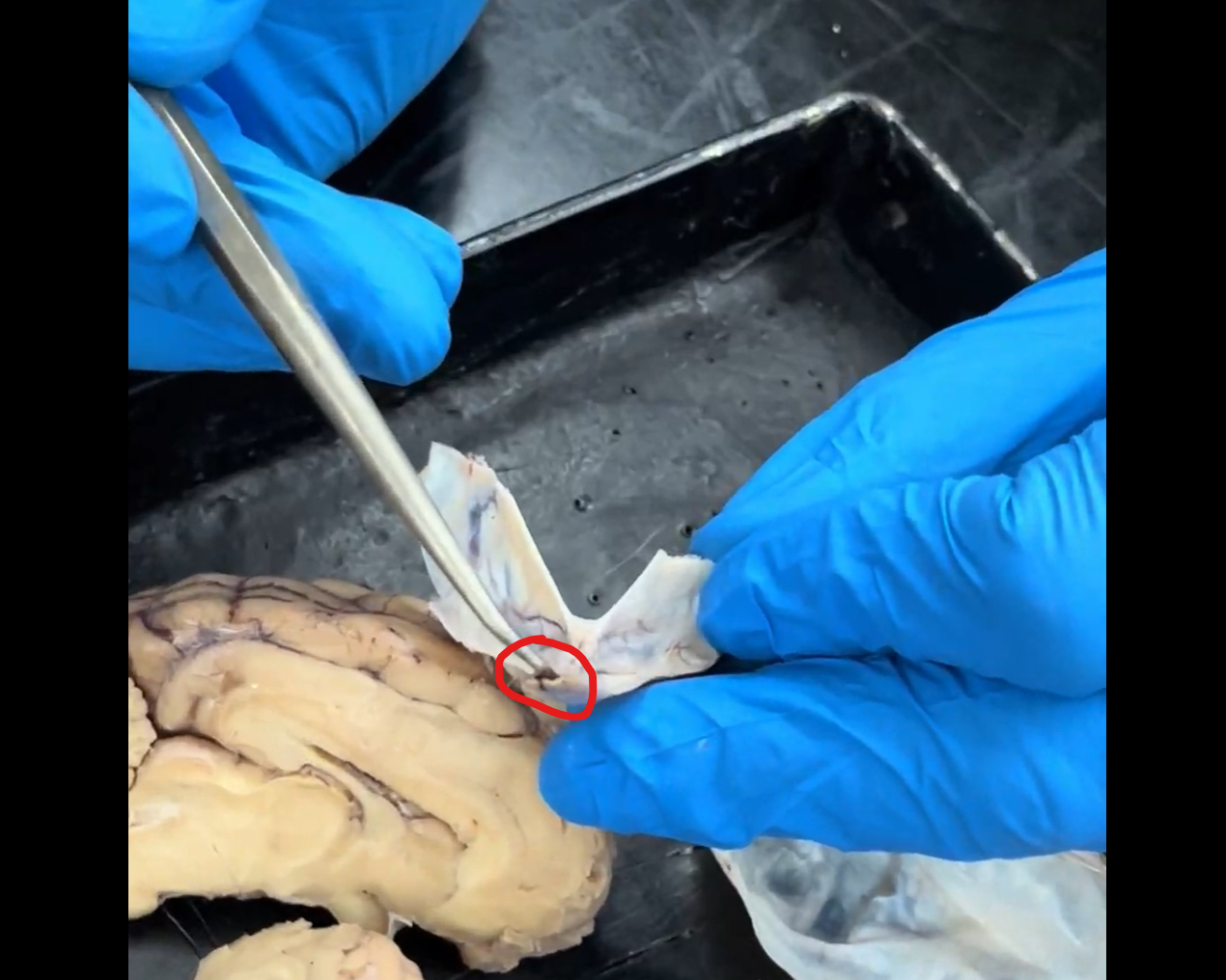
Name the part of the brain within the red circle that the probe is pointing to.
Optic chiasma.
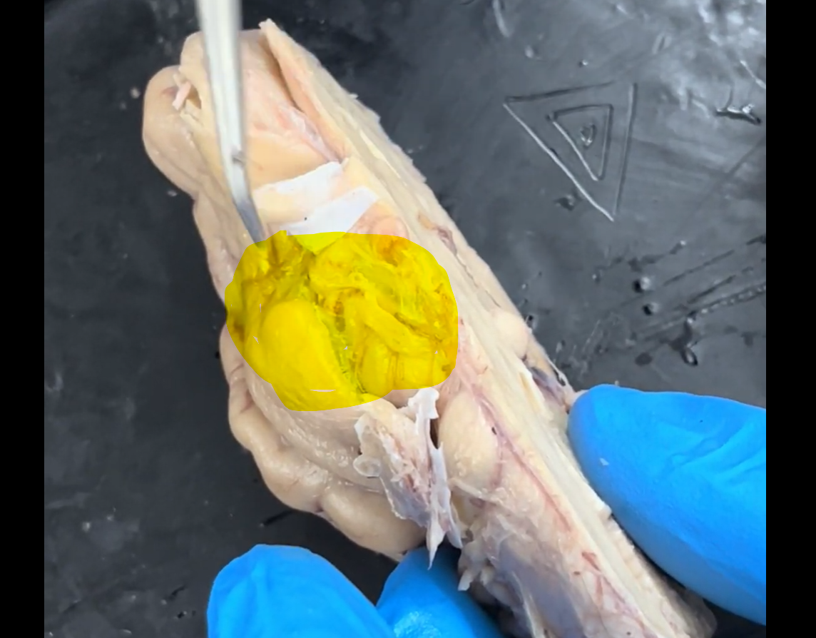
Name the part of the brain that is highlighted.
Optic chiasma.
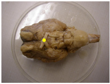
Name the part of the brain marked by the yellow dot.
Optic chiasma.
Name the part of the brain that is highlighted.
Optic chiasma.
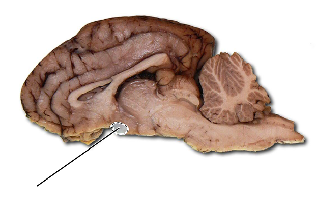
Name the part of the brain the tip of the probe is pointing to.
Optic chiasma.
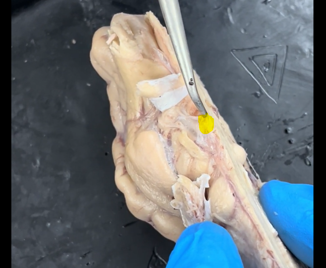
Name the part of the brain marked by the yellow dot.
Oculomotor nerve (cranial nerve III).
III = 3
Name the part of the brain that is highlighted.
Oculomotor nerve (cranial nerve III).
III = 3
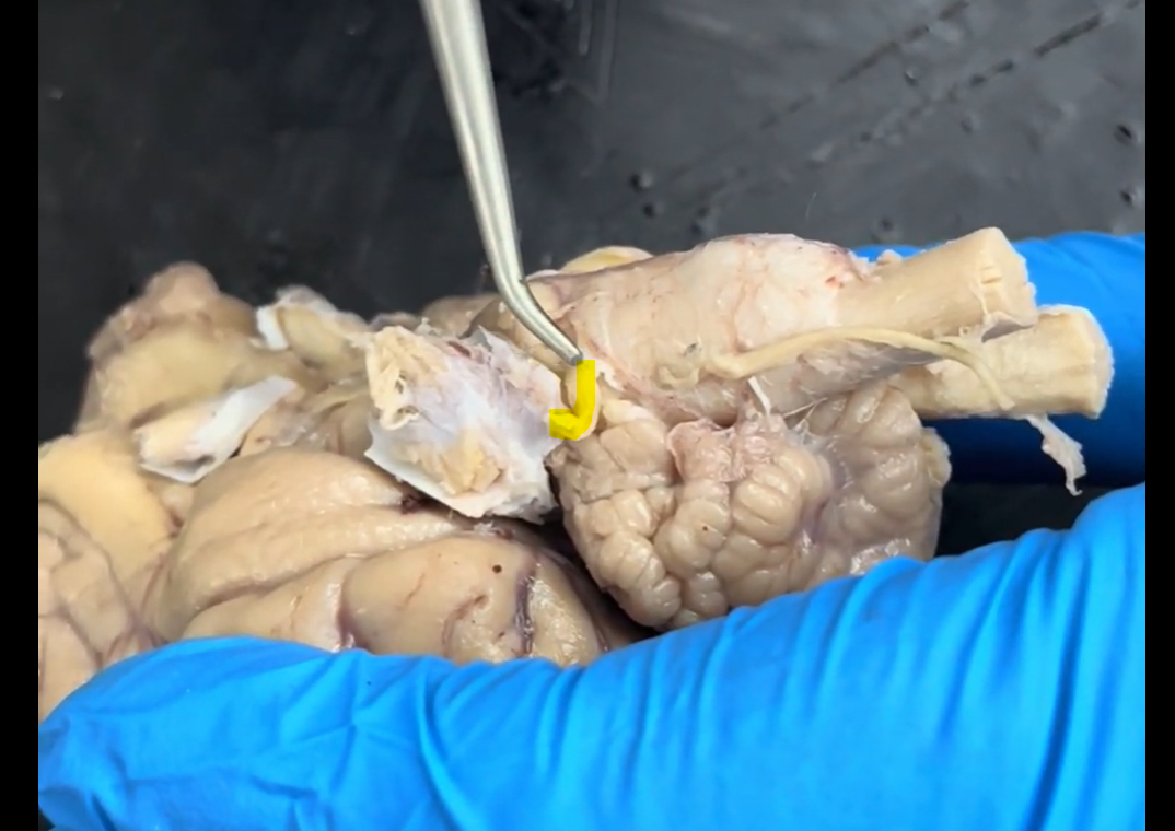
Name the part of the brain being lifted by the probe.
Oculomotor nerve (cranial nerve III).
III = 3
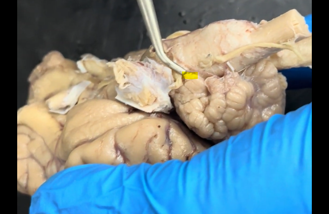
Name the part of the brain being raised by the probe and highlighted in yellow.
Trochlear nerve (cranial nerve IV).
IV = 4
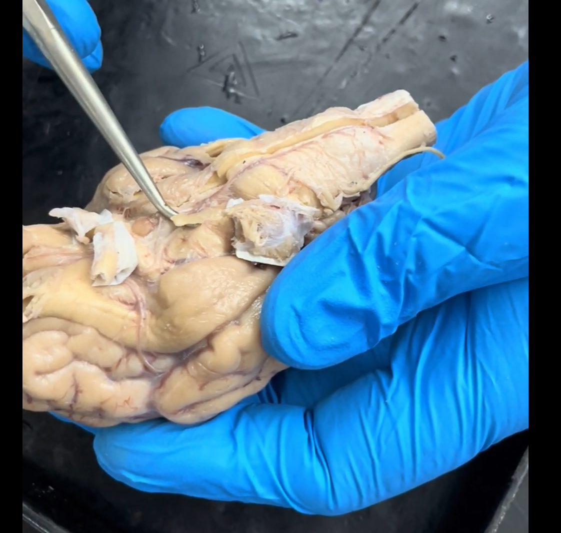
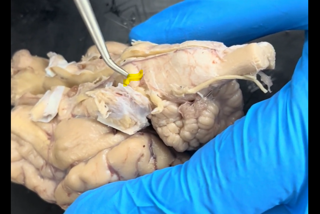
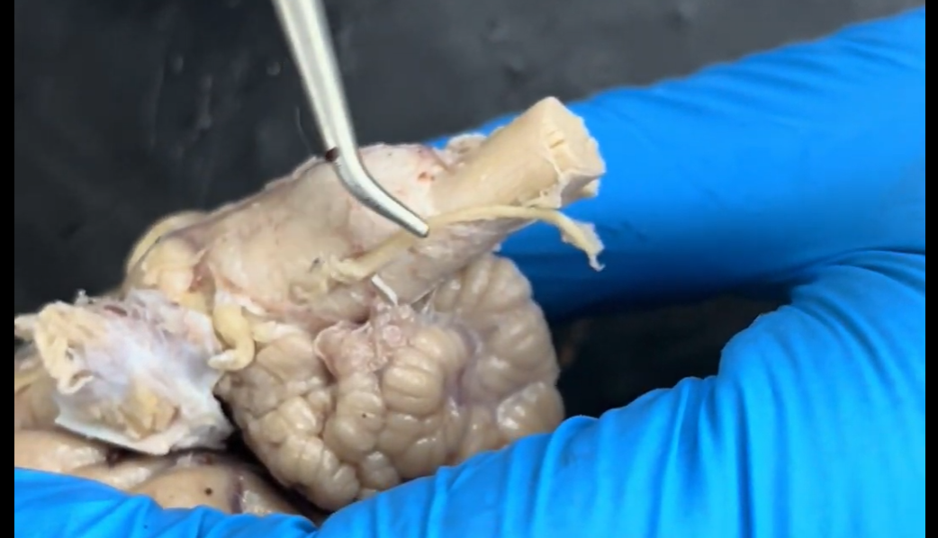
Name the part of the brain being lifted by the probe.
Trigeminal nerve (cranial nerve V).
V = 5
Name the part of the brain highlighted in yellow.
Trigeminal nerve (cranial nerve V).
V = 5
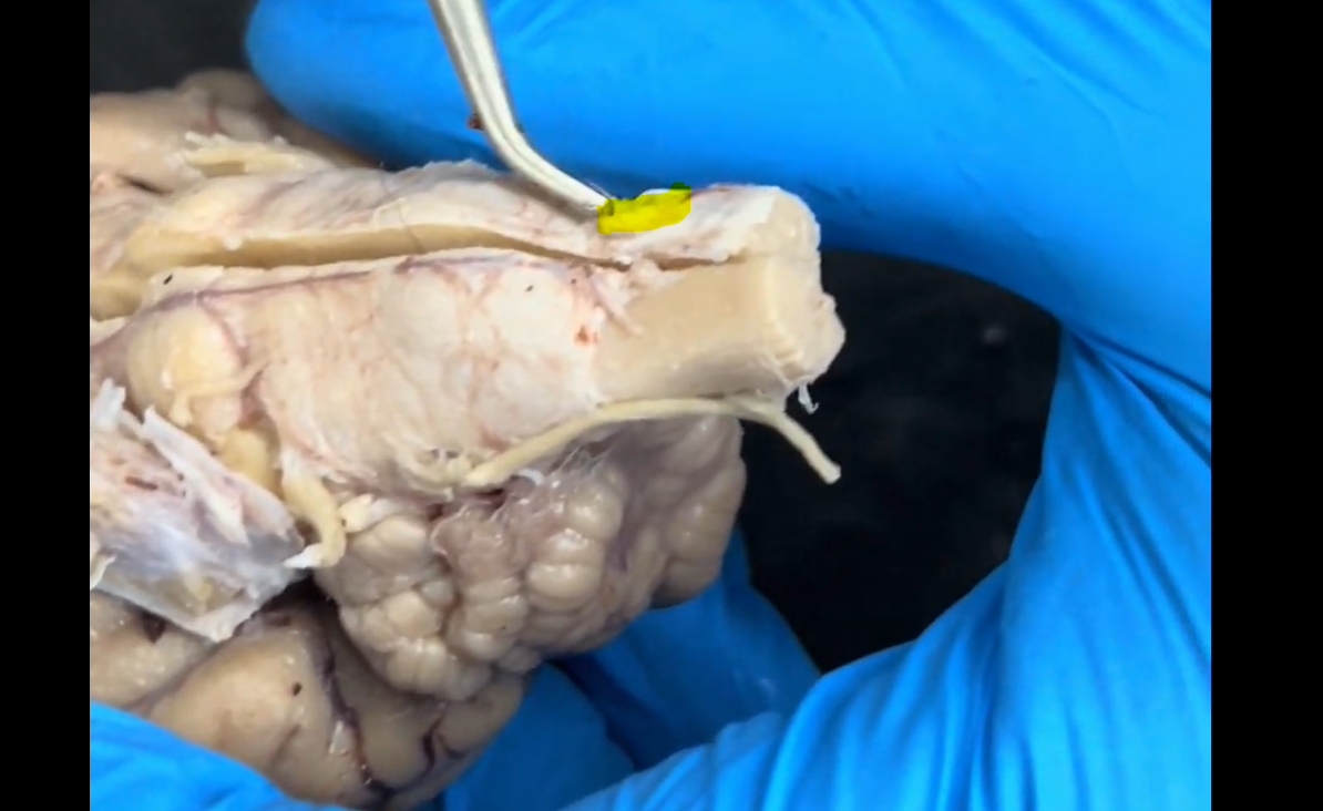
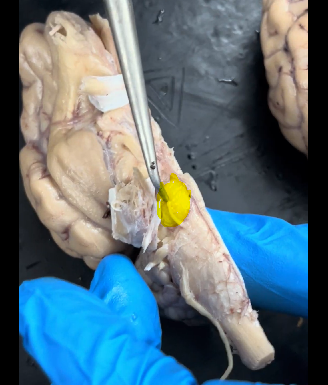
Name the part of the brain at the tip of the probe and highlighted in yellow.
Abducens nerve (cranial nerve VI).
VI = 6
Name the part of the brain at the tip of the probe and highlighted in yellow.
Facial nerve (cranial nerve VII).
VII = 7
Name the part of the brain at the tip of the probe and highlighted in yellow.
Vestibulocochlear nerve (cranial nerve VIII).
VIII = 8

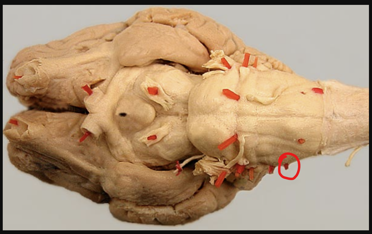
Name the part of the brain in the red circle.
Glossopharyngeal nerve (cranial nerve IX).
IX = 9
Name the part of the brain in the red circle.
Vagus nerve (cranial nerve X).
X = 10
Name the part of the brain at the end of the probe.
Accessory nerve (cranial nerve XI).
XI = 11
Name the part of the brain at the tip of the probe and highlighted in yellow.
Hypoglossal nerve (cranial nerve XII).
XII = 12
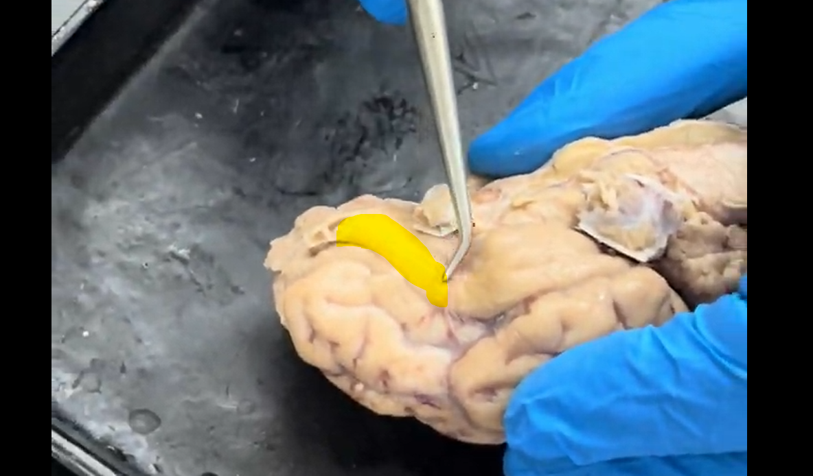
Name the part of the brain at the tip of the probe and highlighted in yellow.
Olfactory tract.
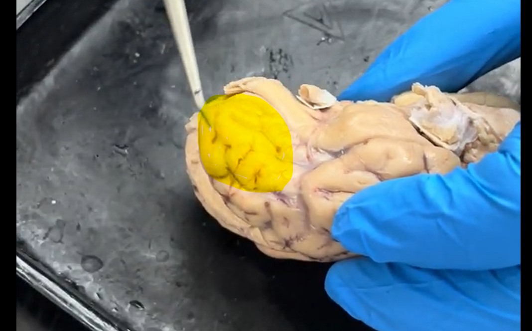
Name the part of the brain at the tip of the probe and highlighted in yellow.
Frontal lobe.
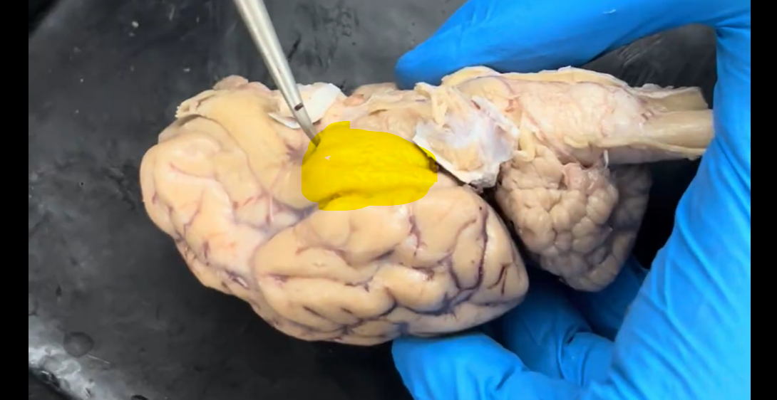
Name the part of the brain at the tip of the probe and highlighted in yellow.
Temporal lobe.
Name the part of the brain at the tip of the probe and highlighted in yellow.
Parietal lobe.
Name the part of the brain at the tip of the probe and highlighted in yellow.
Occipital lobe.
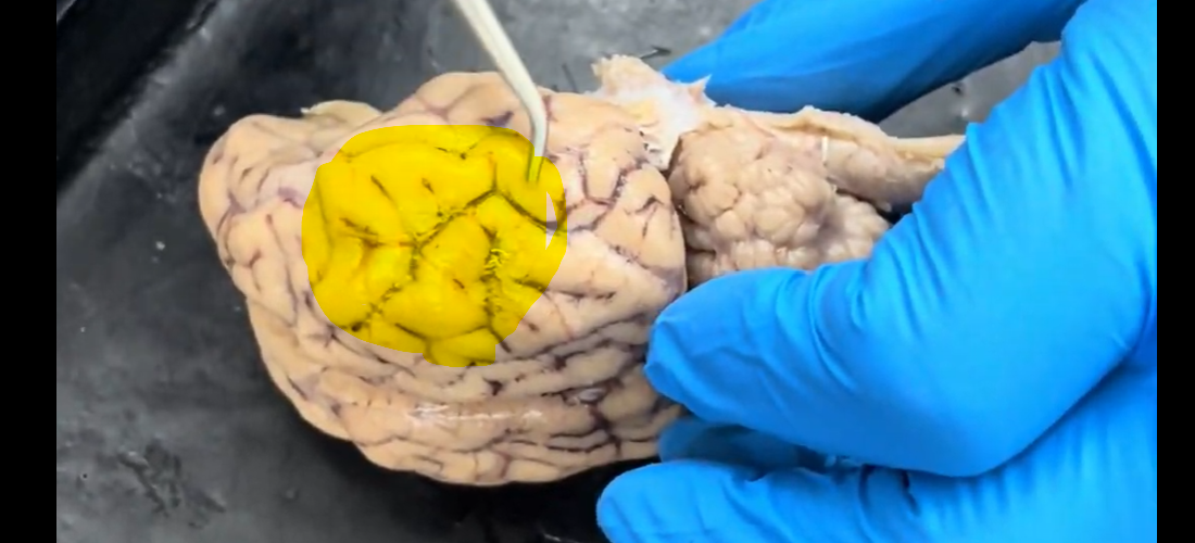
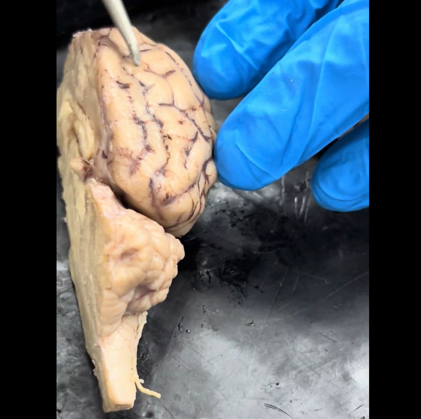
Name the part of the brain being indicated by the probe and highlighted in yellow.
Cerebellum.

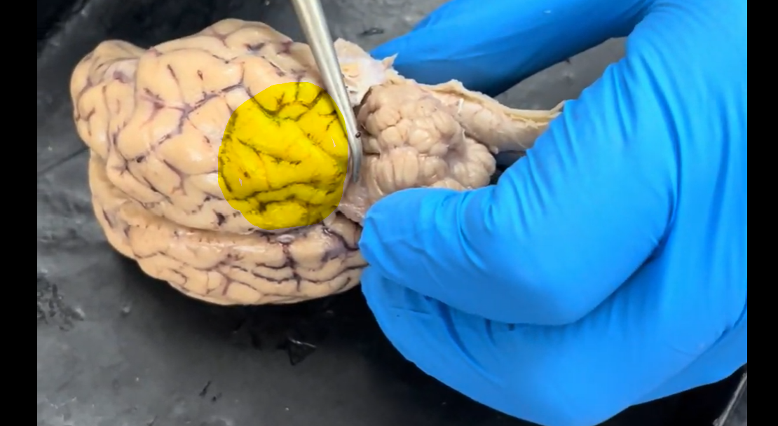
Name the part of the brain that is highlighted.
Cerebellum.
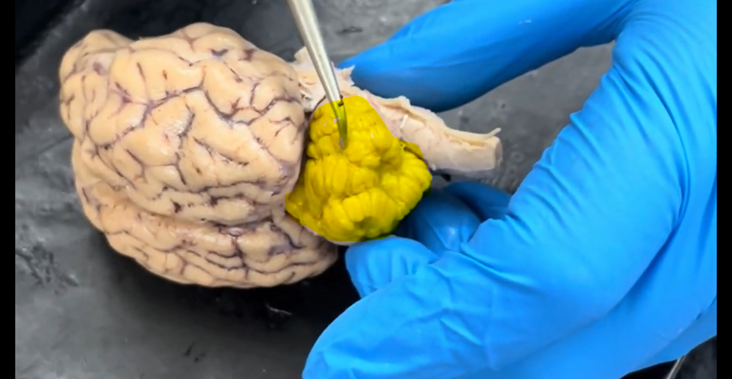
Name the part of the brain that is highlighted.
Cerebellum.
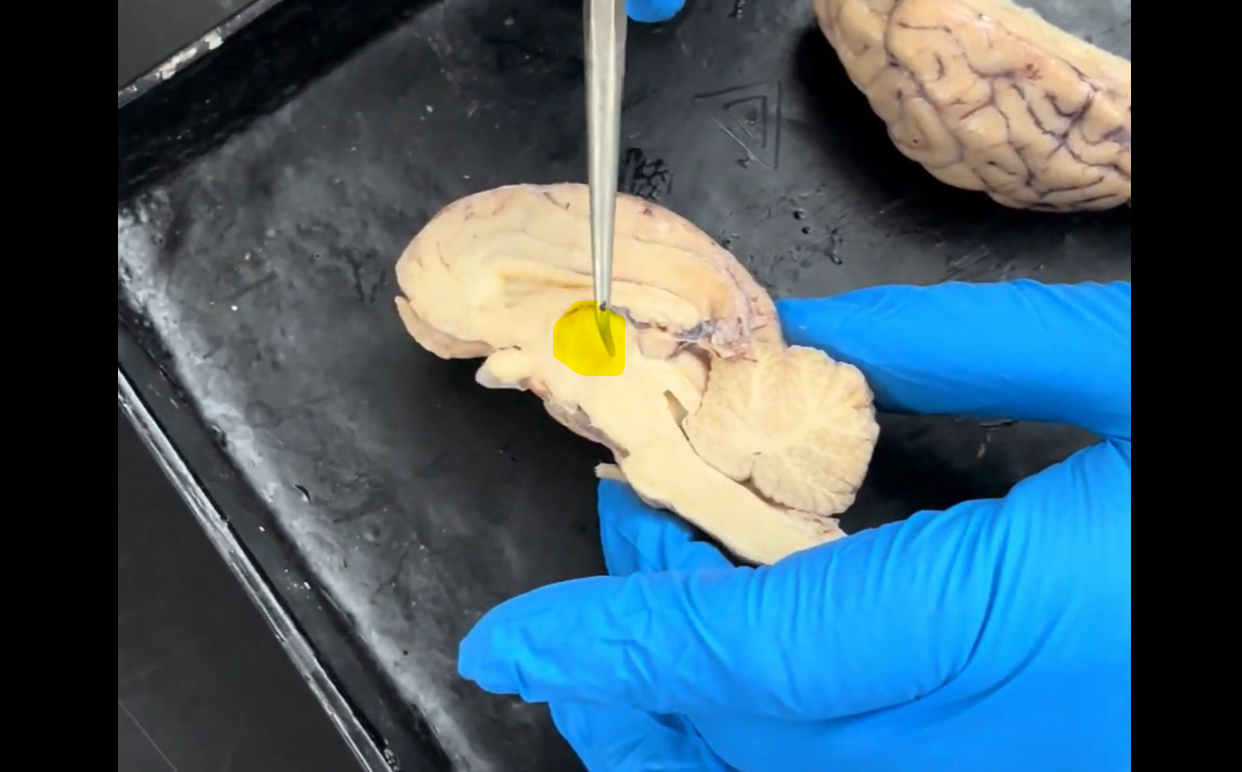
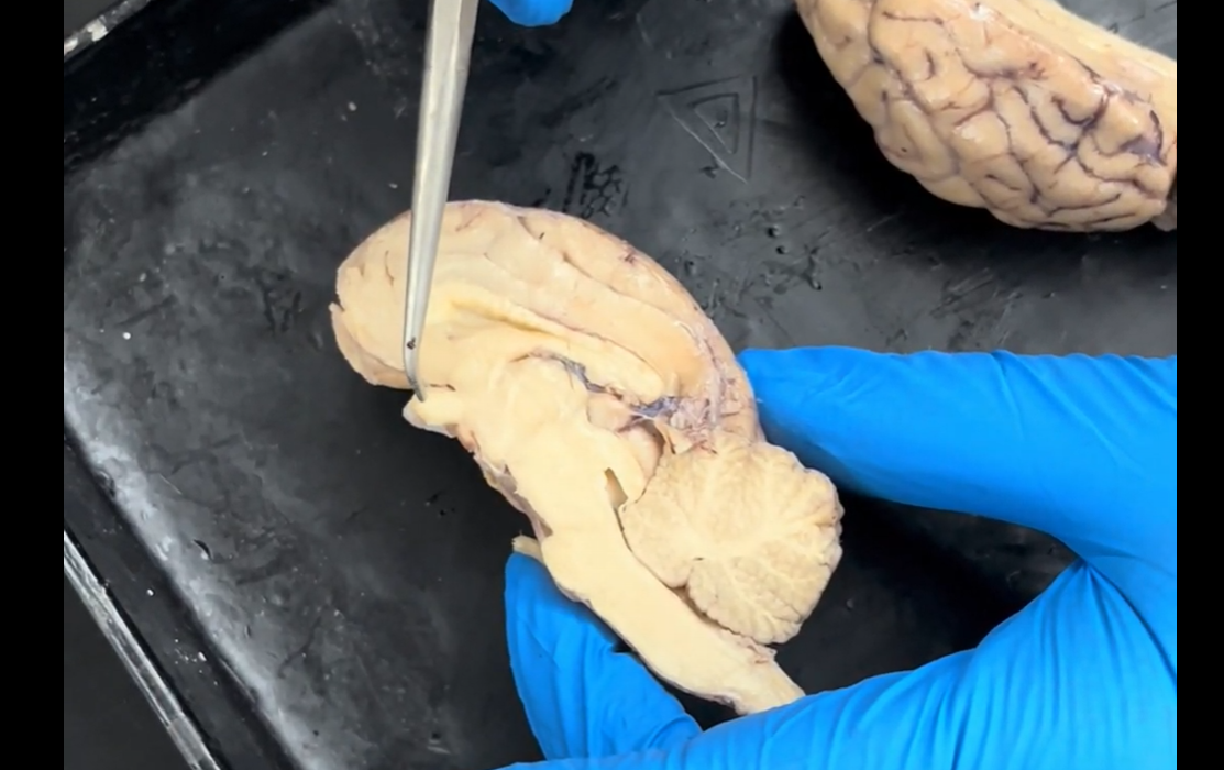
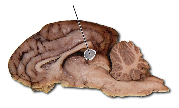
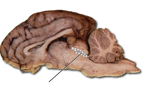
Name the part of the brain highlighted in yellow that the probe is pointing to.
Thalamus.
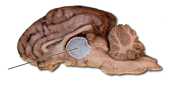
Name the part of the brain that is highlighted.
Thalamus.
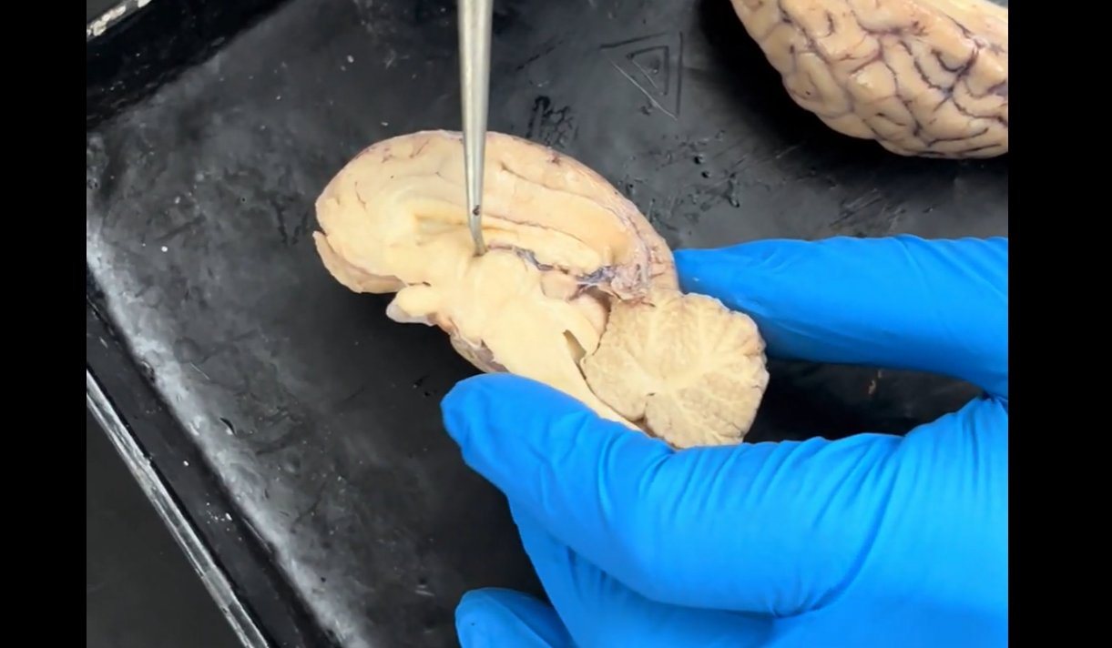
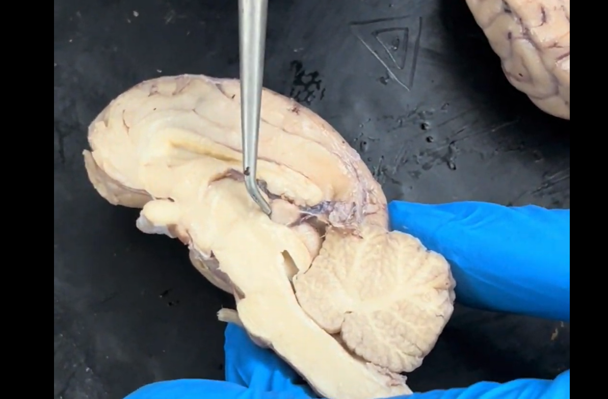
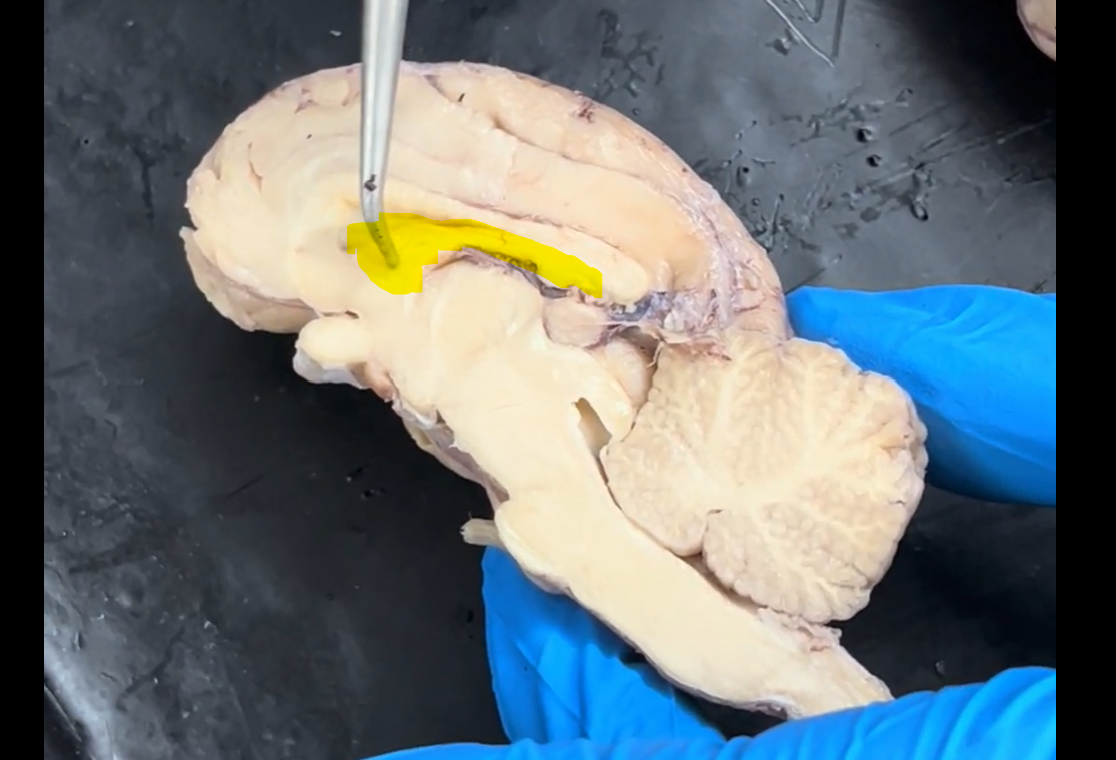
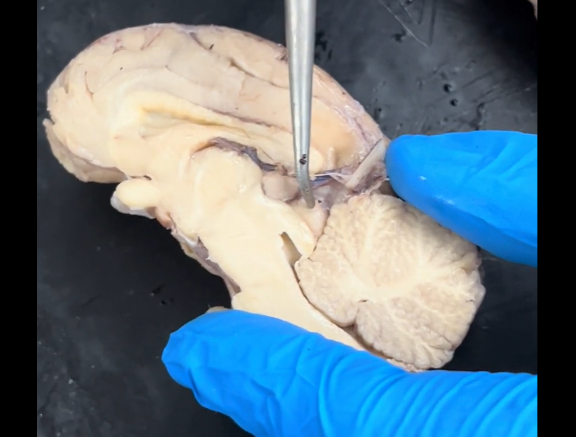
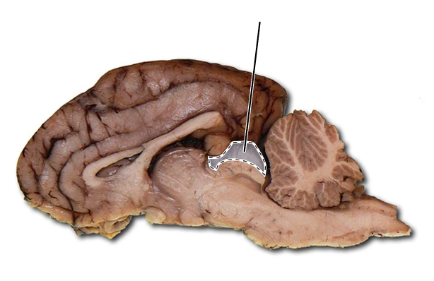
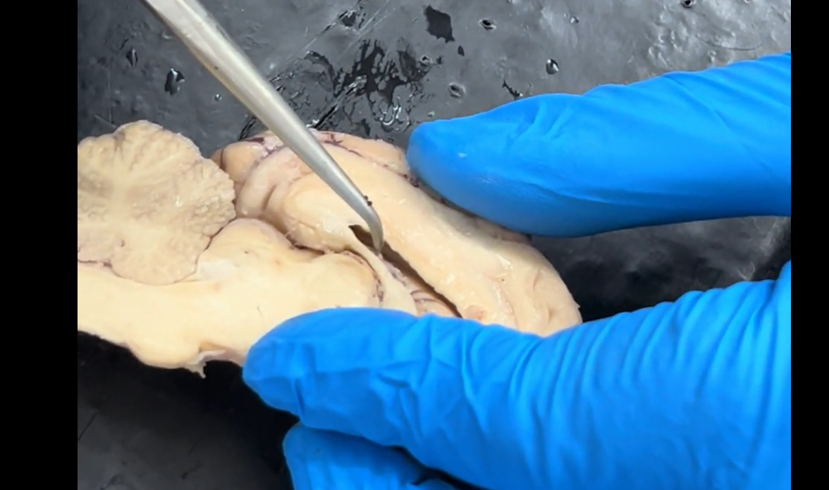
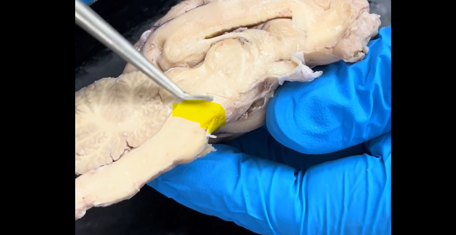
Name the groove that the tip of the probe is resting in.
Third ventricle.
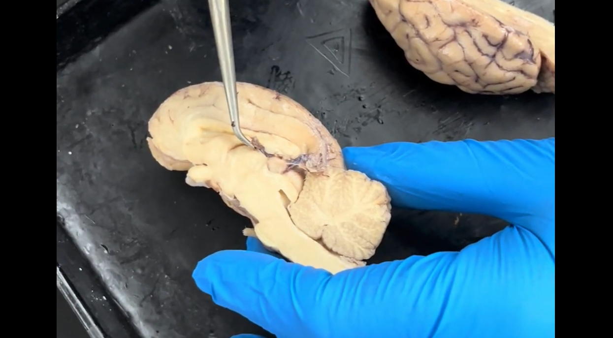
Name the dark brown material that the tip of the probe is pointing to.
Choroid plexus.
Name the round structure the probe is resting on.
Pineal gland.
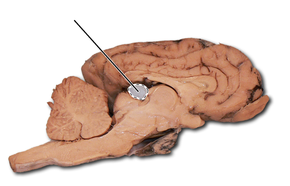
Name the highlighted structure.
Pineal gland.
Name the highlighted structure.
Pineal gland.
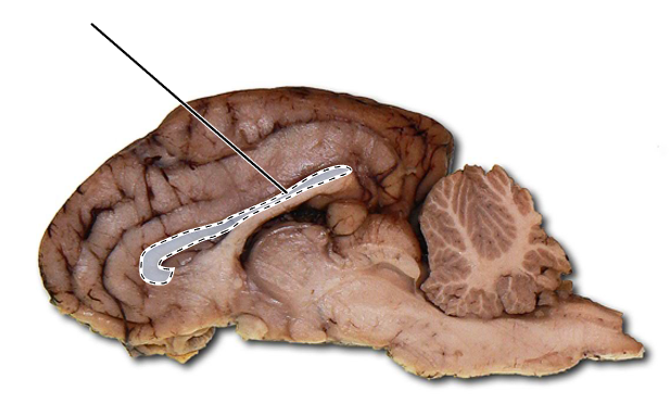
Name the part of the brain highlighted in yellow.
Fornix.
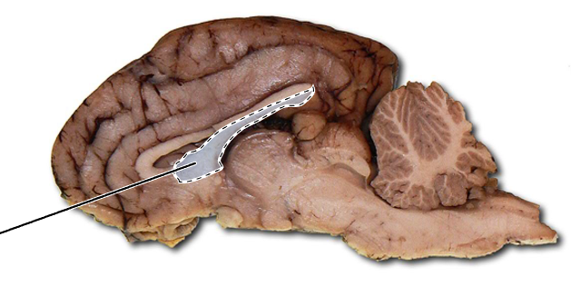
Name the part of the brain that is highlighted.
Fornix.
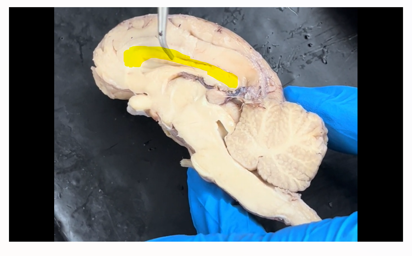
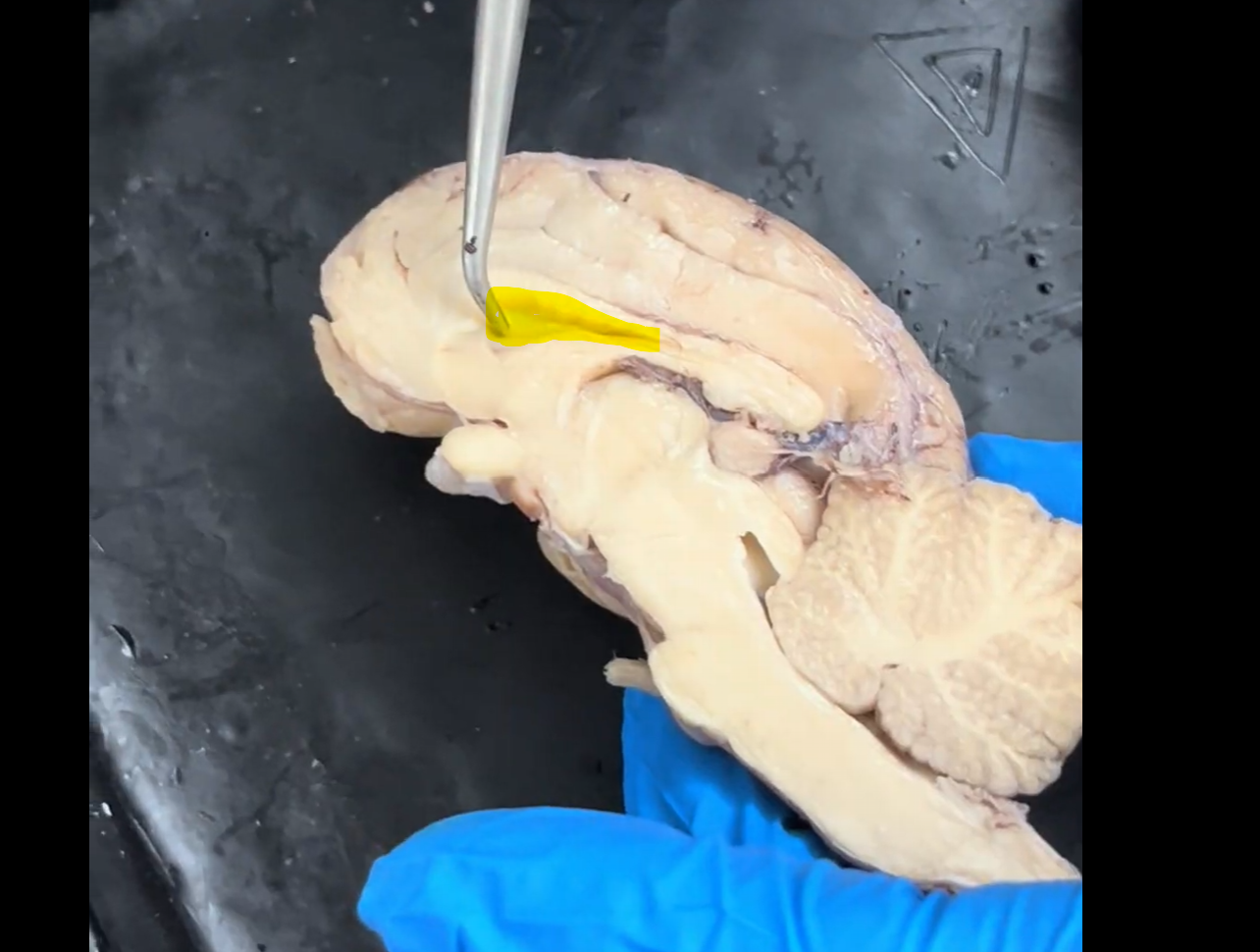
Name the part of the brain highlighted in yellow.
Corpus callosum.
Name the part of the brain that is highlighted.
Corpus callosum.
Name the highlighted membrane.
Septum pellucidum.
Name the structure indicated by the probe.
Superior colliculus (right).
Name the part of the brain that is highlighted.
Superior colliculus (right).
Name the structure indicated by the probe.
Inferior colliculus (right).
Name the part of the brain that is highlighted.
Inferior colliculus (right).
When the two highlighted structures are combined with their matching structures on the other side of the midsagittal section, what structure is formed?
Corpora quadrigemina.
What structures make up the corpora quadrigemina?
2 superior colliculi (right and left).
2 inferior colliculi (right and left).
(A total of four bodies make up the corpora quadrigemina).
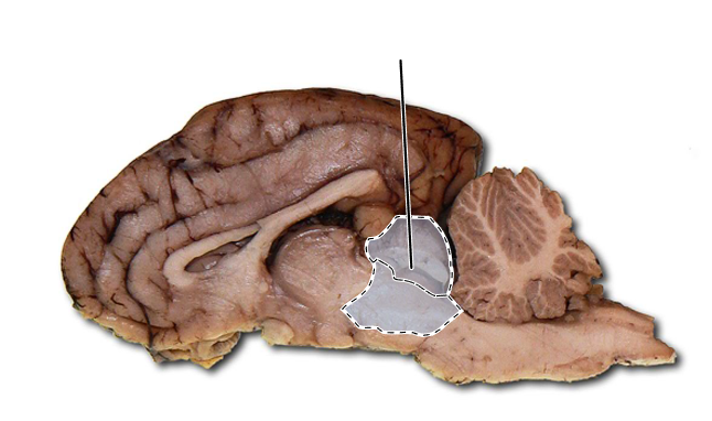
Name the part of the brain that is highlighted in yellow.
Hypothalamus.
Name the part of the brain that is highlighted.
Hypothalamus.
Name the part of the brain that is highlighted.
Hypothalamus.
Name the structure the probe is pointing to.
Mammillary body.

Name the part of the brain that is highlighted.
Mammillary body.
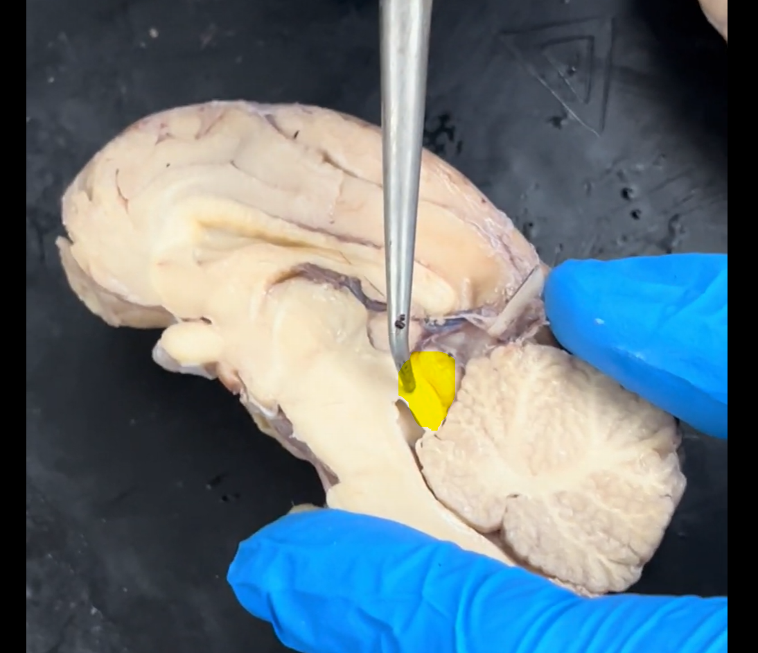
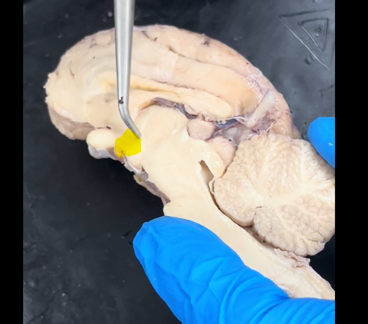
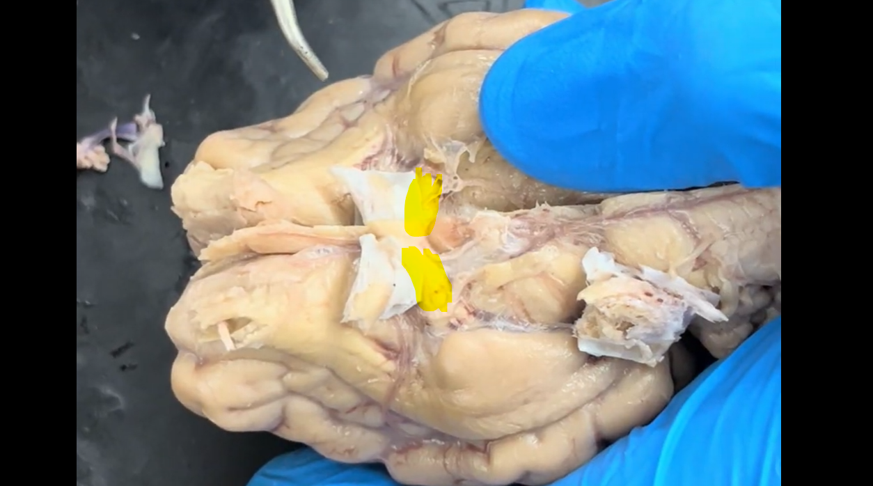
What structure should go where the yellow highlight is?
Infundibulum (pituitary stalk).
Name the part of the brain that is highlighted.
Infundibulum (pituitary stalk).
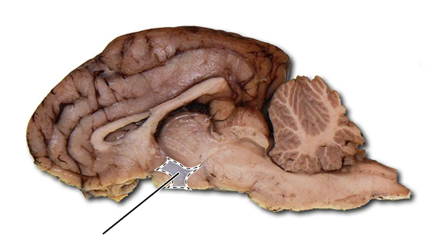
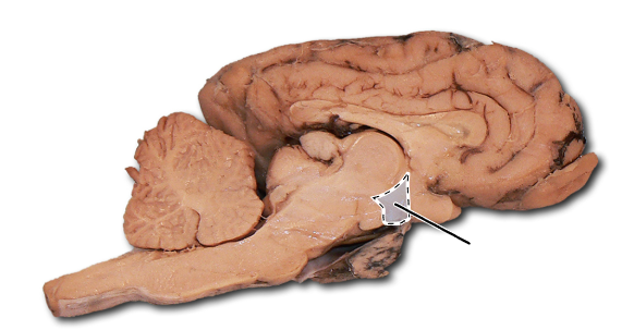
What structure normally sits inferior to the brain in the highlighted area?
Pituitary.
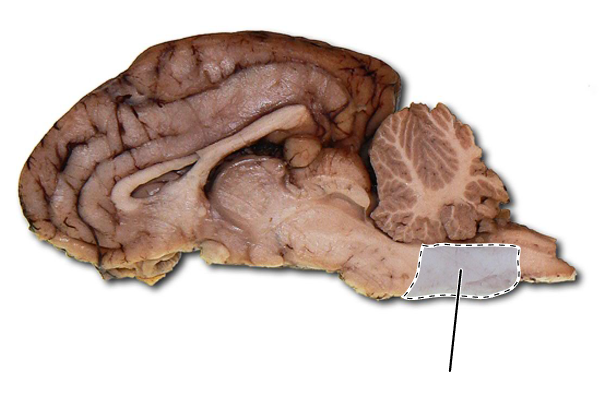
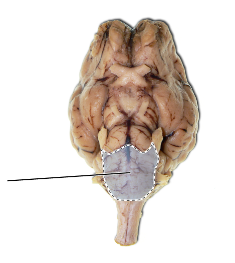
Name the part of the brain that is highlighted in yellow.
Pons.
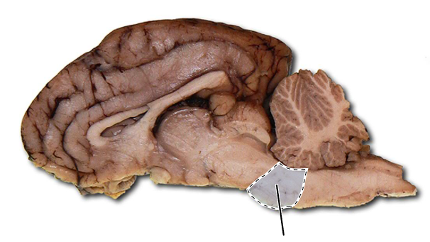
Name the part of the brain that is highlighted.
Pons.
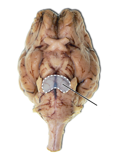
Name the part of the brain that is highlighted.
Pons.
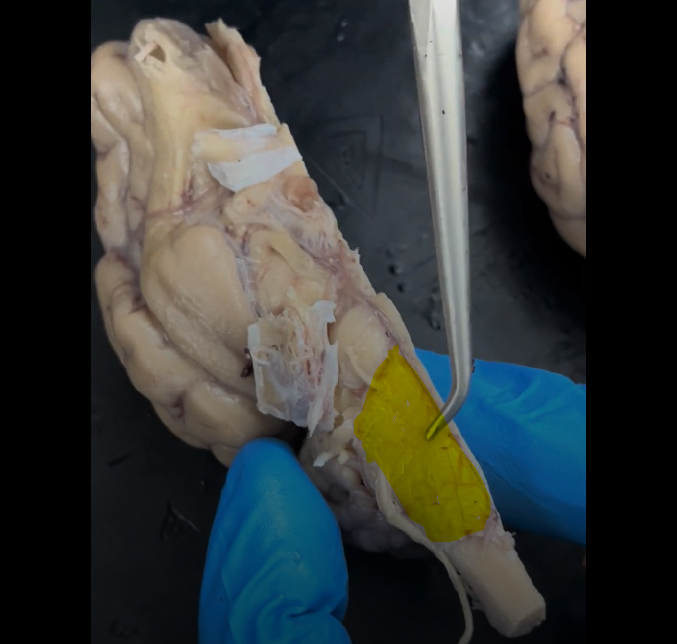
Name the part of the brain highlighted in yellow.
Medulla (oblongata).
Name the part of the brain that is highlighted.
Medulla (oblongata).
Name the part of the brain that is highlighted.
Medulla (oblongata).
Name the groove that the probe is in.
Lateral ventricle.
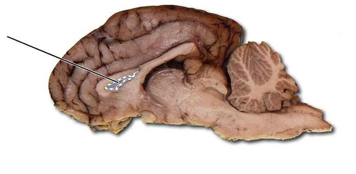
Name the highlighted part of the brain.
Lateral ventricle.
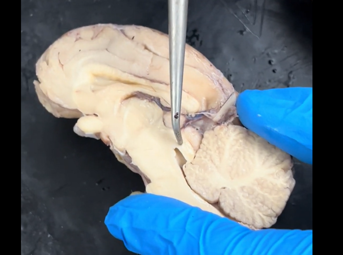
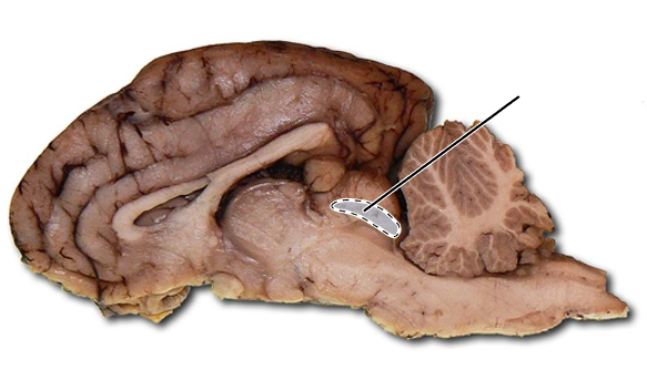
Name the highlighted groove that runs between the third and fourth ventricles.
Cerebral aqueduct.
Name the highlighted part of the brain.
Cerebral aqueduct.
Name the groove that the tip of the probe is in.
Fourth ventricle.
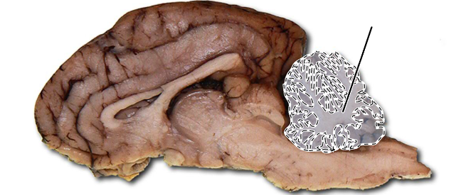
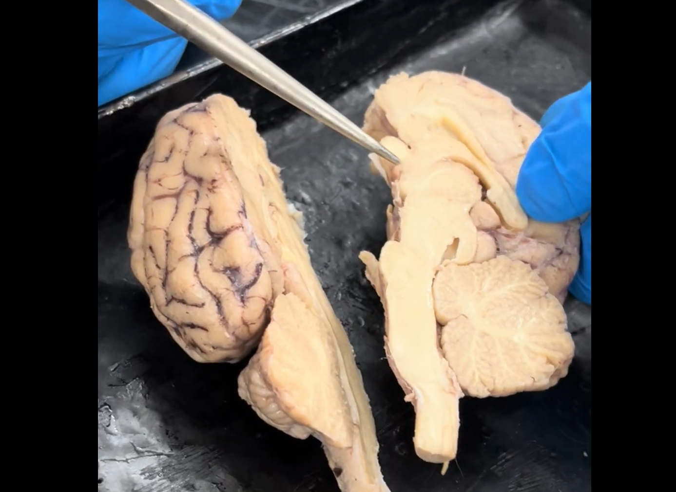
Name the white tendrils found within the cerebellum.
Arbor vitae.
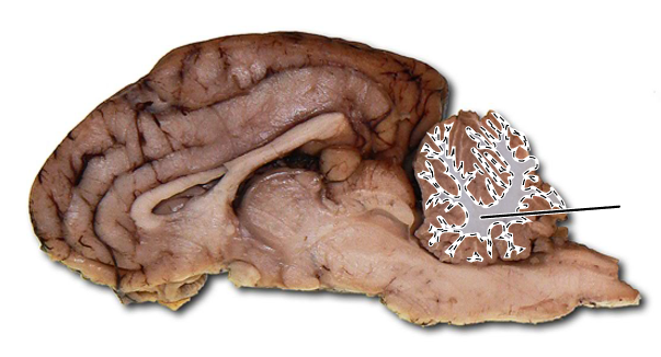
Name the highlighted part of the brain.
Arbor vitae.
Name the part of the brain that is highlighted in yellow.
Cerebral peduncle.
Name the part of the brain that is highlighted in yellow.
Midbrain (mesencephalon).
Name the highlighted part of the brain.
Midbrain (mesencephalon).
What is the highlighted structure within the center of the thalamus called?
Intermediate mass.
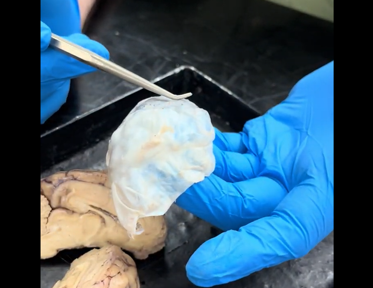
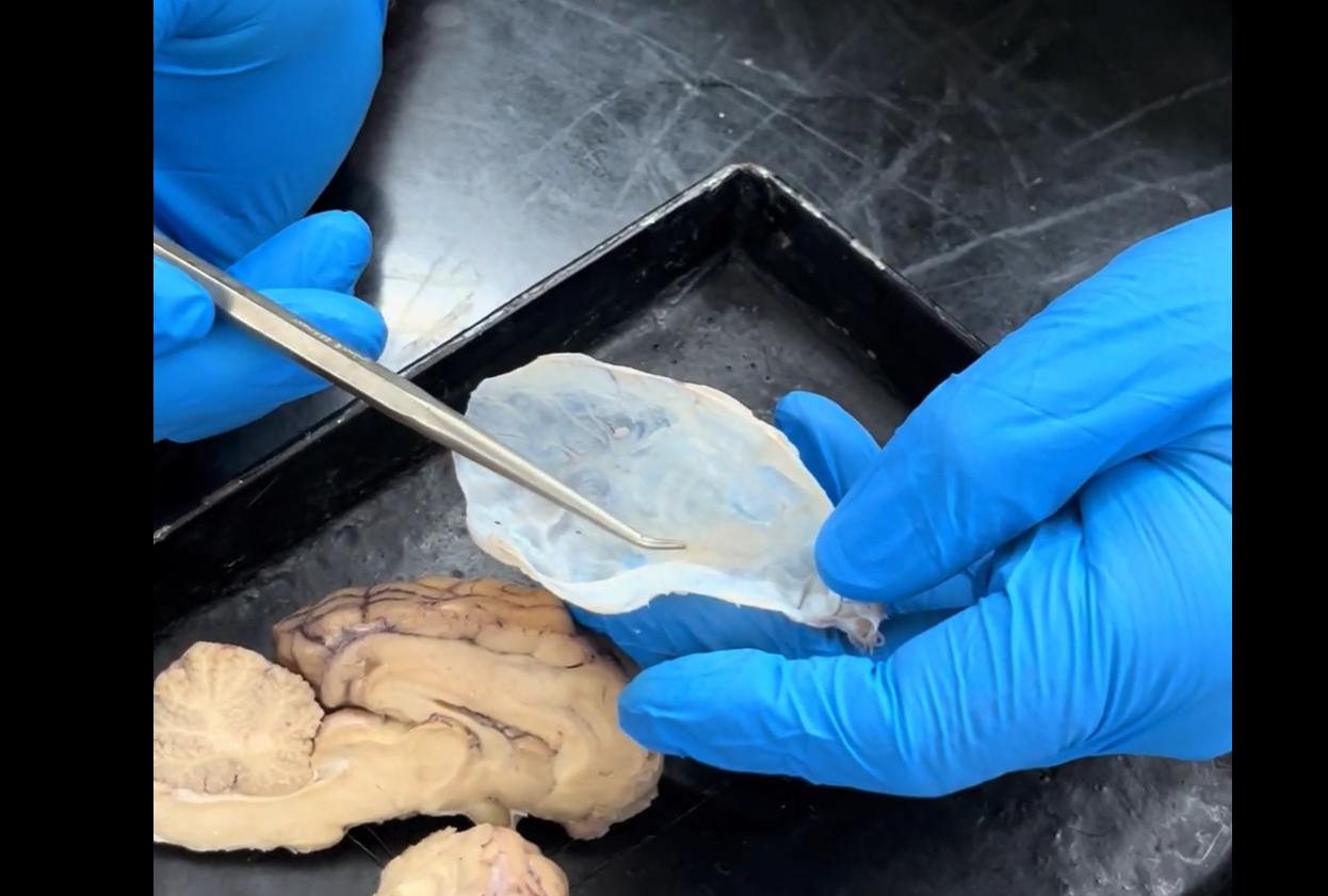
What is this white structure that coats the outside of the brain called?
Dura mater (meningeal dura).
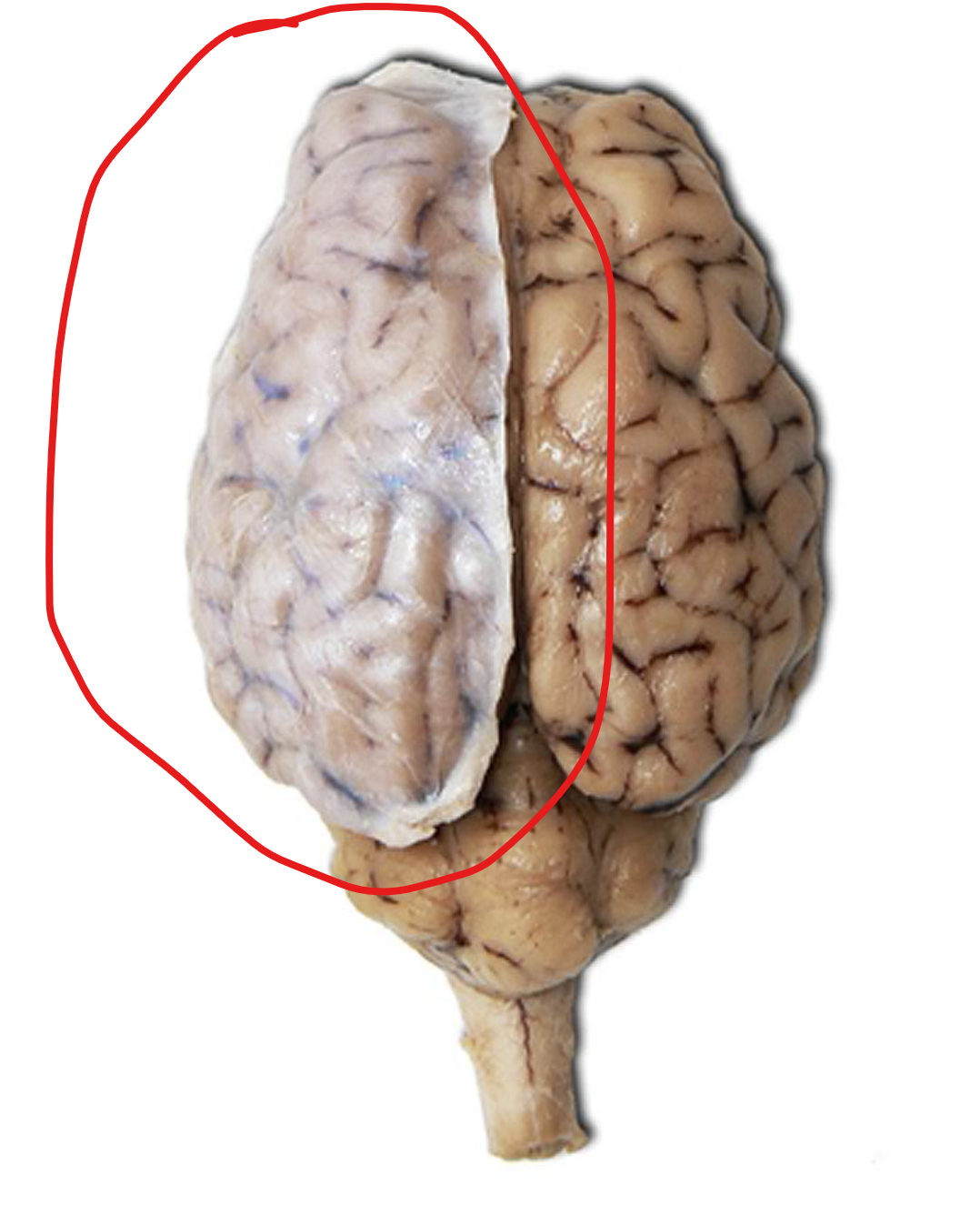
What is the white structure/sheet within the red circle called?
Dura mater (meningeal dura).
What is found on the inside layer of the meninges?
Arachnoid.

What is the space between the meninges and the brain itself called?
Subarachnoid space.
Name the dark spot seen within the red circle called.
Superior sagittal sinus.
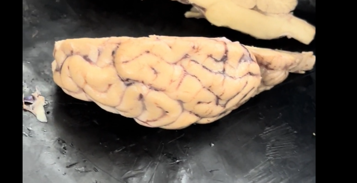
What are the “hills” on the surface of the brain called?
Gyri.

What are the dark “valleys” on the surface of the brain called?
Sulci.
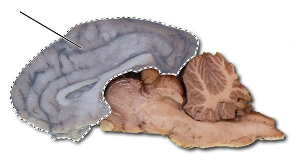
Name the highlighted part of the brain.
Cerebrum.
Name the highlighted structures.
Optic tracts.
What is the clear, shiny membrane on the surface of the brain called?
Pia mater.
What are the “white” or lighter parts of the brain called (seen most clearly in the brainstem).
White matter.
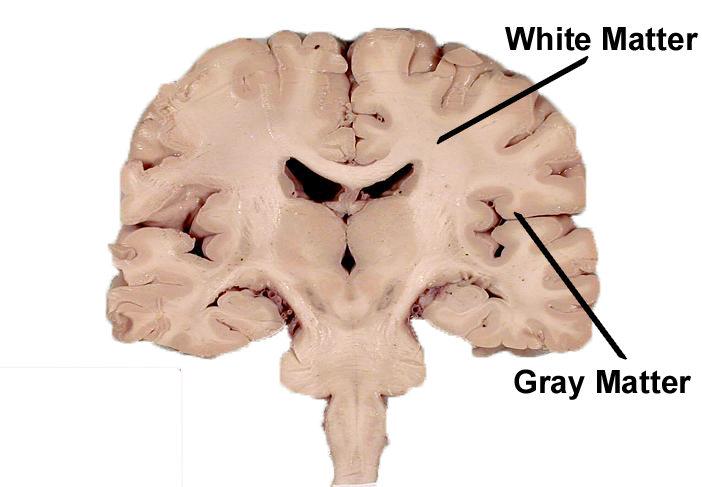

What are the “gray” or darker parts of the brain called (seen most clearly in the cerebellum, in contrast to the arbor vitae.
Gray matter.
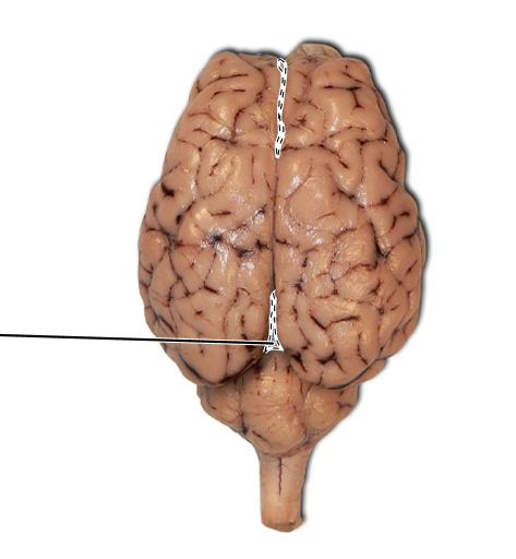
What is the line down the midsagittal plane of the brain called?
Longitudinal cerebral fissure.
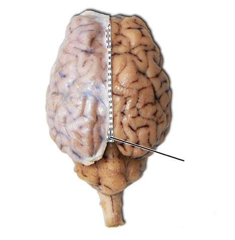
What is the line down the midsagittal plane of the brain called?
Longitudinal cerebral fissure.
What is this portion of the brain called (contains both the thalamus and the hypothalamus)?
Diencephalon.
(The diencephalon can be described as any region of the brain with “thalamus” in its name).