619 8/25 GLA Embryology and Development
 Call Kai
 Learn
Learn Practice Test
 Spaced Repetition
 Match
Match Flashcards
 Knowt Play
1/97

There's no tags or description
Looks like no tags are added yet.
Name | Mastery | Learn | Test | Matching | Spaced | Call with Kai | Chat |
|---|
No analytics yet
Send a link to your students to track their progress
98 Terms
Prenatal Development vs Pregancy
pregnancy: 40 weeks
prenatal development: 38 weeks (fertilization to birth)
Last Menstrual Period
date two weeks before fertilization that most women use to know if they are pregnant
Gestation
the 40-week pregnancy time span in reference to the fetus
Developmental Age
the actual developmental age of the fetus
Gestational Age
the age of an embryo or fetus timed from the first date of the lmp
Divisions of Prenatal Development
weeks 1-2
weeks 3-8
weeks 9-38
Weeks 1-2 (Simple)
fertilization, implantation in endometrium of uterus, formation of two layers, development of primitive streak
Weeks 3-8 Embryonic Period (Simple)
formation of three layers, beginning of development of all orgnas and body parts (aka organogenesis)
Weeks 9-38 Fetal Period (Simple)
growth and maturation of organs and body parts
Overall Impact of Tertogens: Week 1-2
all or none
Teratogens
substances that can harm the fetus
Overall Impact of Tertogens: Week 3-8 Embryonic Period
major morphologic anomalies
Overall Impact of Tertogens: Week 9-38 Fetal Period
physiologic and minor morphologic anomalies
What is the most sensitive time for the development of all organs and body parts?
embryonic period (3-8)
Overall Fetal Development Chart
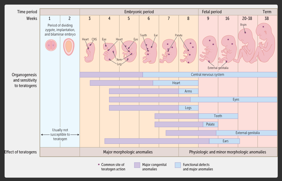
Week 1: Fertilization and Implantation
egg and sperm encounter each other in the distal oviduct/uterine tube
sperm release enzymes that create a hole in the egg covering (zona pellucida) so one sperm enters the egg
diploid zygote is formed by haploid sperm/egg
mitosis begins
morula develops, then blastocyst develops from morula
while the morula and blastocyst form, they are moved through the oviduct into the uterus
in the uterus, the blastocyst implants in the endometrium during the last days of week 1
Distal Oviduct/Uterine Tube
region where egg contacts multiple sperm
Zona Pellucida
covering of the egg that is degraded by sperm enzymes to allow fertilization
Fertilization Diagram
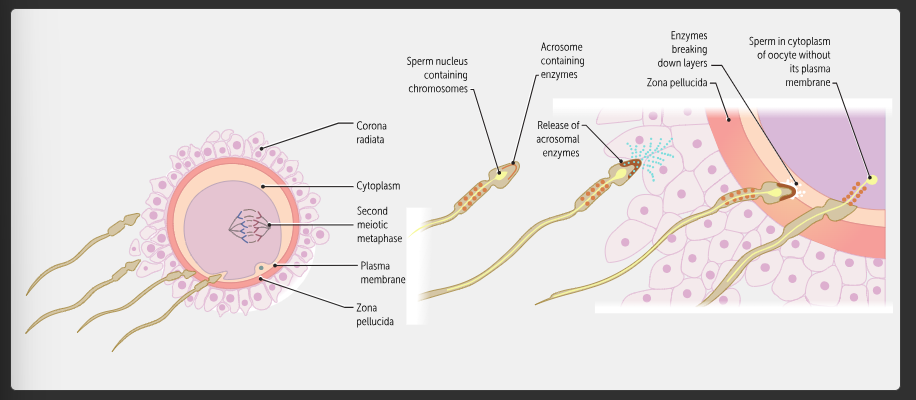
Morula
solid ball of cells created when mitosis happens after diploid zygote formation
Blastocyst
formed when a fluid-filled cavity develops in the morula
2 Regions of Blastocyst
trophoblast
inner cell mass
Trophoblast
circle of peripheral cells of the blastocyst
Trophoblast Develops into
placenta and fetal membranes, aka extraembryonic tissues
Inner Cell Mass
inner ball of cells at one pole of the blastocyst
Inner Cell Mass Develops Into
embryo
Blastocyst Implantation
while the blastocyst is developing from the morula, it is traveling through the oviduct to the uterus where it is then implanted into the endometrium during the last days of week 1
Week 1 Fertilization and Implantation Diagram
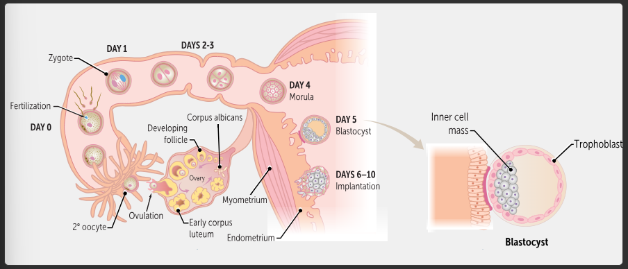
Week 2: Bilaminar Disc
inner cell mass of blastocyst organizes itself into the two layers of the bilaminar disc
layers of bilaminar disc develop cavities
primitive streak develops
Bilaminar Embryonic Disc
formed during week 2 from the inner cell mass
2 Layers of Bilaminar Disc
epiblast
hypoblast
Bilaminar Disc Formation Diagram
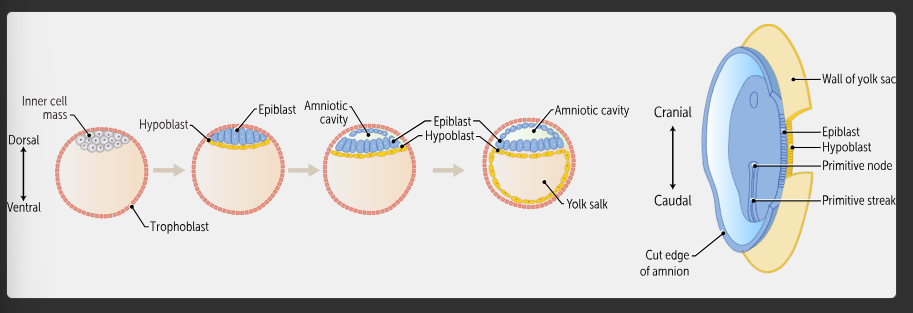
Epiblast
dorsal layer of bilaminar disc that forms the entire embryo
Hypoblast
the ventral layer of the bilaminar disc that forms the lining of the yolk sac but does not form any parts of the embryo
eventually disappears
2 Cavities that Develop from Bilaminar Disc
amniotic cavity
yolk sac
Amniotic Cavity
develops dorsally in the epiblast during week 2
Yolk Sac
develops dorsally in the hypoblast during week 2
Primitive Streak
thickening that arises in the caudal midline of the epiblast at the end of week two
Primitive Node
cranial end of the primitive streak (near the midpoint of the embryonic disc), the organizer and inducer for a variety of embryonic events through release of signaling molecules
What is the primitve streak the source of?
cells that are involved in gastrulation in week 3
Teratogen in Weeks 1 & 2
the conceptus either dies from injury or lives without an effects from the agent, called all-or-none effecta
Conceptus Susceptibilityin Weeks 1 & 2
conceptus is not very susceptible to teratogens in week 1 and 2
Week 3: Gastrulation
bilaminar disc becomes trilaminar (3 layers)
forms ectoderm, mesoderm, endoderm
organogenesis can begin
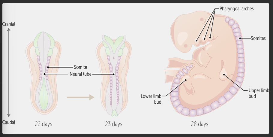
somites develop as soon as the three germ layers have formed in week 3
3 Layers of Trilaminar Disc Formed During Gastrulation (from dorsal to ventral)
ectoderm
mesoderm
endoderm
Trilaminar Disc Diagram
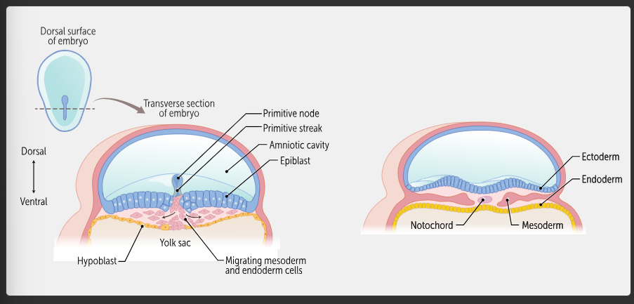
Ectoderm is Associated with
amniotic cavity
Endoderm is Associated with
yolk sac
Somites
42-44 paired blocks of mesoderm, next to the midline, that arise in a craniocaudal sequence
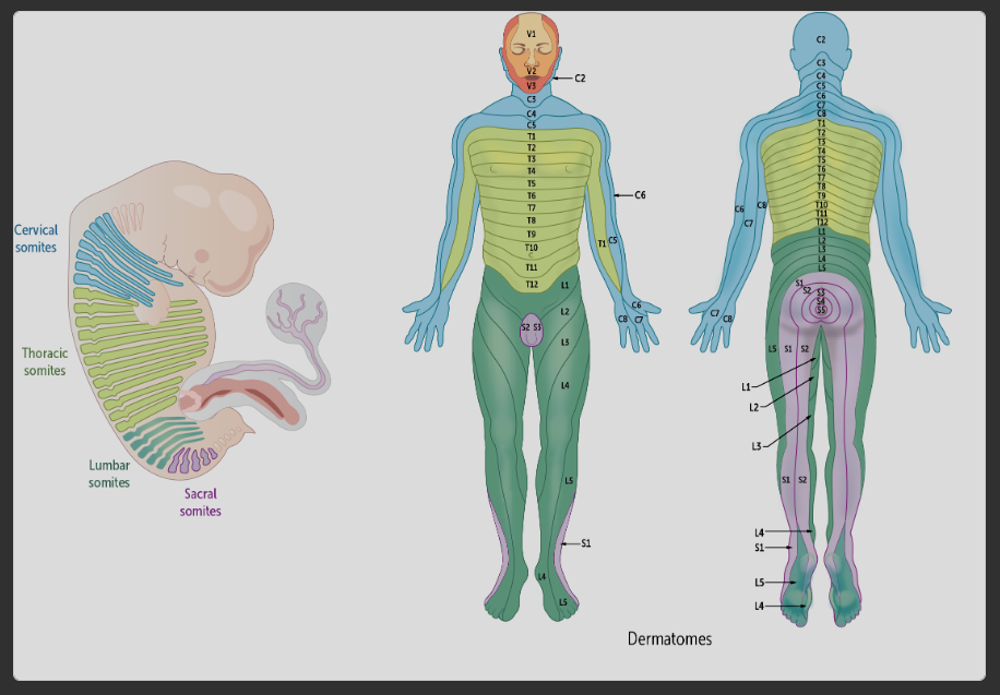
form the body segments, visible in dermatomes
Somites Formation Diagram
Dermatomes Fetus vs Adult Diagram
Somites Development Order
sequentially in the cranial to caudal direction
occurs from proximal to distal
Week 4: Folding
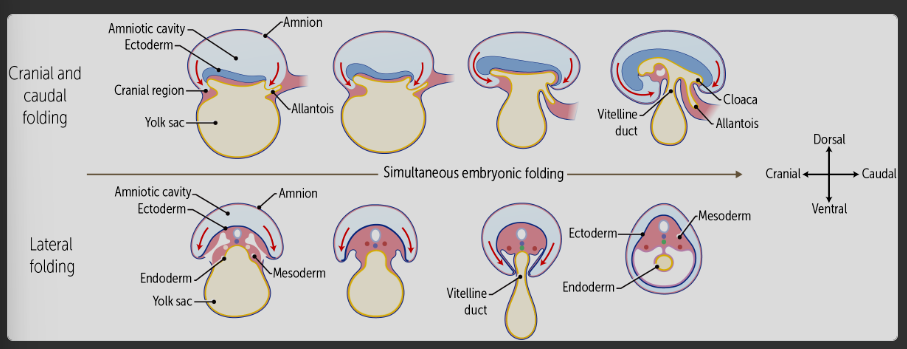
portions of the flat trilaminar embryonic disc fold
the cranial and caudal end folds and lateral sides each fold ventrally to create a 3D trunk
four limbs begin to develop from the trunk as well as gastrointestinal, respiratory, genitourinary, and cardio
neurulation ocurs
Folding of Week 4 Diagram
When does the heart start to beat?
week 4
Neurulation
occurs during week 4 to give rise to the neurological system
Weeks 5-8: Organ Systems
organs and body parts arise and begin to mature
by the end of this period the exterior of the body is nearly complete and all organ systems have begun to form
Why do teratogens in weeks 3-8 cause major atomic anomalies?
because all organs and body parts are just beginning to form, so an injury would have a profound impact on that structure
Weeks 9-38: Fetal Period Development
each body part and organ continues to mature and grow, and by week 38 all organs and body parts are present and mature enough to allow for life outside of the uterus
Why do teratogens have a minor effect on weeks 9-38?
in these later weeks, each body organ has already been formed
8 Families of Genes Important for Human Development
homeobox
hox
sox
pax
wnt
hedgehog
transforming growth factor b
fibroblast growth factor (fgf)
Homeobox
family of genes important for human development, encode transcription factors and are involved in body positioning and patterning
Hox
family of genes important for human development, encode transcription factors and are involved in reproductive and nervous system development
Pax
family of genes important for human development, encode transcription factors
Wnt
family of genes important for human development, encode signaling proteins and are involved in cell fate and patterning
Hedgehog
family of genes important for human development, includes the sonic the hedgehog (SHH) gene which encodes a signaling protein that has roles in cell growth, cell specialization, and the normal shaping/patterning of the body
Transforming Growth Factor b
family of genes important for human development, encode signaling proteins and are involved in cell mitosis, differentiation, motility, adhesion, and apoptosis
includes bone morphogenic protein for skeletal and organ development
Fibroblast Growth Factor
family of genes important for human development, encode signaling proteins and are involved in mitosis, cell growth, shaping, and other functions
Induction
the process by which one group of cells, the inducing tissue, directs the development of another group of cells, the responding tissue
Division of Somities
4 occipital
8 cervical
12 thoracic
5 lumbar
5 sacral
2-3 coccygeal
Where do the somites develop?
either side of the neural tube and notochord
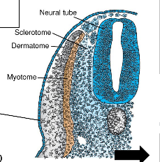
3 Parts of Somite
dermatome
myotome
sclerotome
Dermatome
part of somite, deep dermis (deep to the ectoderm) of the body
Myotome
part of somite, muscle of the body wall and limbs
Sclerotome
part of somite, vertebrae and axial bones
Somite Layers Diagram
Origin of the Typical Spinal Nerve
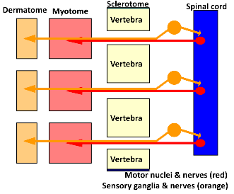
neurons in the spinal cord anterior horn send motor axons to skeletal muscles
sensory axons from the posterior root ganglia extend laterally towards dermatomes and medially to reach the posterior horn of the spinal cord
the motor and sensory axons tether together as the spinal nerve
Spinal Nerve Diagram

What innervates the myotome?
anterior horn axons
What innervates the dermatome and posterior horn?
posterior root ganglion axons
Sclerotome to Vertebra
they split in half as fissure develop so axons can pass, then remnants fuse with their neighbors to become vertebrae
Origin of Spinal Nerve Diagram
Vertebral Development
intervertebral discs and the intervertebral foramina will remain at the site of the fissures
annulus fibrosus develops from the sclerotome (one dermatome separates, top of one connects to bottom of other)
nucleus pulposus is the only mature remnant of the notochord
Sclerotome Mesenchyme
surrounds the neural tube and notochord, forming a loose model of the vertebra and ribs
Chondrification Centers
form in the sclerotome and start to replace it with cartilage in the 6th week
When does bone being replaxing cartilage in the vertebra?
week 7
Vertebral Ossification
occurs in two steps, primary ossification during development and secondary ossification during adolscence
Primary Ossification Centers of Vertebral Ossification
form in the vertebra body, pedicles, and lamina
the bone replaces the mesenchymal cartilage expanding outward from the primary ossification centers, so in the intermediary stage the vertebra are both bone and cartilage
Secondary Ossification Centers in Vertebral Ossification
in the transverse and spinous processes (and annular epiphyes) fuse to the rest of the vertebra
Spina Bifida/Dysraphism
occurs when ossification of the spinous processes of the last part of the vertebra fails, or when the spinous processes do no form at all
How is severe spina bifida detected?
in utero by the presence of a-fetoprotein in materal blood
5 Types of Spina Bifida
occulta
meningocele
meningomyelocele
myeloschesis
rachischesis
Spina Bifida Occulta
failure of the neural arch (spinous process) to ossify
the resulting defect is not large and is sometimes marked by a tuft of dense hair or a small depression in the lower back
Meningocele
occurs when meninges and CSF herniate through the hole left by the failed neural arch
Meningomyelocele
occurs when meningies, CSF, spinal cord and/or roots herniate through the hold left by the failed neural arch
Myeloschesis
failure of the neural tube to fold properly, the neural arch cannot form
Rachischesis
typically occurs when multiple neighboring segments are affected by myeloschesis
Abnormalities in Hox Gene Expression
may cause lumbarization of the S1 vertebrae or sacralization of the L5 vertebrae
Ascent of the Spinal Cord
embryo (3 months): spinal cord runs full length of vertebral column
end of 5th month: conus medullaris ascends to the S1 level
newborn: conus is now at the L3 level