Unit 11- Benign & Malignant Primary Neoplasms (Elie)
1/23
There's no tags or description
Looks like no tags are added yet.
Name | Mastery | Learn | Test | Matching | Spaced |
|---|
No study sessions yet.
24 Terms
What are the 3 benign primary neoplasms?
Cavernous Hemangioma
Hamartoma
Lymphangioma
What is a Cavernous Hemangioma?
Mass of large blood-filled cystic spaces
Cavernous hemangiomas are most commonly:
Asymptomatic
US appearance of a Cavernous Hemangioma?
Varied appearance
Inhomogeneous echogenic mass with multiple, small hypoechoic areas
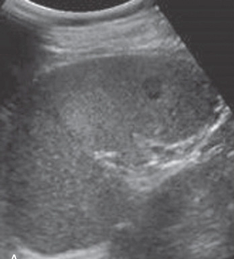
What is this image showing?
Cavernous Hemangioma
Hamartomas are typically ___________ & made of _______ _______?
Asymptomatic
Lymphoid tissue
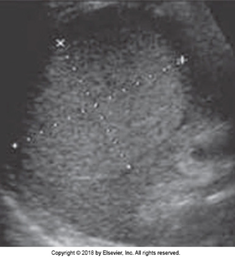
US appearance of a Hamartoma:
Solitary or multiple
Well defined but NOT encapsulated
Solid & cystic components
Hyperechoic
What is this image showing?
Hamartoma
What is a Lymphangioma?
Rare malformation of lymphatics
Endothelium-lined cystic spaces
May involve multiple organ systems
US appearance of a Lymphangioma:
If cysts are large enough, appear anechoic
If cysts are multiple & grouped closely together, appear as solid lesion
Cystic Lymphangioma = Mass with extensive cystic replacement of splenic parenchyma
What are the 2 Malignant Primary Neoplasms?
Lymphoma
Hemangiosarcoma
What is the most common malignant involvement of the spleen?
Lymphoma
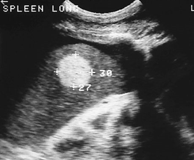
US appearance of Lymphoma?
Focal or diffuse lesions
Typically, HYPOECHOIC
Splenomegaly may or may not be present
What are the 4 appearances of a Lymphoma?
1) Diffuse (low grade lymphomas & Hodgkin’s)
2) Focal small nodules (low grade lymphomas & Hodgkin’s)
3) Focal large nodules
4) Bulky disease
What is a Hemangiosarcoma?
A rare, malignant, vascular endothelium mass
US appearance of a Hemangiosarcoma:
Mixed cystic appearance
Resembles cavernous hemangioma
May be hyperechoic
How is Mets spread to the spleen?
Hematogenous through the blood stream from the primary site
Mets is the _____ most common site in the spleen?
10th
Where do mets occur from?
Breast
Lung
Ovary
Stomach
Colon
Kidney
Prostate
Melanoma
Mets to the spleen are usually ________
Asymptomatic
US appearance of Mets:
Well-defined
Isoechoic, hypoechoic, hyperechoic
Target or halo lesions
If mets are hypoechoic, what should you suspect it is?
Melanoma
Lymphoma
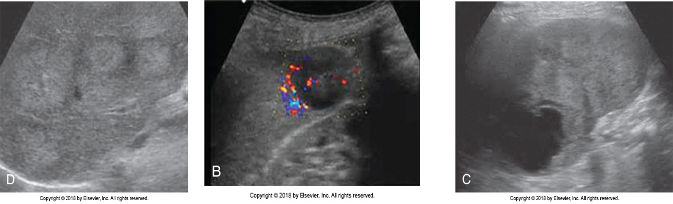
What are these images showing?
Metastases
What should you be careful when evaluating mets?
Be careful in evaluating echogenicity