Ch 5: Peritoneal Cavity
 Call Kai
 Learn
Learn Practice Test
 Spaced Repetition
 Match
Match Flashcards
 Knowt Play
1/49

There's no tags or description
Looks like no tags are added yet.
Name | Mastery | Learn | Test | Matching | Spaced | Call with Kai | Chat |
|---|
No analytics yet
Send a link to your students to track their progress
50 Terms
Addison Lines
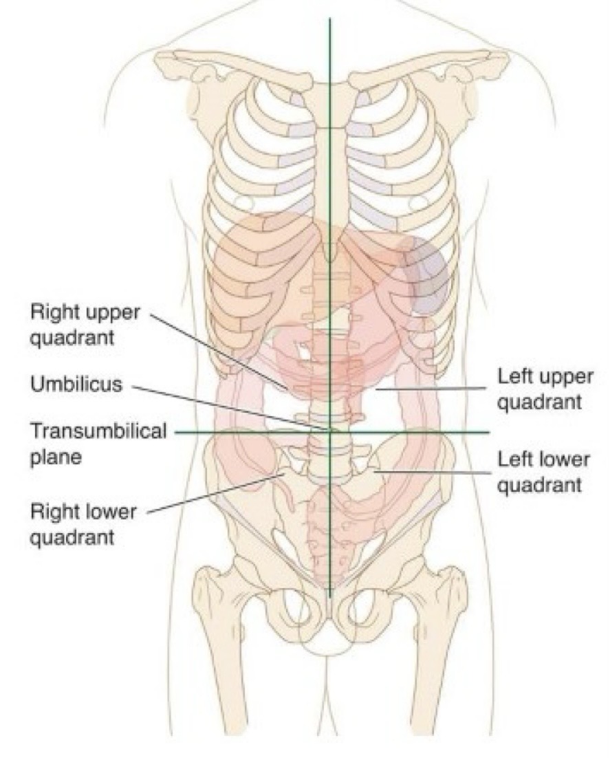
Divides abdominopelvic cavity into 9 regions. has 2 sagittal and 2 transverse lines

Quadrants
Know the organs in each quadrant:
Right Upper Quadrant (RUQ): Liver Gallbladder Right kidney Part of the stomach Pancreas (small portion)
Left Upper Quadrant (LUQ): Stomach Spleen Left kidney Pancreas Parts of the stomach
Right Lower Quadrant (RLQ): Appendix Cecum (first part of the large intestine) Right reproductive organs Right ureter
Left Lower Quadrant (LLQ): Descending colon and sigmoid colon (parts of the large intestine) Left reproductive organs Left ureter
What does Peritoneal Membrane do
divides abdominal cavity into peritoneal and retroperitoneal compartments and is formed by 4th embryonic week
Visceral peritoneum
layer of membrane that covers organs in peritoneal cavity
Bare area
part of retroperitoneum/area of peritoneal organ that isnt overed by membrane. typically hilum of organs where vessels/ducts/nerves enter
Parietal Peritoneum
Part of membrane that lines the walls of abdominal cavity
serous fluid
produced and sits between visceral and parietal membrane layers to prevent friction
what organs does the greater sac contain
GLISSO: gallbladder, liver, intestines, stomach, spleen, and ovaries
Lesser sac
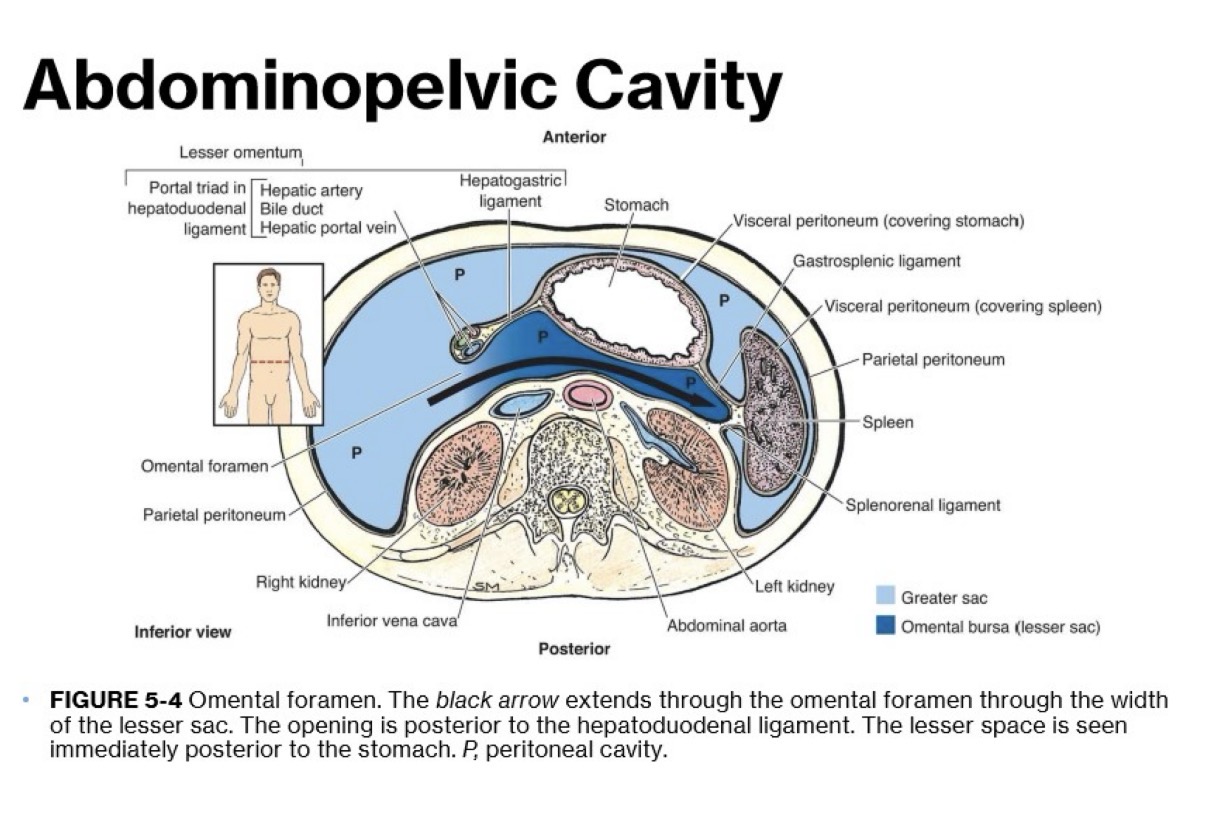
also called omental bursa. sits right behind the stomach and does not contain any organs. (could contain fluid if there is inflammation or swelling of pancreas)

What is the foramen of Winslow
also called omental foramen. it connects lesser sac to greater sac and allows movement/extra space (for stomach expanding during digestion/swelling/inflammation)
What are the greater and lesser omentum
double sheet of peritoneum and acts as abdominal cushion (protects intestines) and helps connect stomach to other organs
Where is the greater omentum located
at the bottom edge of the greater curvature of the stomach and drops down in front of the intestines and attaches to portion of colon. prevents parietal peritoneum from fusing to the visceral peritoneum
Where is lesser omentum located
stretches from lesser curvature of stomach and the liver and then the first part of the small intestine (duodenum)
What is potential space and why is it sonographically significant
when peritoneal layer is reflected between 2 organs or an organ and the peritoneal wall. it is an empty space and in the presence of disease, fluid, or other materials can collect in these spaces. sonographers should examine this spaces since many pathologies present with excretions
Location of right subphrenic space and why it matters
It is an extension of the greater sac between the right hemidiaphragm and anterior superior liver rightward of the falciform ligament.
It is a primary site for fluid accumulation and an anatomical landmark for identifying pathology
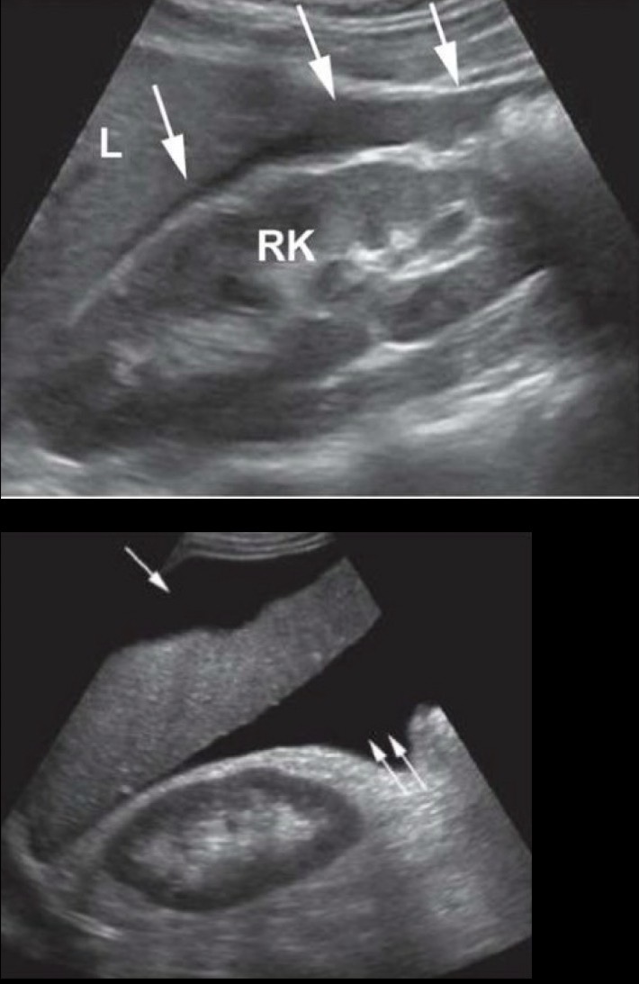
Image Sagittal shows fluid right anterior subphrenic space indicated by single arrow

Location of hepatorenal space (Morrison pouch) and why it matters
This space is created by the peritoneum reflecting from the liver over the right kidney and right posterior peritoneal wall.
When a patient is in supine position, this becomes most gravity dependent potential space in abdominal cavity collecting fluid from supracolic area and lesser sac
2nd image Sagittal shows fluid in hepatorenal space indicated by double arrow (note the sharp fluid filled corners created at organ interface and how organs project into the fluid that fills the space)
Location of anterior subphrenic space and why it matters
Its an extension of the greater sac between the diaphragm and the anterior superior liver leftward the falciform ligament.
It is a key potential pocket where free fluid, pus, or blood can collect and ultrasound helps distinguish these collections from plural effusions
Image shows transverse view of right (*) and left(**) anterior subphrenic space with fluid, separated by falciform ligament

Plural Effusion
Fluid around the lung

Location of posterior subphrenic space and why it matters
It is an extension of the lesser sac between diaphragm and posterior superior liver.
This is a major potential space in trauma scans (FAST exams)
Location of omental bursa (lesser sac) space and why it matters
Between the posterior stomach and parietal peritoneum covering the anterior pancreas (front to back) and splenorenal and gastrosplenic ligaments and epiploic foramen (side to side).
Fluid or pseudocyst may be identified if patient had posterior gastric wall perforation or inflammation or trauma to the pancreas
Location of right and left paracolic gutters space and why it matters
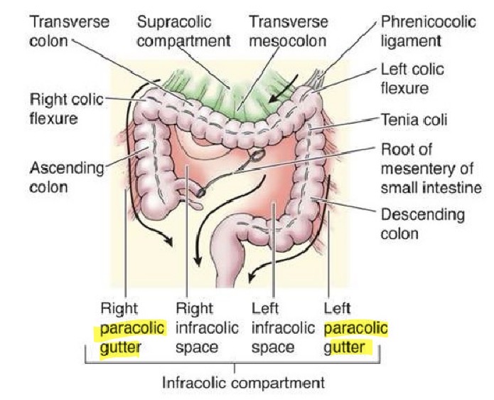
Found along the lateral ascending and descending colons that conduct fluid between supracolic compartment of abdomen and the infracolic compartment of interior abdomen and pelvis.
it is important in determining the extension of disease
Location of pouch of Douglas (rectouterine or posterior cul-de-sac) space and why it matters
Created by parietal peritoneum draping over anterior rectum and posterior vaginal wall and uterus.
when in supine position, this space is the most gravity dependent potential space of pelvic cavity draining fluid from infracolic area
1st image shows fluid in pouch of Douglas (D)

Location of uterovesical space (anterior cul-de-sac) and why it matters
Created by peritoneal reflection over the uterine fundus, anterior uterus, broad ligament, and posterior urinary bladder.
its a primary site for detecting free fluid, inflammatory changes, and invasive tissue growth (endometriosis)
Image shows fluid in uterovesical space (UV)

Location of vesicorectal space and why it matters
Created by peritoneal reflection over the rectum and posterior bladder wall.
when a male is in supine position, this space is the most gravity dependent potential space of pelvic cavity draining fluid from infracolic area
Location of space of retzius and why it matters
Extra peritoneal space located between anterior wall of urinary bladder and pubic symphysis.
Pathologies, traumatic fluids, or surgical implants could be in this area
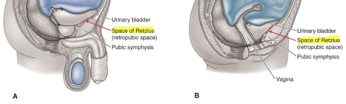
is peritoneum generally evaluated as primary focus of an exam
no
FAST Protocol (why and what is scanned)
A scanning technique for scanning peritoneal cavity. typically for patient who has sudden swelling, blunt abdominal trauma, ruptured ovarian cyst, etc. (something where fluid will be accumulating in the abdomen). Requires sagittal and transverse images of Morrisons pouch (hepatorenal space), posterior right hemidiaphragm/liver interface (subphrenic space), spleen/left kidney interface, pouch of Douglas
Free fluid (ascites)
follows contour of peritoneal organs and must be differentiated from cystic masses of the peritoneum (cystic masses may demonstrate a mass effect)
Loculated ascites
Ascites that demonstrates multiple septa with apparent sequestered pockets of fluid

How will ascites demonstrate bowel
bowel moving freely within it
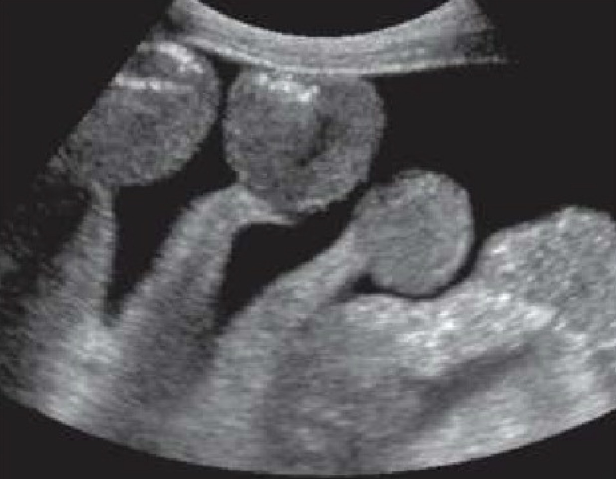
Common places to see ascites build up
Morrisons pouch, paracolic gutters, and pouch of Douglas
Transudative Ascites
Comes from fluid/electrolyte shift due to high pressure in blood vessels. there is no proteins/cellular material which gives it a simple appearance. typically associated with portal hypertension, liver cirrhosis, and injective heart failure
Exudative Ascites
Comes from damage, inflammation, or leaky blood vessels. fluid from blood vessels contain proteins/cellular material which causes more complex appearance. associated with renal failure, bowel disease, malignancy’s and parotiditis
Image appears grainy because of high protein level

Peritoneal Abscess
is a collection of infectious debris identified in potential space or adjacent to inflamed or perforated organ. most common location is subphrenic space.
Sonographic appearance: thick walled fluid collection. complex in appearance and may have blood flow around edges
Image: note rounded edges of perisplenic abscess indicated that it’s not free fluid

Hemoperitoneum
Bleeding into peritoneal cavity caused by blunt trauma or caused iatrogenically, such as following a biopsy, angioplasty, or other surgical intervention. requires FAST exam
Image shows transverse hemoperitoneum in a pouch of Douglas and uterovesical pouch (D)

Hemoperitoneum sonographic appearance
anechoic or echogenic depending on the age of the hemorrhage
Hematoma definition, cause
blood clot/focal area of coagulated blood (bruise on the inside) caused from post surgical complications, trauma such as car crash, or occurring spontaneously in patients with hemophilia or other coagulation diseases or those taking anticoagulant medications.
Hematoma sonographic appearance
variable appearance. initial stage, blood will be anechoic but as clotting starts, it will become echogenic. as it heals/breaks down, it will get anechoic areas (complex look) and go away
Top Image: spleen ruptured due to abdominal trauma and left fluid around kidney

Pseudomyxoma Peritonei (PMP)
Is a metastatic spread of malignant tumor or when is ruptures and fills peritoneal cavity with mucusy material. ascites associated with it that has gelatinous texture (will have debris/will look more abnormal). malignant tumors implant into peritoneum and produce adhesions where tissues get stuck together. common secondary to malignancy of the appendix
Pseudomyxoma Peritonei (PMP) Sonographic Appearance
appears as simple or multiloculated ascites but may present as several thin walled cysts varying in sizes scattered throughout peritoneum. rarely appears as hypoechoic solid mass
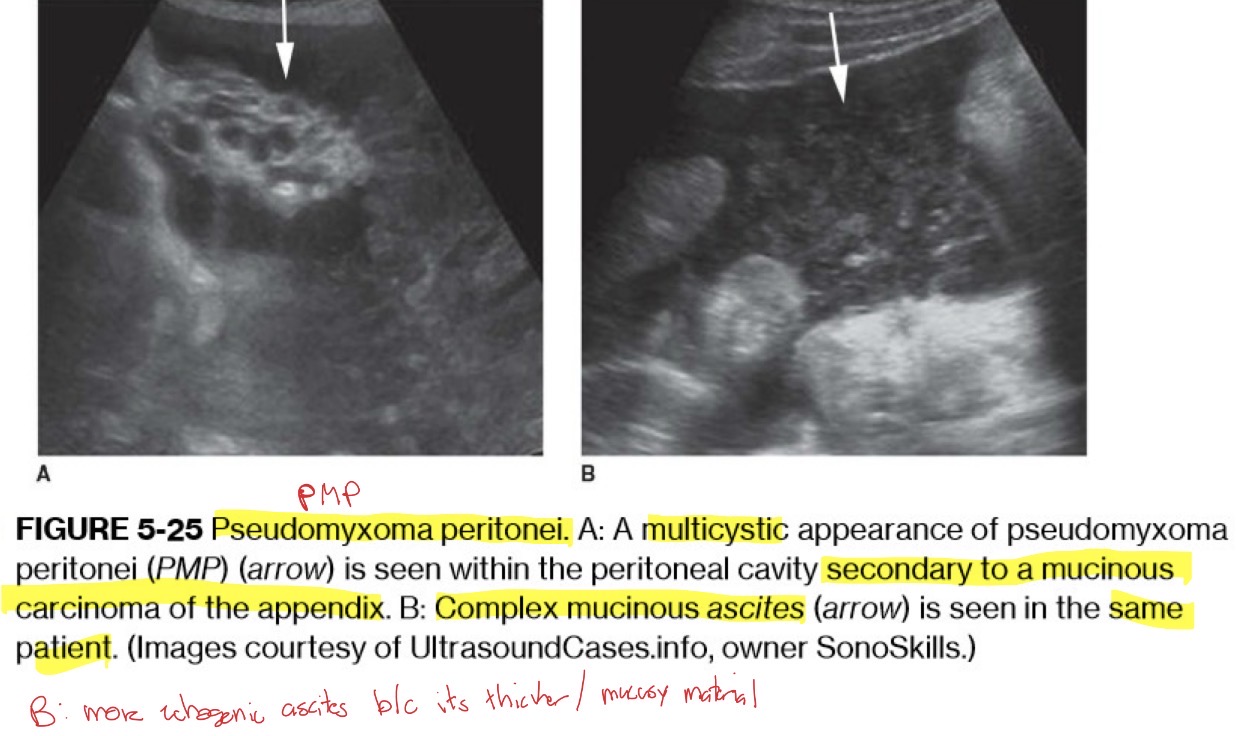
Seroma
Fluid collection composed of blood products located adjacent to or surrounding transplanted organs in early postsurgical period. typically anechoic but may contain septa (has internal walls/division)
Lymphocele
Collection of lymphatic fluid outside lymph system due to disruption of lymphatic vessels or lymph node resection. collections are usually simple but may contain septations. they are slow to develop after surgery and typically present 4-8 weeks after. in peritoneal cavity lymphocele may be seen following prostatectomy and lymph node dissection. body may reabsorb small collections but larger may require intervention
Biloma
Anechoic collection of bile located within peritoneal cavity outside of the biliary tree. typically associated with hepatic transplant due to biliary leak or biliary tree ischemia but may also be secondary (sequela) of trauma, biopsy, or cholecystectomy

Urinoma
Free urine in peritoneal cavity (appropriately called ascites) associated with rupture of urinary tract. typically found adjacent to the kidney in the retroperitoneal perirenal space. urine will appear as simple anechoic fluid and urinary wall should be evaluated for discontinuity in cases of urine ascites
Peritoneal Mesothelioma
A rare primarily malignant tumor of peritoneum that’s associated with asbestos exposure. it metastasizes by direct invasion into adjacent organs and appears as general thickening of peritoneum and mesentery or as a discrete nodule (hard to pick up on ultrasound)
Peritoneal implants or caking
A tumor from an organ, primarily from ovaries, stomach, or colon, metastasizes to peritoneum (peritoneal metastasis). appears as multiple polypoid masses. other findsing are ascites and omental caking
Omental caking
thickening of greater omentum due to malignant infiltration
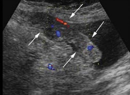
Mesenteric Lymphadenopathy

What is a Paracentesis and its purpose
Aspiration of ascitic fluid from peritoneal cavity that could be for diagnostic or theraputic purposes. typical drainage location is right paracolic gutter and uses Yueh catheter (are ultrasound guided)
Percutaneous Abscess Drainage (PAD)
Flexible catheter placed into abscess for aspiration and sent to lab for analysis