Functional Anatomy 1 - Lecture 13: Knee
 Call Kai
 Learn
Learn Practice Test
 Spaced Repetition
 Match
Match Flashcards
 Knowt Play
1/58

There's no tags or description
Looks like no tags are added yet.
Name | Mastery | Learn | Test | Matching | Spaced | Call with Kai | Chat |
|---|
No analytics yet
Send a link to your students to track their progress
59 Terms
tibiofemoral joint
distal femur on proximal tibia
largest joint cavity in the body
knee joint
two condyloid joint surfaces (medial and lateral)
“modified hinge” joint
no articulation with fibula
movements of tibiofemoral joint
flexion and extension
internal and external rotation (minimal)
abduction and adduction (minimal)
tri-compartmental joint
two tibiofemoral articulation
medial femoral condyle and medial tibial condyle
lateral femoral condyle and lateral tibial condyle
one patellofemoral joint
posterior patella with trochlea of distal femur (middle of femoral condyles)
joint stability of knee
rely on
active stability of surrounding musculature
passive stability from surrounding ligaments, menisci, and joint capsule
anterior knee osteology
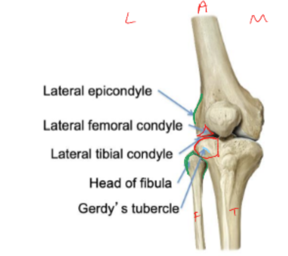
posterior knee osteology
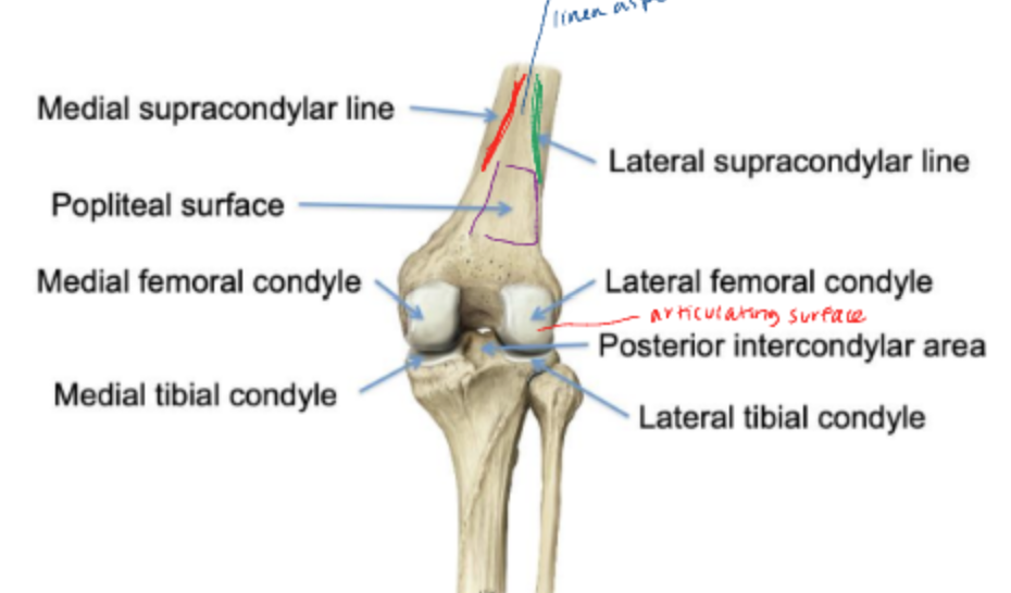
trochlea
intercondylar groove
medial and lateral facets articulate with patella
lateral facet has a steeper slope (in sagittal plane) to help stabilize patella
intercondylar fossa
attachments for cruciate ligaments
possible risk factor for cruciate ligament injury
narrower intercondylar fossa increases risk ACL injury
patella
sesamoid bone embedded in quadriceps tendon
quadriceps tendon → (this) → patellar ligament → tibial tuberosity
posterior surface has thick articular cartilage
thickest in the body
posterior medial patella
medial facet and odd facet
posterior lateral patella
lateral facet
patellar chondromalacia
softening and swelling of the articular cartilage of the patellar articular cartilage
leads to anterior knee pain
condyles
(medial and lateral)
articulate with femoral condyles
tibial plateau
intercondylar region (attachment for ACL/PCL)
menisci (medial and lateral)
tibial tuberosity
soleal line
soleus attachment
references for muscle attachments superior and inferior
frontal plane alignment
genu varum and valgum
sagittal plane alignment
genu recurvatum, hyperextension
patellar positioning
patella alta, patella baja
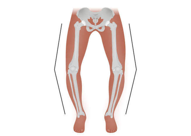
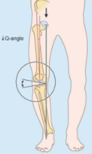
genu varus
bow leg, distal segment toward midline
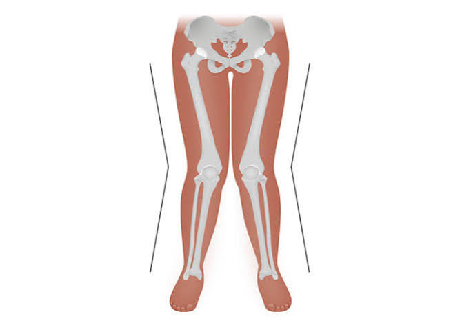
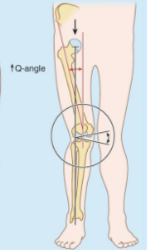
genu valgus (excessive)
knock-knee, distal segment
force of quadriceps line of pull
direction of patellar tracking is influenced by
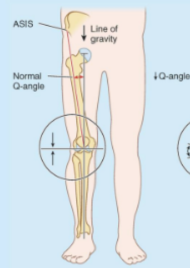
Q-angle measurement
line 1: ASIS to mid-patella
line 2: mid patella to tibial tuberosity
normal: 13-18 degrees
males < females
normal alignment
genu varum
genu valgum
5-10 degrees of hyperextension of the knee joint
normal knee alignment in sagittal plane
genu recurvatum
greater than 10 degrees of hyperextension
patella alta
patella position excessively superior
longer patellar tendon
increased forces through patellofemoral joint
decreased medial/lateral soft tissue stability earlier in knee flexion ranges
increased risk of patellofemoral pain symptoms
patella baja
patella position excessively inferior
shorter patellar tendon
external fibrous layer of knee joint capsule
thickest anterior, thinner posterior
internal synovial later
reflects anterior in intercondylar space
plicae: fold in synovial membrane can be irritated
patellar ligaments
distal to quadriceps tendon
apex of patella to tibial tuberosity
strong, thick, fibrous band
helps form anterior border of fibrous capsule of knee joint
retinacula
medial and lateral patellar retinacula
superficial, thinner, non-specific fiber direction
patellofemoral ligaments
thickening of retinacula
medial and lateral patellofemoral ligaments
deeper, transverse fiber direction
intra-capsular, extra-synovial
cruciate ligaments are described as
anterior cruciate ligament
proximal: posterior medial surface of lateral femoral condyle
distal: anterior medial intercondylar area of tibia
ACL
the weaker of two cruciate ligaments
10x more likely to rupture ACL than PCL
ACL resists
posterior movement of femur on tibia
anterior movement of tibia on femur
internal rotation of tibia
hyperextension of the knee joint
posterior cruciate ligament
proximal: anterior lateral surface of medial femoral condyle
distal: posterior lateral intercondylar area of tibia
posterior cruciate ligament resists
anterior displacement of femur on tibia
posterior displacement of tibia on femur
hyperflexion of the knee joint
medial collateral ligament
proximal: medial epicondyle of the femur
distal (superficial): superior medial tibia
distal (deep): knee fibrous capsule and medial meniscus
extra-capsular, broad/flatter band
valgus force (lateral to medial)
medial collateral ligament resists
lateral collateral ligament
proximal: lateral epicondyle of the femur
distal: head of fibula
extra-capsular, rounded/thinner/cord-like structure
varus force (medial to lateral force)
lateral collateral ligament resists
meniscofemoral ligaments
posterior meniscofemoral ligament (Ligament of Wrisberg)
anterior meniscofemoral ligament (Ligament of Humphrey)
lateral meniscus to medial femoral condyle
added stability to PCL
menisci
crescent-shaped fibrocartilage on proximal tibia
menisci function
increase contact area with distal femur
shock absorption
menisci shape
thicker at external margin, thinner medially
attached at margin (coronary ligaments) and roots
not attached at thin inner border
transverse ligament
joints anterior edges of medial and lateral meniscus
medial meniscus
larger than lateral
elongated C-shape
attachments to MCL (deep)
very little motion on tibia
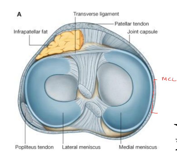
lateral meniscus
smaller than medial
almost a complete circle
no attachment to LCL
more mobile
unhappy triad
ACL, MCL, medial/lateral meniscus injured simultaneously
often from forceful valgus force
knee bursae
pre-patellar
infrapatellar
suprapatellar
pes anserine
blood supply from popliteal artery
superior lateral genicular and superior medial genicular
middle genicular
through fibrous capsule to supply cruciate ligaments, synovial membranes and outer meniscus
blood supply from anterior tibial artery
inferior lateral genicular and inferior medial genicular
meniscus blood supply
middle genicular artery, only on outer edge
popliteal fossa
diamond-shaped space posterior to knee
boundaries of popliteal fossa
semimembranosus, biceps femoris, medial and lateral heads of gastrocnemius
contents of popliteal fossa
small saphenous v.
popliteal a./v.
branches of sciatic n.
tibial n. (medial)
medial sural cutaneous n.
common peroneal n. (lateral)
lateral sural cutaneous n.
popliteal lymph nodes