Biopsychology
1/29
There's no tags or description
Looks like no tags are added yet.
Name | Mastery | Learn | Test | Matching | Spaced | Call with Kai |
|---|
No analytics yet
Send a link to your students to track their progress
30 Terms
What is spatial resolution
Refers to the ability of a brain imaging technique to distinguish between two separate point in the space
Indicates how precisely a scan can locate brain activity within different areas or structures of the brain
High spatial resolution scans (FMRI) can pinpoint where activity is occurring with great activity
What is temporal resolution
Refers to the ability of a brain imaging technique to track changes in brain activity over time
Indicates how quickly a scan can detect shifts in neural activity
Can capture rapid changes in brain function from movement to structure
What is the nervous system
A complex network of nerves in the human body
It communicates via fast electrical signals (action potentials)
2 parts: CNS, Peripheral nervous system
2 Functions:
To collect, process, and respond to information from the world
To co-ordinate the various organs and cells in the body
What is the the peripheral nervous system
Somatic nervous system
Receives incoming sensory information
Movement of skeletal muscles
The autonomic nervous system
Governs bodily functions such as heart rate and breathing
Controls the endocrine system (glands and hormones)
2 parts:
Sympathetic - fight or flight
Para-sympathetic - rest and digest
The endocrine system
Regulates the activity of cells and organs throughout the body
Uses slower, longer lasting chemical messengers called hormones
Made up of glands and hormones
Each gland secretes a specific hormone into the bloodstream
Hormones stimulate specific chemicals
What are the parts of the Neurone cell
The Soma: cell body which contains the nucleus
Dendrites: receive information from other cells
Axons: carry action potentials away from the soma, towards other cells
Axon terminal: end of the axon where they pass neural information on to other cells
Myelin sheath: fatty insulated layer which speeds up action potentials
What are sensory neurons
Structure:
Myelin sheath present
Function:
To carry information towards the nervous system
Location:
In the peripheral nervous system
What are the parts of a sensory neurone
What are relay neurones
Structure:
No myelin sheath
Soma and dendrites together
Function:
To relay neural information between sensory and motor neurones
Location:
In the CNS and visual system
What are the parts of the relay neurone
What are motor neurones
Structure:
Myelin sheath present
Soma and dendrites together
Function:
To relay neural information from the CNS to effectors (muscles and glands)
Location:
Soma is in the central nervous system and axon extends into peripheral nervous system
What are the parts of the motor neurone
What is synaptic transmission
1) Action potential arrives at the pre-synaptic neurone
2) Vesicles containing neurotransmitters release the neurotransmitters into the synaptic gap via exocytosis
3) Neurotransmitters diffuse across the synaptic gap
4) Neurotransmitters bind to receptors
5) Excitatory NT increase the internal voltage, Inhibitory NT decreases the internal voltage
At rest -70mV Action Potential happens at -55mV
Summation = Excitatory + Inhibitiory effects are summed to determine
What is the excitation threshold
Neurones are kept in a polarised state - this means their internal, voltage is around -70mV
If the internal voltage increases to around -55mV, there will be an potential inside the neurone - this is the excitation threshold
What do Excitatory Neurotransmitters do
They increase the voltage inside the neuron - this makes it more likely that the neurone will reach the excitation threshold
What do Inhibitory Neurotransmitters do
Decrease the voltage inside the neurone
By decreasing the internal voltage, it is more difficult to reach the excitation threshold
What is summation
A neurone can receive a combination of excitatory or inhibitory neurotransmitters at the same time
This means the internal voltage might change without crossing the excitation threshold
Whether a neurone fires an action potential is determined by the sum of excitation and inhibition
What is localisation of function
Refers to the idea that different areas of the brain are specialised to perform specific functions
Opposed by the holistic view - suggests the whole brain actually works together
Who is phineas gage
An explosion launched a steel rod through Phineas’ skull, causing massive damage to his frontal lobe
Afterwards, he showed a lack of inhibition and became prone to intense anger
The frontal lobe is responsible for behaviour
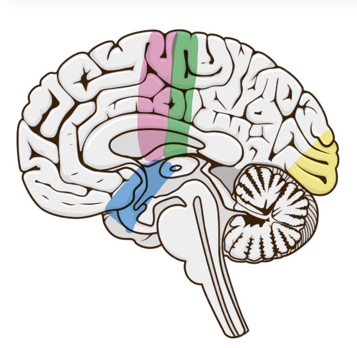
Name the labelled parts of the brain
Pink: Motor Cortex
Green: Somatosensory cortex
Yellow: Visual cortex
Blue: Auditory cortex
What is the motor cortex
Located in the back of the frontal lobe and is responsible for voluntary movement
What is the somatosensory cortex
Located in the front of the parietal lobe, and is responsible for sense of touch
What is the visual cortex
What is the Auditory cortex
What is the Broca’ Area
Located in the left frontal lobe
Thought to be responsible for speech production
Broca studied the Brain of a patient who only says Tan
What is the Wernickes’s area
Located in the left temporal temporal lobe
Thought to be
How do the two hemispheres connect to the body
Each hemisphere is connected to the opposite side of the body - contralateral
The visual fields are also contralateral - the left field is processed by the right hemisphere and vice versa
The two hemispheres are joined together by a bundle of nerve fibres called the corpus callosum
What is hemispheric lateralisation
It is the idea that each hemisphere of the brain is specialised to carry out certain tasks
What is split brain research
Split brain research involves studying patients who have had the corpus callosum severed in order to treat severe epilepsy
By severing the corpus callosum, the two hemispheres cannot communicate – meaning we can assess the functions of each hemisphere separately!
What is the Sperry research
Sperry conducted research on 11 patients who had their corpus callosum severed
He compared the patients to 11 healthy controls (who did not have epilepsy)
Describing what you see: patients asked to verbally name objects presented to either the right or left visual field. Objects in the LVF could not be named but objects in the RVF could be named. This suggests language centres are in the left hemisphere
Drawing task: Patients flashed various objects to the visual fields and asked to draw them. Patients drew more effectively with the left hand, despite being right handed.
Composite words: Patients shows composite words with the word split across the visual fields, e.g. KEY + RING. Patients would say the word ring and then draw a key