Walls and connections of the orbit
1/7
There's no tags or description
Looks like no tags are added yet.
Name | Mastery | Learn | Test | Matching | Spaced | Call with Kai |
|---|
No analytics yet
Send a link to your students to track their progress
8 Terms
orbit
it is a pyramidal shaped structure, consisting of a base, an apex and 4 walls.
The orbit protects the eye from mechanical injury
the base
Superior margin: frontal bone.
Inferior margin: Maxilla and zygomatic bone
Medial margin: frontal bone and frontal process of maxilla
Lateral margin: Zygomatic and zygomatic process of the frontal bone
The apex
Where the 4 walls join at the optic canal
Superior wall
This separates the orbot from the anterior cranial fossa.
The roof of the orbit is formed by the:
Orbital plate of the frontal bone.
Lesser wing of the sphenoid bone.
Structures in the superior wall:
Superior orbital fissure: for the passage of CN 3, 4, V1, 6 and ophthalmic vein.
Optic canal: passage of Optic nerve (CN 2) and opthalmic artery.
Supraorbital foramen (lateral side) and or frontal notch (medial side)
Fossa for the lacrimal gland- at the lateral side of the wall
Inferior wall
It separates the orbit from the maxillary sinus. The floor of the orbit is formed by the:
Orbital surface of the body of maxilla.
Orbital process of palatine bone.
Sturtures:
Inferior orbital fissure
Infraorbital groove and canal
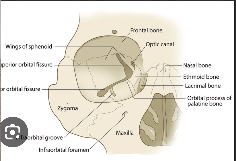
Medial wall:
This separates the orbit from the nasal cavity.
It is formed by the:
frontal process process of maxilla
Lacrimal bone
Orbital plate of the ethmoid bone
Body of sphenoid bone
Stuctures:
Anterior and posterior ethmoidal foramen
Anterior and posterior lacrimal crest.
Lacrimal fossa- between the crest for the lacrimal sac.
Nasolacrimal canal
Lateral wall
Separates the orbit from the temporal and infratemporal fossa.
Formed by the:
Orbital surface of zygomatic bone
Greater wing of sphenoid bone.
Structures:
Zygomatico-orbital foramen: transmits the zygomatic nerve and vessels into the zygomatic bone.
Within the bone, the zygomatic nerve gives 2 branches- the zygomaticofacial and zygomaticotemporal nerves which run in their corresponding foramens and canal.
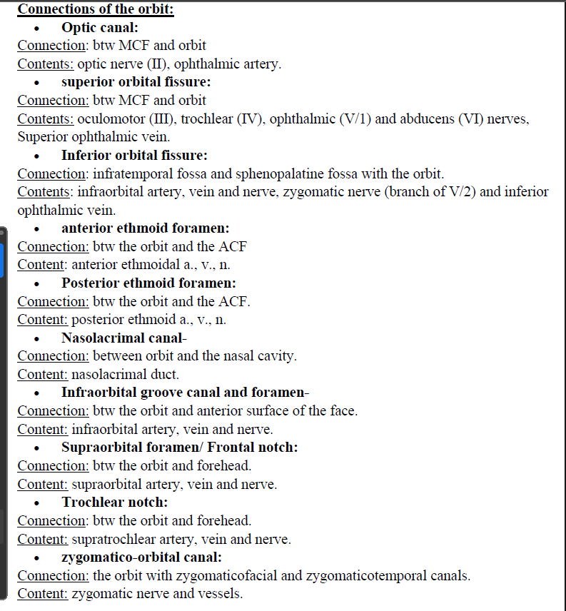
Connections of the orbit:
Optic canal
Superior orbital fissure
Inferior orbital fissure
Anterior ethmoidal foramen
Posterior ethmoid foramen
Nasolacrimal canal
Infraorbital groove, canal and foramen
Supraorbital foramen/ frontal notch
Trochlear notch
Zygomatico orbital canal