Ch.7-cellular basis of inhertience
1/46
There's no tags or description
Looks like no tags are added yet.
Name | Mastery | Learn | Test | Matching | Spaced | Call with Kai | Chat |
|---|
No analytics yet
Send a link to your students to track their progress
47 Terms
diploid dominant
in which multicellular diploid stage is the most obvious life stage)
halploid dominant
multicellular halploid life stage
alternation of generations
in which the two stages, halploid and dominant are apperant to one degree or another depending on the group, as with plants and some algaee
gern cells
a special cell line that only produces gamates
fertillization
occurs with the two fusion of two gamates, usually from different individuals, restoring the diploid state
-a union of two cells from two individual prhamos,s
gametophytes
-halploid multicellular plants are this due to producing gamates
sporophyte
zygote will undergo many rounds of mitosis and give rise to diploid multicellular plant
interphase
-conists of the G1, S, and G2 phases
G1-focuses on cell growth
S phase- DNA of the chromsomes is replicated
G2 phase- the cell undergoes the final perpartions for meiosis
-during the S phase, each chromosome becomes of two identical copies and are held together at the centromere until they are pulled apart during meiosis II
-in an animal cell the centrosomes that organize the mircotubles of the meiotic spindle also replicate
-prepares cell for the first meiotic phase
Meiosis I
-Nuclear envelope begins to break down, the proteins with homologous chromosomes bring the pair close to each other
-tight pairing of the homologous chromosomes are synapisis
-in synapsis the genes on the chromatids of the homologous chromosomes are precisly aligned with each other
-an exchange of chromosomes segments between non-sister homologous chromatids occurs and is crossing over
-this process is revealed visually after the exchange as chiasmata
-as prophase I progresses, the close association between homologous chromsomes begins to break down, and the chromosomes continue to condense, although the homologous chromsomes remain attached to each other at chismata
-at the end of prophase I, the pairs are held together only at chaismata which is the tetrads because the four sister chromatids of each pair of homologous chromosomes now visible
-The crossover events are the first source of genetic variation produced by meiosis
-a single crossover event between between homologous non-sister chromatids leads to a reciprocal exchange of equivalent DNA between a maternal chromosome and a paternal chromosome
-Now, when that sister chromatids is moved into a gamete, it will carry some DNA from one parent of the individual and some from the DNA from the other parent
-the recombinant sister chromatid has a combination of maternal and paternal genes that did not exist before the crossover
the key event is prometaphase I attachment of the spindle fiber mircotubles to the kinetochore proteins at the centromeres
-the mircotubles assembled to the kinetochore proteins at the centromeres
-The mircotubles assembled from centrosomes at opposite poles of the cell grow toward the middle of the cell
-at the end prometaphase 1, each tetrad is attached to mircotubles from both poles, with one homologous chromsomes are still held together at chaismata
-In addition, the nuclear membrane has broken down entirely
-during metaphase I, the homologous chromsomes are arranged in the center of the cell with the kinetochores facing opposite poles
-The orientation of each pair of homologous chromosomes at the center of the cell is random
-the randomness is independent assortoment, is the physical basis for the generation of the second form of genetic variation in offspring
-In metaphase I, these pairs line up at the midway point between the two poles of the cell
-because there is equal chance that a mircotuble fiber will encounter or paternally inhertied chromsome, the arrangement of the tetrads at the metaphae plate is random
-any materally inhertied chromsome may also face either pole
-each cell that undergoes meiosis, the arrangement of tetrads is different
-the variation depends on the chromsomes making up set
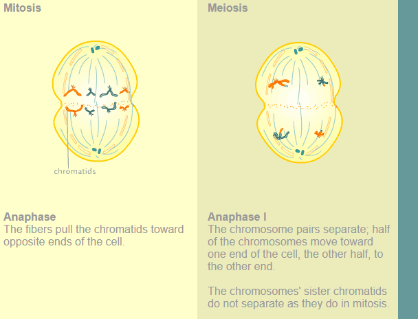
-in anaphase I the spindle fibers pulled the linked chromsomes apart. The sister chromatids remain tightly bounded together at the centrosome
in the chsisma connections that are broken down in anaphase I as the fibers attached to the fised linetochores pull the homologous chromsomes
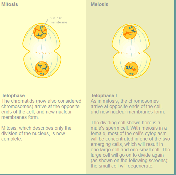
-In telophase I, the separated chromosomes arrive at opposite poles. The remainder of the typical telophase events may or may not occur depending on the species. In some organisms, the chromosomes decondense and nuclear envelopes form around the chromatids in telophase
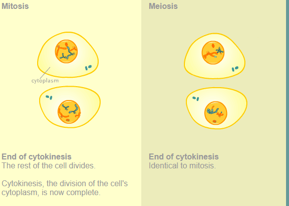
Cytokinesis, the physical separation of the cytoplasmic components into two daughter cells, occurs without reformation of the nuclei in other organisms. In nearly all species, cytokinesis separates the cell contents by either a cleavage furrow (in animals and some fungi), or a cell plate that will ultimately lead to formation of cell walls that separate the two daughter cells (in plants). At each pole, there is just one member of each pair of the homologous chromosomes, so only one full set of the chromosomes is present. This is why the cells are considered haploid—there is only one chromosome set, even though there are duplicate copies of the set because each homolog still consists of two sister chromatids that are still attached to each other. However, although the sister chromatids were once duplicates of the same chromosome, they are no longer identical at this stage because of crossovers.
do the number of chiasmata vary with the species?
Yes the number of chiasmata varies with the species and length of chromosomes
what are the two possiblties for orientation (for tetrad)?
-possible number of alignments equals 2n where n is the number of chromsomes per set
-humans have 23 chromosome pairs which results in over eight million (2²³) possiblities
Meiosis II
separates sister chromatids to produce four haploid gametes
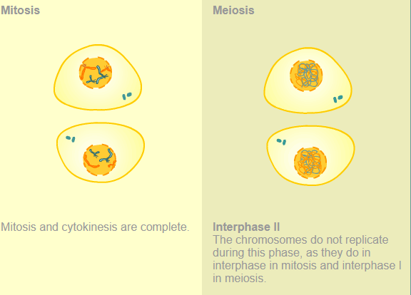
In meiosis II, the connected sister chromatids remaining in the haploid cells from meiosis I will be split to form four haploid cells. In some species, cells enter a brief interphase, or interkinesis, that lacks an S phase, before entering meiosis II. Chromosomes are not duplicated during interkinesis. The two cells produced in meiosis I go through the events of meiosis II in synchrony. Overall, meiosis II resembles the mitotic division of a haploid cell.
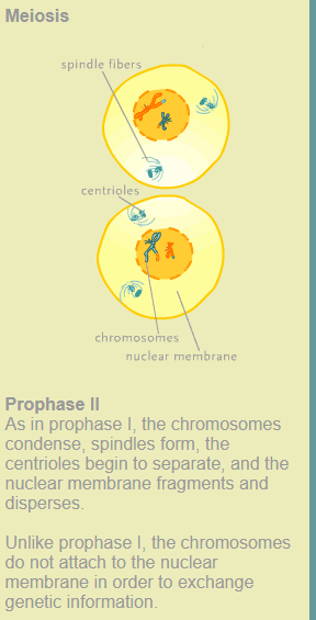
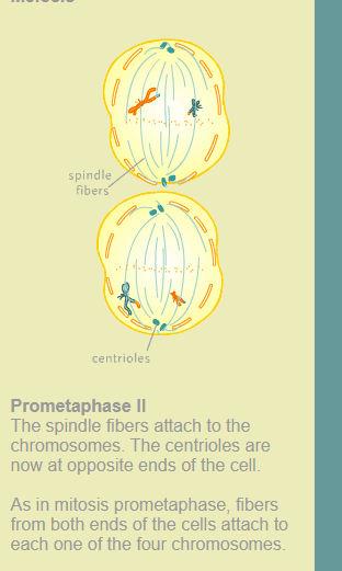
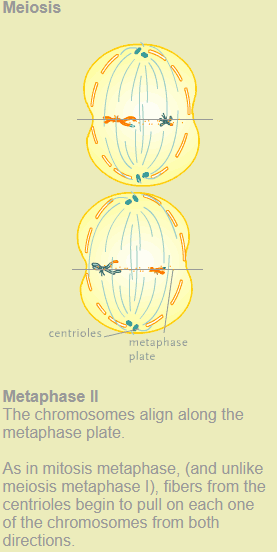
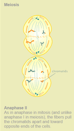
In prophase II, if the chromosomes decondensed in telophase I, they condense again. If nuclear envelopes were formed, they fragment into vesicles. The centrosomes duplicated during interkinesis move away from each other toward opposite poles, and new spindles are formed. In prometaphase II, the nuclear envelopes are completely broken down, and the spindle is fully formed. Each sister chromatid forms an individual kinetochore that attaches to microtubules from opposite poles. In metaphase II, the sister chromatids are maximally condensed and aligned at the center of the cell. In anaphase II, the sister chromatids are pulled apart by the spindle fibers and move toward opposite poles.
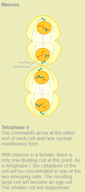
In telophase II, the chromosomes arrive at opposite poles and begin to decondense. Nuclear envelopes form around the chromosomes. Cytokinesis separates the two cells into four genetically unique haploid cells. At this point, the nuclei in the newly produced cells are both haploid and have only one copy of the single set of chromosomes. The cells produced are genetically unique because of the random assortment of paternal and maternal homologs and because of the recombination of maternal and paternal segments of chromosomes—with their sets of genes—that occurs during crossover.
Meioisis I
produces two haploid cells each containing one chromomsome from the homologous pairs
synapsis
pairing of homologous chromosomes
Tetrad
4 sister chromatids of paired homologous chromosomes
crossing over
-recombination of alleles in homologous chromsomes
-results of crossing over-gentic variety in gametes
difference in compaeing meiosis and mitosis
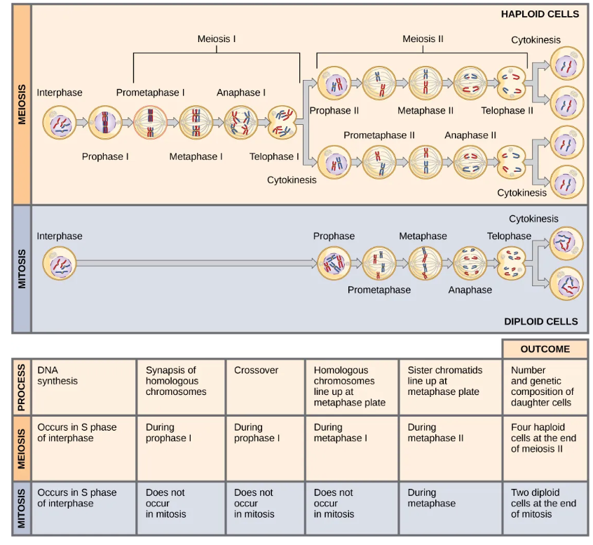
Mitosis and meiosis, which are both forms of division of the nucleus in eukaryotic cells, share some similarities, but also exhibit distinct differences that lead to their very different outcomes. Mitosis is a single nuclear division that results in two nuclei, usually partitioned into two new cells. The nuclei resulting from a mitotic division are genetically identical to the original. They have the same number of sets of chromosomes: one in the case of haploid cells, and two in the case of diploid cells. On the other hand, meiosis is two nuclear divisions that result in four nuclei, usually partitioned into four new cells. The nuclei resulting from meiosis are never genetically identical, and they contain one chromosome set only—this is half the number of the original cell, which was diploid (Figure 7.6).
The differences in the outcomes of meiosis and mitosis occur because of differences in the behavior of the chromosomes during each process. Most of these differences in the processes occur in meiosis I, which is a very different nuclear division than mitosis. In meiosis I, the homologous chromosome pairs become associated with each other, are bound together, experience chiasmata and crossover between sister chromatids, and line up along the metaphase plate in tetrads with spindle fibers from opposite spindle poles attached to each kinetochore of a homolog in a tetrad. All of these events occur only in meiosis I, never in mitosis.
Homologous chromosomes move to opposite poles during meiosis I so the number of sets of chromosomes in each nucleus-to-be is reduced from two to one. For this reason, meiosis I is referred to as a reduction division. There is no such reduction in ploidy level in mitosis.
Meiosis II is much more analogous to a mitotic division. In this case, duplicated chromosomes (only one set of them) line up at the center of the cell with divided kinetochores attached to spindle fibers from opposite poles. During anaphase II, as in mitotic anaphase, the kinetochores divide and one sister chromatid is pulled to one pole and the other sister chromatid is pulled to the other pole. If it were not for the fact that there had been crossovers, the two products of each meiosis II division would be identical as in mitosis; instead, they are different because there has always been at least one crossover per chromosome. Meiosis II is not a reduction division because, although there are fewer copies of the genome in the resulting cells, there is still one set of chromosomes, as there was at the end of meiosis I.
Cells produced by mitosis will function in different parts of the body as a part of growth or replacing dead or damaged cells. They may even be involved in asexual reproduction in some organisms. Cells produced by meiosis in a diploid-dominant organism such as an animal will only participate in sexual reproduction.
Interphase
Mitosis:
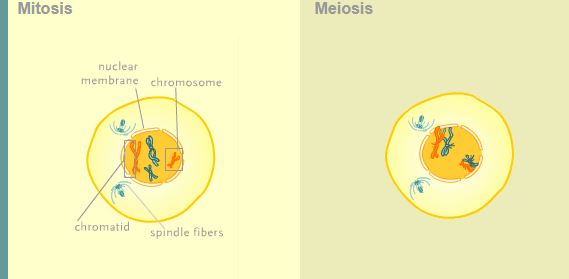
Interphase is the period between cell divisions. During this time, chromosomes replicate—each DNA strand unzips into two strands while free-floating bases attach to the unzipped strands. The chromosomes are loosely packed and not visible with a microscope.
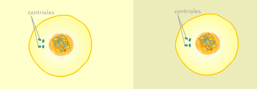
Two pair of centrioles lie just outside the nucleus, next to each other. A centriole is a cylindrical structure within the cell that plays a part in cell reproduction.
Meiosis Interphase I
The activities within this cell are the same as in the mitosis-dividing cell.
Prophase
Mitosis:
Chromosomes begin to condense, taking on the form that they are usually depicted in: four arms connected at a point. Each chromosome is, at this time, actually two identical copies. Each copy is called a chromatid.
A spindle begins to form from the centrioles. This spindle is made of fibers. The centrioles begin to separate.
Also, the membrane of the nucleus, or nuclear envelope, fragments and disperses.
Prophase I (Meiosis)
The activities are the same as in mitosis, except that in this cell the chromosomes attach to the membrane of the nucleus and then pair up with their corresponding chromosome.
While paired up, enzymes cut sequences of DNA (genes) from the chromosomes. These sequences are exchanged between the chromosomes, which allows for an exchange of genes between the two
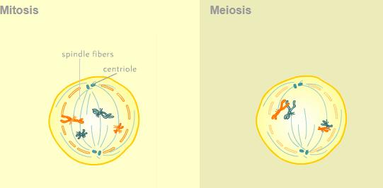
Prometaphase
Mitosis
The centrioles are now at opposite ends of the cell.
The spindle fibers from both of the centrioles attach to each one of the chromosomes.
Meiosis: ProMetaphase I
Same as in mitosis, except that the spindle fibers from each centriole attach to one chromosome of a matching chromosome pair.
In other words, the fibers from one centriole attach to 23 chromosomes, and the fibers from the other centriole attach to the other 23 chromosomes. (Again, only four chromosomes are shown here in order to simplify the illustration.)
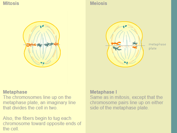
Metaphase
The chromosomes line up on the metaphase plate, an imaginary line that divides the cell in two.
Also, the fibers begin to tug each chromosome toward opposite ends of the cell.
anaphase
telophase
cytoskinesis
Meiosis- Interphase II
Meiosis-Prophase II
meiosis-Prometaphase II
meiosis-Metaphase II
Meiosis anaphase II
meisosis-Telophase II
end of cytoskines

Karyotype
number ans apperence of chromsomes, includes their lengtj, banding pattern, and centromere position
Translocation
the process by which one segment of the chromosome dissociates and reattaches to a different nonhomologous chromosome
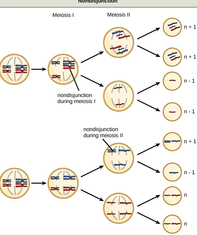
Nonsdisjunction
-which occurs when pairs of homologous chromosomes or sister chromatids fail to separte during meisis
-risk of nondisjunction increases with the age of parents
euploid
an individual with the appropriate number of chromosomes for their species
autosome
any of the non-sex chromosomes
aneuploid
an individual with an error in chromosome number; includes deletions and duplications of chromosome segments
monosomy and trisomy
an otherwise diploid genotype in which one chromosome is missing
an otherwise diploid genotype in which one entire chromosome is duplicated
X inactivation
the condensation of X chromosomes into Barr bodies during embryonic development in females to compensate for the double genetic dose
polyploid
an individual with an incorrect number of chromosome sets
chromsome inversion
the detachment, 180° rotation, and reinsertion of a chromosome arm
What is a likely evolutionary advantage of sexual reproduction over asexual reproduction?
sexual reproduction results in greater variation in the offspring
Meiosis produces ________ daughter cells.
four haploid
At which stage of meiosis are sister chromatids separated from each other?
anaphase II
The part of meiosis that is similar to mitosis is ________.
Meiosis II
The genotype XXY corresponds to:
Klinefelter syndrome
Abnormalities in the number of X chromosomes tend to be milder than the same abnormalities in autosomes because of ________.
X inactivationn