Skeletal Muscles
1/193
There's no tags or description
Looks like no tags are added yet.
Name | Mastery | Learn | Test | Matching | Spaced |
|---|
No study sessions yet.
194 Terms
What are the 3 primary tissue types found in muscles
Cardiac muscle (heart)
Smooth muscle - walls of hollow organs
Skeletal muscle - makes up half of the nody’s mass and most muscle in the body
4 Properties of Muscle Tissue
Excitability
Contractility
Extensibility
Elasticity
Excitability
muscle cells can respond to signals that are sent from the nervous system, allowing them to generate electrical impulses and leading to muscle contractions.
Contractility
muscles can contract, so this allows for the muscle fibres to shorten and it generate force, and that’s what causes movement
Extensibility
muscles can also stretch, so this is important for the muscle to be able to extend its fibres.
Elasticity
this allows the muscle to return to their original shape and length after theyve been stretched out or contracted
Primary Function of Muscle Tissue
movement
Posture and Stability
Heat Production
Support and protection
Control of openings
What percentage of body mass is made up of skeletal muscles?
Skeletal muscles make up about 40% of body weight
How do muscle cells respond to signals from the nervous system?
Muscle cells respond to signals from the nervous system, which triggers electrical impulses and leads to muscle contraction
4 components to the skeletal muscles
Skeletal muscle tissue
Connective tissue
Blood vessels
Nerves
Skeletal muscle
attach to the bony skeleton
Produced movement
Each muscle is an organ
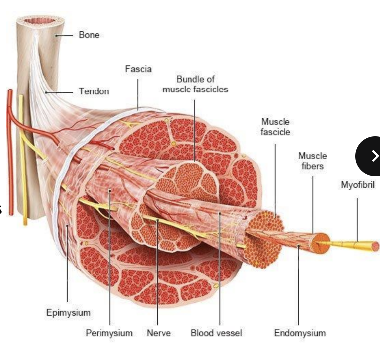
Muscle structure Components and what they are
Muscle: a bundle of fascicles
Fascicles: bundles of muscle fibres and they’re wrapped in a layer called perimysium
Muscle fibre: bundles of myofibrils. They are long, contract and what creates movement
Myofibril: responsible for contraction
Actin & Myosin are proteins found in myofibril
Structural Connective tissue compnents
Epimysium
Perimysium
Endomysium
Bone
Tendon
Fascia
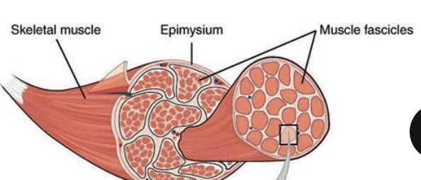
Epimysium
a dense regular connective tissue around the entire muscle and in the outer layer
Perimysium
a fibrous connective tissue around each fascicle
Endomysium
a fine connective sheath around each muscle fibre on the inside
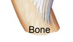
Bone
a framework for muscle attachment and enables movement
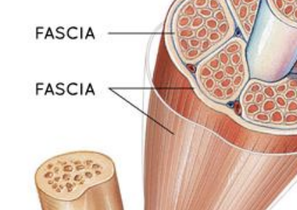
Fascia
a connective layer surrounding muscles and organs and provides support
Support Systems for Muscle
Nerves
Blood vessel
Nerves
transmit electrical signals to muscle fibres and trigger contractions
Blood vessel
delivers oxygen and nutrients and removes waste
Innervation of skeletal Muscle
Motor neurons
Neuromuscular junction
axon terminal’
Synaptic cleft
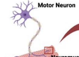
Motor Neurons
innervate the skeletal muscle tissue
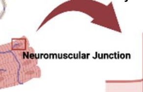
Neuromuscular junction
where nerve ending meets muscle fiber
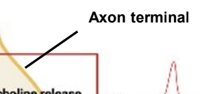
Axon terminal
stores neurotransmitters
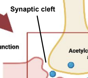
Synaptic cleft
the space between the axon terminal and sarcolemma
What three factors influence muscle action?
Fascicle arrangement
lever mechanics
muscle position relative to the joint
Fascicles arrangement in circular muscles
Fascicles are arranged in concentric rings
surrounding external body openings (sphincter)
Examples of circular muscles
Orbicularis oris and orbicularis oculi
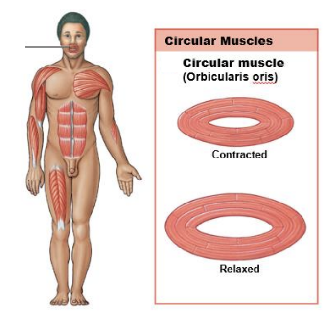
How do circular muscles function?
They work like a drawstring; when they relax, the opening gets larger, and when they contract, it closes
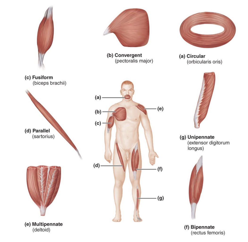
What are the types of Fascicle Arrangement
circular
convergent
parallel
Pennate
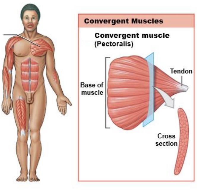
Fascicle Arrangement on Convergent muscles
Broad origin, with fascicles converging toward the tendon of insertion
ex: pectoralis major
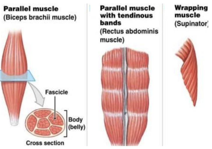
Fascicle Arrangement in Parallel muscles
Fascicles run along the muscle’s long axis
2 types of Parallel muscle
Fusiform: biceps
Straplike: supinator
Fascicle Arrangement in Pennate Muscle
Fascicles are arranged on an angle to a central tendon and run through the middle of the msucle
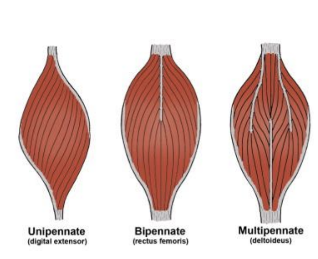
3 types of Pennate Muscle
Unipennate
Bipennate
Multipennate
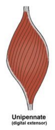
Unipennate
the fascicle insert into one side of tendon
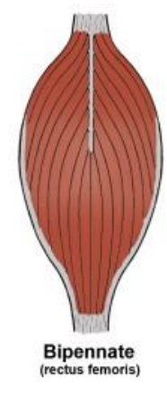
Bipennate
inserts on both sides
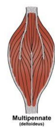
Multipennante
the fasicles inserts into tendon from all sides
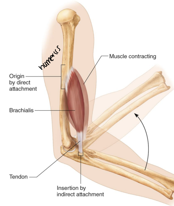
Origins & Insertions
Origin: less movable attachment
Insertion: more movable attachment
most skeletal muscles span between two bones
Muscle Attachment
muscles are attached by connective tissue
There are direct and Indirect attachments
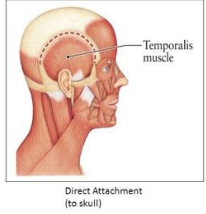
Direct Attachment
Connective tissue fuses to the bone
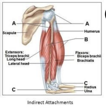
Indirect attachment
Connective tissue forms a tendon or aponeurosis
Aponeurosis
a sheet of pearly white fibrous tissue that takes the place of a tendon in flat muscles having a wide area of attachment.
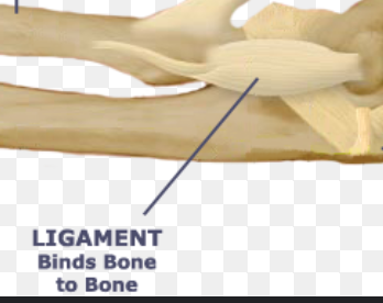
Ligaments
Connects bone to bone
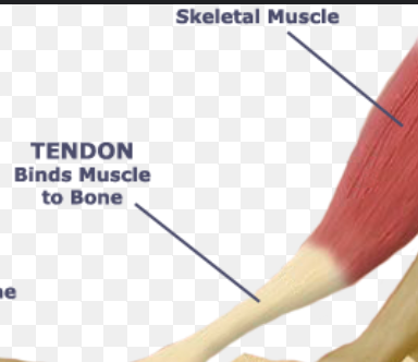
Tendon
Connects muscle to bone
4 Lever System
lever - a rigid bar that moves
fulcrum- a fixed point
effort - applied force
load - resistance
Function of Levers
move a heavier load
move a load farther
What is the mechanical advantage of levers?
effort arm > (longer) load arm
make it easy to lift heavier things
What are the mechanical disadvantage of levers?
effort arm is < load arm
What are the lever systems in the body?
Bones: levers
Joints: fulcrums
Muscle contraction: provides effort at muscle
Load: body part moved
Describe a first-class lever
effort at one end and load at the other
Fulcrum between load and effort.
Depending on where the fulcrum is it can either cause a mechanical advantage or disadvantage
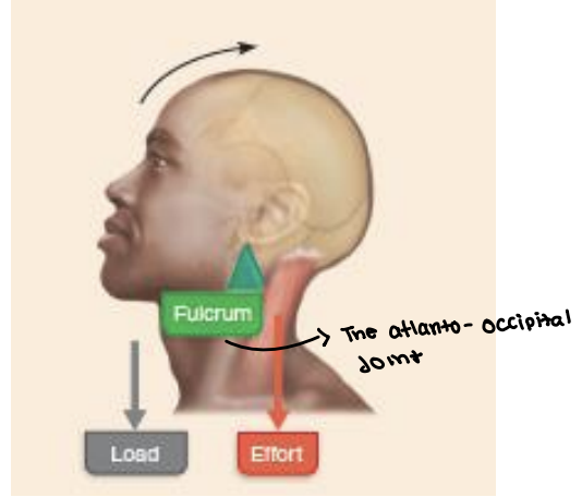
Examples of a First-Class lever
seesaws, scissors, lifting your head of your chest
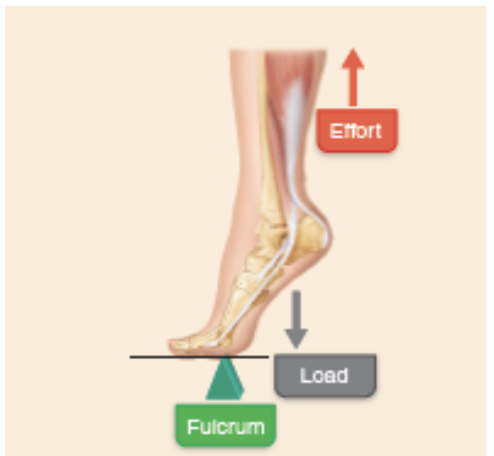
Describe the Second-Class Lever
Effort at one end and Fulcrum at the other
The load is between the effort and fulcrum
a Mechanical advantage
Example of Second-class Lever
wheelbarrow
standing on your tiptoe
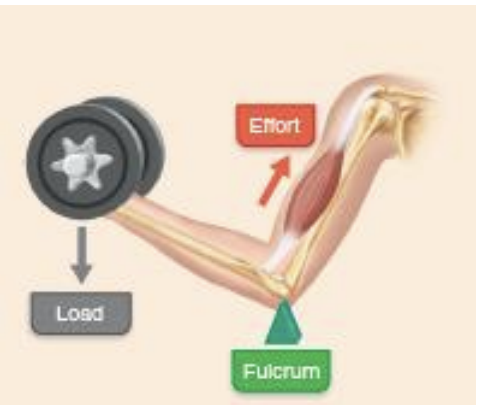
Describe a Third-Class Lever
Effort is closer to the fulcrum than the load
Fast
Always at a mechanical disadvantage
Examples of Third-Class lever
biceps brachii
Muscle Action and Interactions
muscles can’t reverse their own movements
Require opposing muscles
Muscles with opposite action on opposite sides of a joint
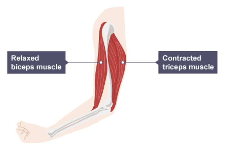
Roles in Muscle Movement
Prime mover (agonist)- a major role in the movement
Antagonist- opposes or reverses movement
Synergist - helps prime mover, and adds force or reduces unwanted movement
Fixator: sygernist that stabilizes
Prime mover
a major role in the movement
has an opposing muscle compartment with the antagonist
Antagonist
opposes or reverses the movement
has an opposing muscle compartment with an agonist
Synergist
helps prime mover and adds force or reduces unwanted movement
has the same muscle compartment
Fixator
synergist that stabilizes the bone
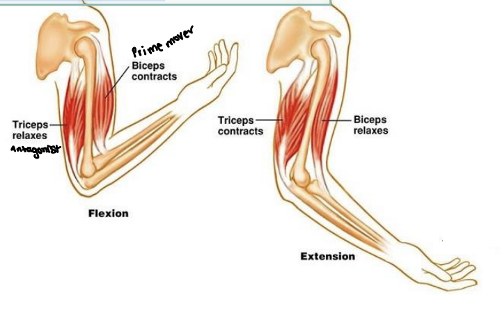
Example of the prime mover and Antagonist
When you bend your elbow the biceps brachii contracts, So it lifts the forearm, That makes It the prime mover for the action. While we're bending our elbow, the triceps brachii acts as the antagonist. So it's responsible for straightening that elbow, and it has to be relaxed for
the bicep to be able to bend.
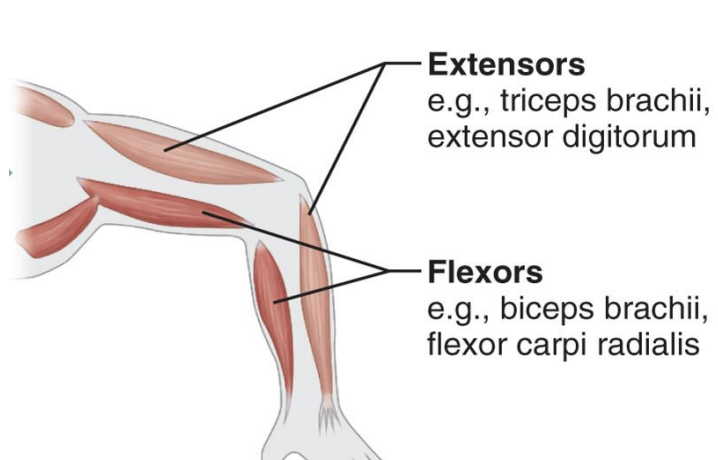
Limb Muscles
Extensors - dorsal to limb bones
Flexors - ventral to limb bones
Extensor and Flexor in the Upper limb
Extensors are posterior and Flexors are anterior
Extensors and Flexors in the Lower limb
Extensors are anterior, and flexors are posterior
What divides limb muscles into compartments?
Dense connective tissue
4 Muscle Compartments of the Upper Limb
Anterior arm component
Anterior forearm compartment
Posterior arm compartment
Posterior forearm compartment
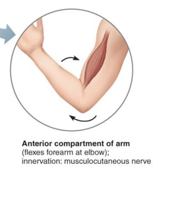
Anterior arm Component
Flexes shoulder/arm
Innervation is the musculocutaneous nerve
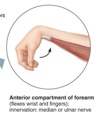
Anterior forearm compartment
Flexes wrist and digit
The Innervation is the median and ulnar nerve
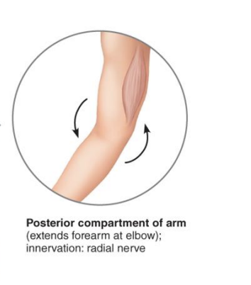
Posterior Arm compartment
Extends the elbow
Innervation is the radial nerve
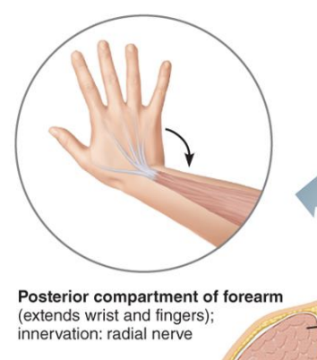
Posterior Forearm Compartment
Extends the wrist and digits
Innervation is the radial nerve
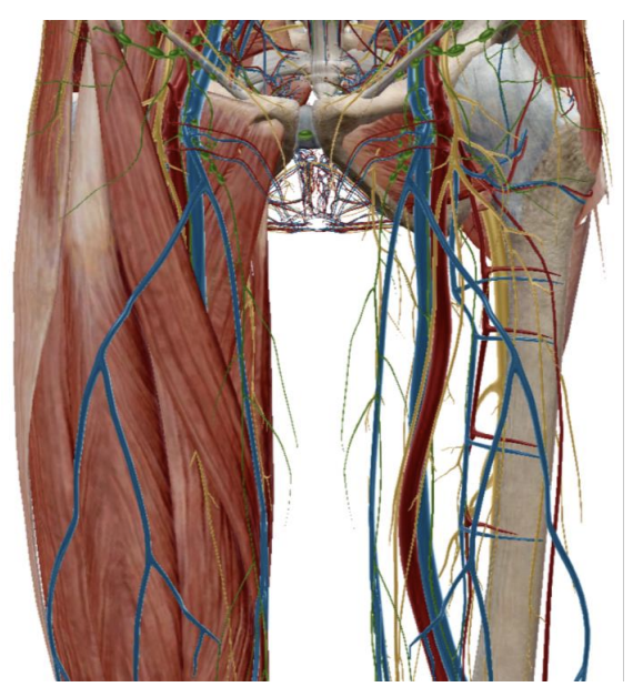
3 Muscle Compartments of the Thigh
Posterior compartment
Anterior Compartment
Medial Compartment
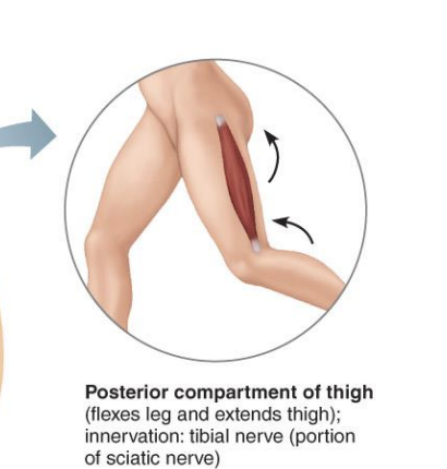
Posterior Compartment of the thigh
Extends the hip and Flexes the knee
Innervation is the tibial branch of the sciatic nerve
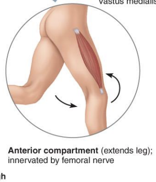
Anterior Compartment of the thigh
Flexes the hip and extend the knee
Innervation in the femoral nerve
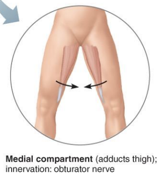
Medial Compartment of the thigh
Adduct Thigh
Innervation is the obturator nerve
3 Muscle Compartment of the leg
Posterior compartment
Anterior compartment
Lateral compartment
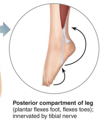
posterior compartment of the leg
Digital and plantar flexors
help point the foot down and flex the toes
Innervation is the tibial nerve
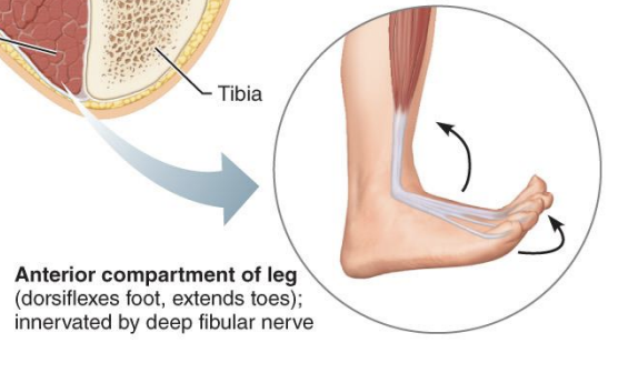
Anterior compartments of the leg
Digital extensors and dorsiflexors
lifts the foot and extends the toes
Innervation is the deep fibula nerve
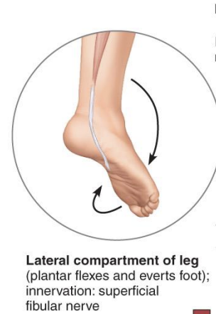
Lateral Compartment of the Thigh
Plantar flexes and everts foot
turns the foot outwards, and points it down
Innervation is the superficial fibular nerve
7 Facial expression muscles
Epicranius
Corrugator Supercilii
Zygomaticus major
Orbicularis oris
Orbicularis oculi
Mentalis
Platysma
What are the two components of Epicranius
Frontalis - at the front of your head
Occipitalis - Base of skull
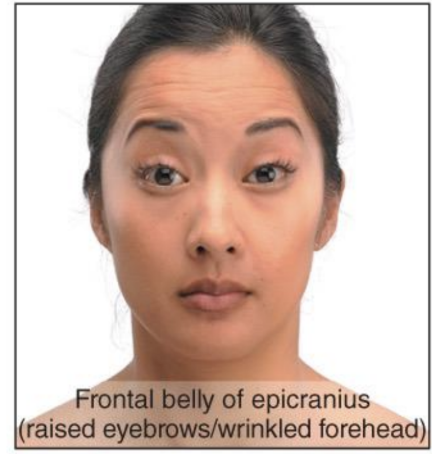
Epicranius - Frontalis- Action
Raises eyebrows, wrinkles forehead and assists in lifting eyelids
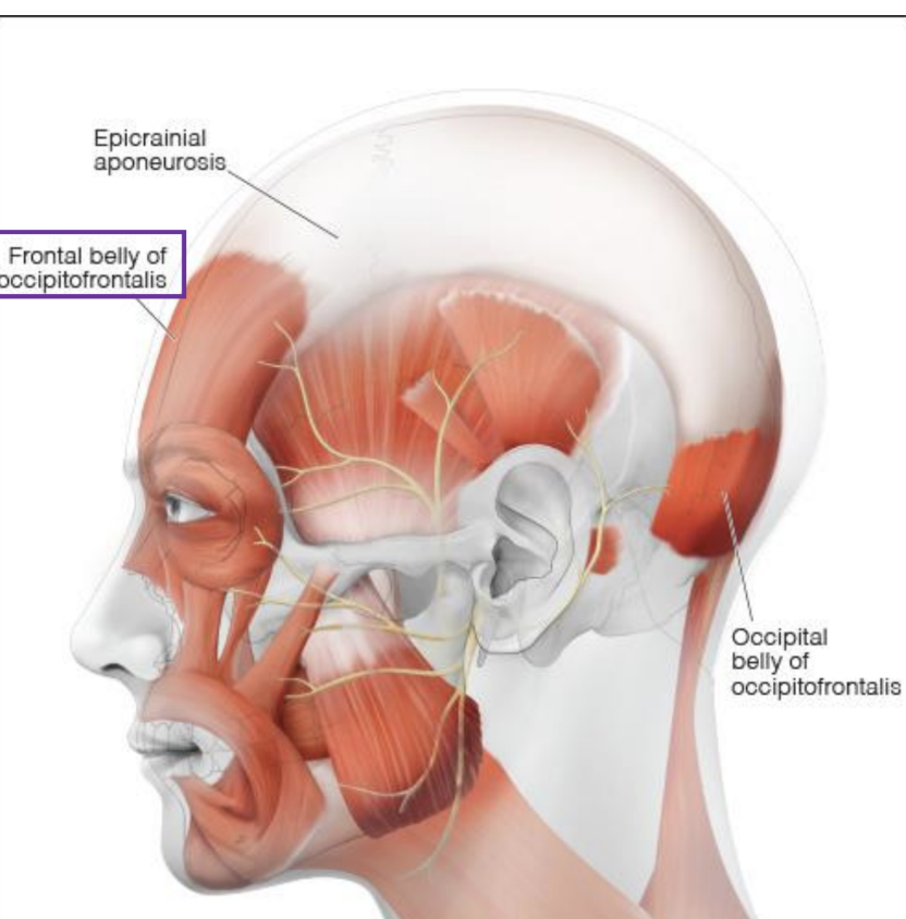
Frontalis - Origin & insertion
Origin - Galea aponeurotica
Insertion: Skin of eyebrows and bridge of noes
Epicranius - occipitalis, Action
Pulls scalp (flatten forehead) and raises eyebrows
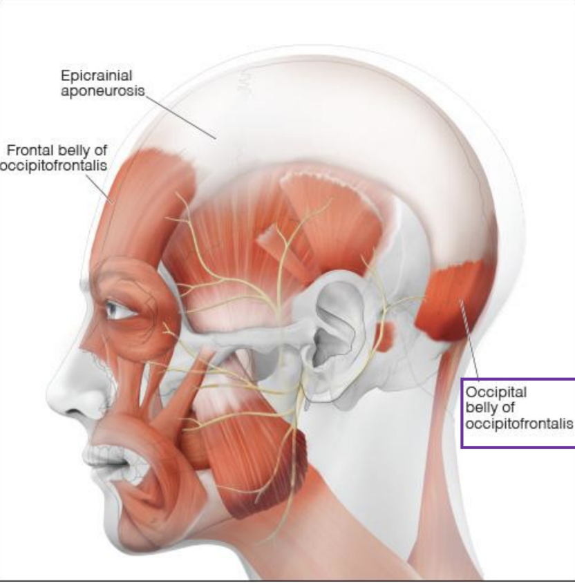
Occipitalis - Origin & Insertion
Origin- Superior nuchal line, and mastoid process
Insertion: Galea aponeurotica
Galea aponeurotica
a fibrous, tough sheet of fascia that connects the muscles of our scalp
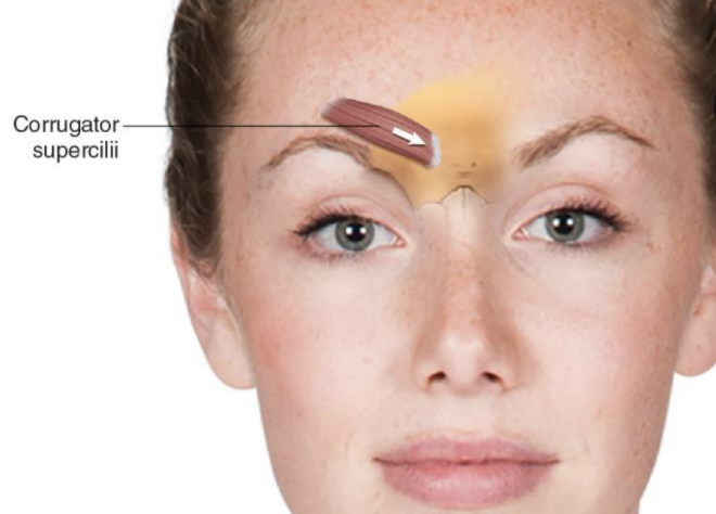
Corrugator Supercilii
Deep to the frontalis
helps with facial expressions
Action of the Corrugator Supercilii
pulls the eyebrow down and medially, which creates forehead wrinkles
Origin and Insertion of the Corrugator Supercilii
Origin- at the medial end of the superciliary arch
Insertion- Skin of eyebrow and orbital fascia
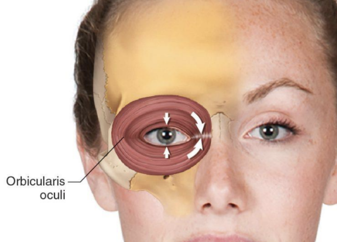
Orbicularis Oculi
a circular muscle around the eye, and it closes the eyelids
Origin and Insertion of the Orbicularis Oculi
Origin- frontal bone, maxilla and the orbit ligaments
Insertion- Skin around the eyelids and lateral palpebral raphe (the ligamentous band near the eye)
Zygomaticus Major
A diagonal muscle from the cheek (zygomatic bone) to the mouth corners
Origin and Insertion of the Zygomatic Major
Origin- zygomatic bone
Insertion- skin and muscles at an angle of the mouth
Action of the Zygomatics Minor
elevates the upper lip and exposes teeth
draws the upper lip backward and up
used for smiling and sneering
Origin and Insertion of Zygomaticus minor
Origin: Zygomatic bone (anterior part)
• Insertion: Skin of upper lip (medial to the zygomaticus major)
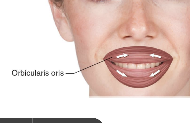
Orbicularis Oris
Surrounds the mouth and forms most of the lips
action of the Orbicularis Oris
closes and protrudes lips for speech
could be used for kissing and whistling
Origin and Insertion of Orbicularis Oris
Origin: Maxilla and mandible;
Insertion: Skin and mucous membrane at the lips