DAT Biology: Reproduction and Development Biology
 Call Kai
 Learn
Learn Practice Test
 Spaced Repetition
 Match
Match Flashcards
 Knowt Play
1/90

There's no tags or description
Looks like no tags are added yet.
Name | Mastery | Learn | Test | Matching | Spaced | Call with Kai | Chat |
|---|
No analytics yet
Send a link to your students to track their progress
91 Terms
Asexual Reproduction
Organism produces genetically identical offspring
Four Types: binary fission, budding, regeneration + fragmentation, parthenogenesis
Binary Fission
Form of asexual reproduction where DNA replicates, septum forms in the middle, separating the cell
Done by prokaryotes and mitochondria
Budding
Form of asexual reproduction where DNA is replicated and deposited into a bud, which forms a new organism
Done by hydra and yeast
Regeneration
Form of asexual reproduction where a Piece of an organism breaks off, and can regenerate the broken segment
Done by hydra, planaria, fungi
Fragmentation
Form of asexual reproduction where an organism breaks into pieces, and each piece grows into a brand-new, compelte individual
Parthenogenesis
Form of asexual reproduction where unfertilized eggs develop into a viable organism
Done by honeybees which exhibit haplodiploidy (males haploid, females diploid)
Sexual Reproduction
Gametes (male sperm and female egg) combine to form zygotes—genetically distinct offspring
Germ Cells
in males, known as male spermatogonia, and in females, known as female oogonia; specialized biological cell producing gametes via meiosis (can do both mitosis and meiosis)
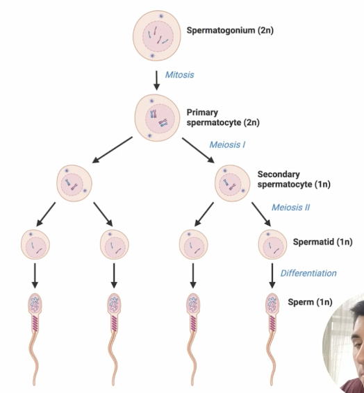
Spermatogenesis
Formation of haploid spermatoza (sperm cells) from diploid germ cells (spermatogonia)
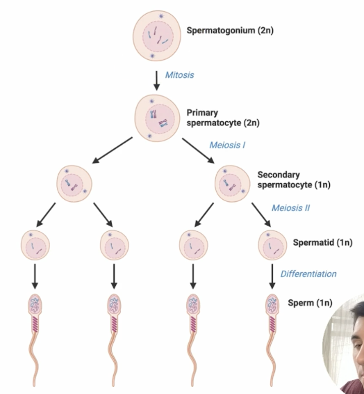
Spermiogenesis
Final stage in spermatogenesis in which spermatid differentiates into spermatoza
Seminal Vesicles
Secrete fructose (nutrient), viscous mucus (cleans and lubricates urethra), and prostaglandins (stimulate urethral contraction)
Viscous Mucus in Spermatogenesis
Cleans and lubricates urethra
Prostaglandins in Spermatogenesis
Stimulates urethral contraction
Prostate Gland
Alkaline secretions counteract uterine acidity
Bulbourethral Glands
Viscous mucus
Follicle Stimulating Hormone (FSH)
Stimulates sperm production in seminiferous tubules
Stimulates follicles in the ovary to develop
Activates Sertoli cells: nourish sperm cells, produce inhibin (negative feedback on FSH)
Activaes estrogen and progesterone production
Inhibin
Produced by Steroli cells upon activation by FSH, performs negative feedback on FSH
Lutenizing Hormone (LH)
Stimulates Leydig cells to produce testosterone
Stimulates ovulation of egg, corpus luteum formation
Testosterone
Primary male sex hormone responsible for regulating sperm maturation within the seminiferous tubules and driving the development of male secondary sex characteristics (deepening voice, facial hair growth, increased muscle mass)
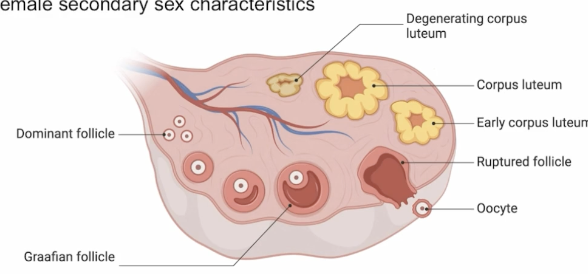
Ovary
Produces egg cells (ovum, ova)
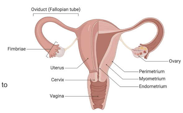
Fimbriae
Receives eggs released from the ovary
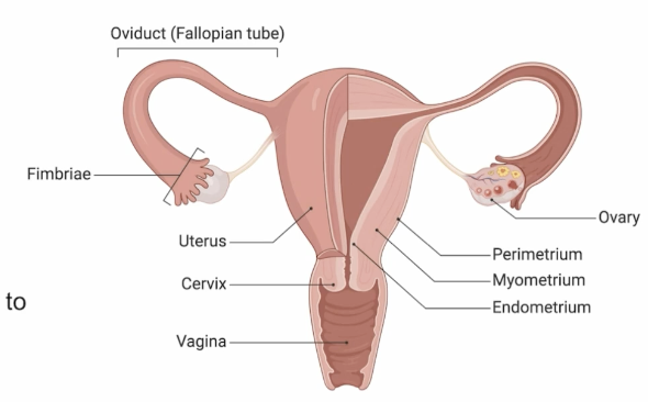
Oviduct/Fallopian Tube
Path between ovaries and uterus that the egg takes, egg has the opportunity to be fertilized here
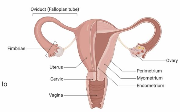
Uterus
Provides ideal environment for fertilized egg (if fertilized) to implant and develop, having 3 layers:
Perimetrium (outer), Myometrium (middle, muscular), and Endometrium (inner epithelial)
Cervix
Narrow opening between uterus and vagina
Vagina
Opening to external environment, where sperm can enter and birth occurs
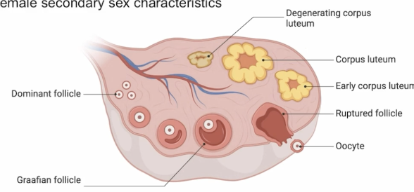
Follicle
Fluid-filled sac containing immature eggs (arrested in prophase I) prior to ovulation, upon ovulation, transforms into corpus luteum
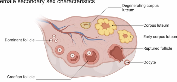
Corpus Luteum
Temporary endocrine gland forming in the ovary from an empty follicle after an egg is released during ovulation, secretes progesterone/estrogen to thicken and maintain the uterine lining for potential pregnancy
If fertilization doesn’t occur, it degenerates within about 2 weeks, causing hormone levels to drop and trigger menstruation
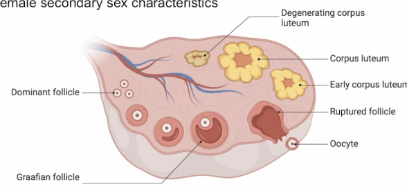
Ovulation
A mature egg (oocyte) released from one of the ovarian follicles into the abdominal cavity, swept into the fallopian tube by the fimbriae
Event triggered by a sudden surge in LH and typically occurring around the midpoint of the menstrual cycle (14 of 28-day cycle) marking the fertile window when pregnancy is most likely to occur
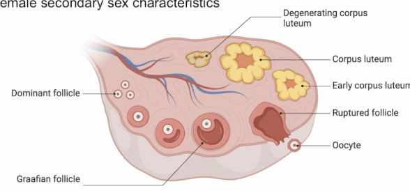
Estrogen
Drives female secondary sex traits like breasts, hips, and thickens uterine lining during the first half of the cycle
Progesterone
Released by the corpus luteum after ovulation (day 14), maintains that lining for pregnancy; if fertilization doesn't occur, its drop triggers menstruation.
Lactation
When infants sucking increases prolactin, stimulating milk production (lactation)
Oxytocin is also produced, which releases milk, where milk is let down
Follicular Phase
Estrogen leads to the thickening of the endometrium
Ovulation
Egg is released from the Graafian follicle, and the fimmbriae receive the egg, cilia sweep egg travels through oviduct awaiting fertilization
Luteal Phase
Follicle develops into corpus luteum, which releases progesterone and some estrogen and is maintained by FSH and LH, and estrogen and progesterone exhibit negative feedback on FSH and LH
Implantation
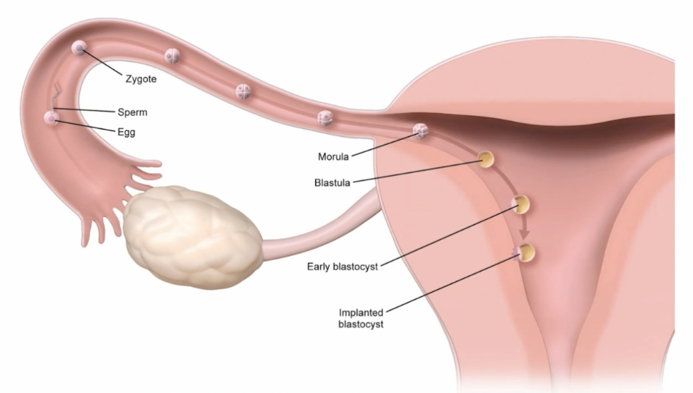
process where a fertilized egg (now called a blastocyst) embeds itself into the thickened, vascular uterine lining (endometrium), typically occurring about 6–10 days after ovulation (around cycle days 20–24). If successful, the embryo releases hCG to rescue the corpus luteum, ensuring it keeps producing progesterone to sustain the lining; if implantation doesn't occur, the corpus luteum degenerates, progesterone plummets, and the lining is shed as menstruation
No Implantation
FSH and LH decrease until the corpus luteum can no longer be maintained
Estrogen and Progesterone drop
Endometrium sloughs off (menstruation), and cycle repeats
Fertilization
Joining of a haploid sperm and haploid egg cell to form a diploid zygote
Capacitation
Final maturation step for sperm prior to encountering the egg, triggered by secretions from the uterine wall
Destabilizes plasma membrane proteins and lipids, preparing sperm tip for acrosomal reaction (meeting egg), and increases calcium permeability resulting in a hyperactive state where the sperm is more active in hopes of meeting an egg cell
Acrosomal Reaction
When the egg and sperm make contact
Corona Radiata
Outermost layer nourishing developing egg in mammalina cells
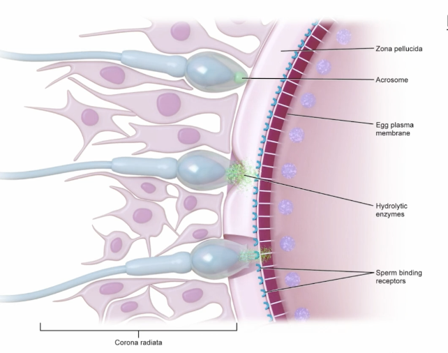
Vitelline Layer
layer of glycoprotein (jelly coat) above the plasma membrane, known as zona pellucida in mammals
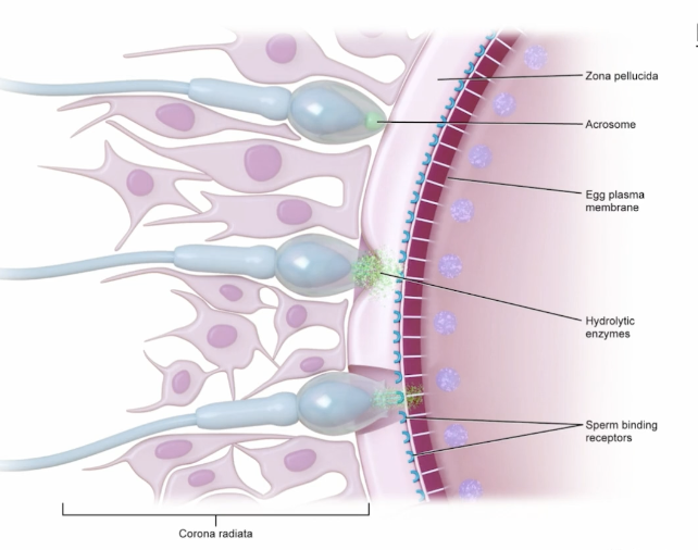
Zona Pellucida
Vitelline layer in mammals, layer of glycoprotein/jelly coat above the plasma membrane
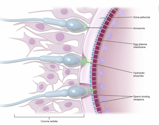
Polyspermy Blocks
Ways to prevent more than one sperm from entering one egg cell
Cleavage
Rapid cell divisions without changing the total mass of cells, to create resulting cells known as blastomeres
3 Types of Cleavage: axis of cleavage, fate of cells, evenness of embryo division
Blastomeres
Cells resulting from rapid cell divisions
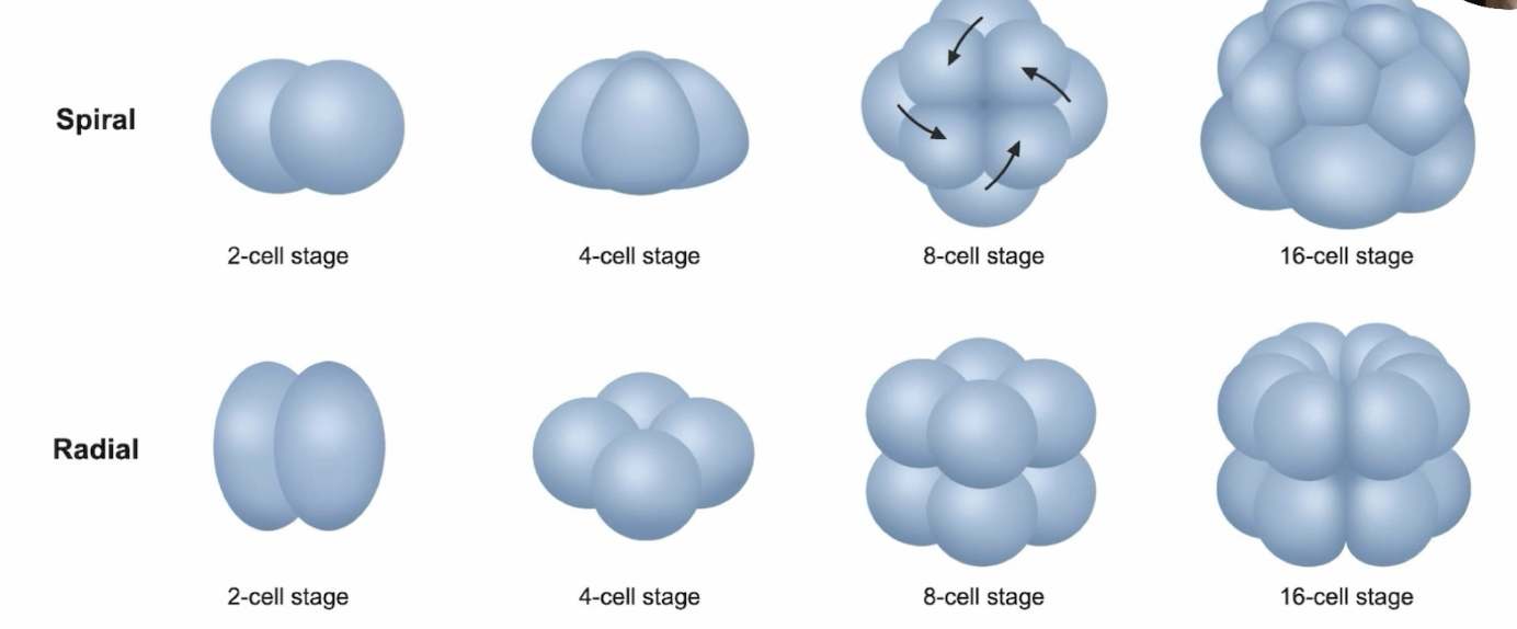
Axis of Cleavage
Spiral: cells deviate from the axis (protostomes)
Radial: cells aligned in vertical axis (deuterostomes)
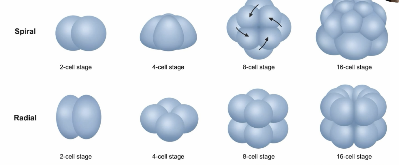
Spiral Axis of Cleavage
cells deviate from the axis (protostomes)
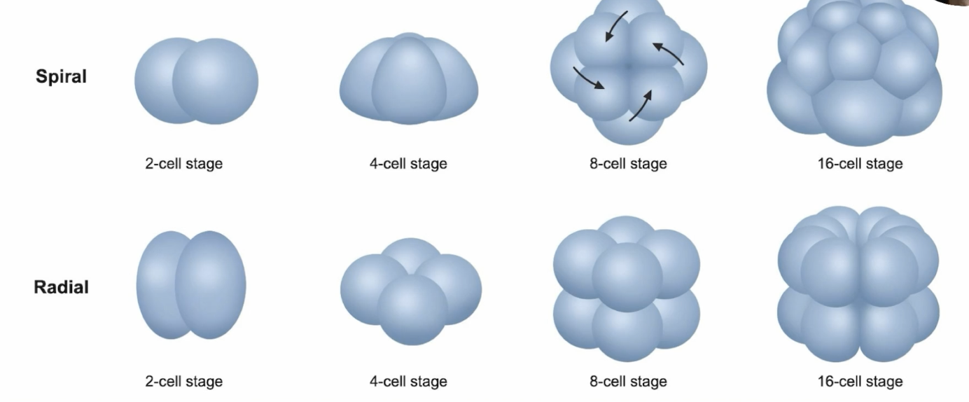
Radial Axis of Cleavage
cells aligned in vertical axis (deuterostomes)
Fate of Cells
Regulative (Indeterminate): blastomeres are totipotent, don’t have pre-determined fate (ex. deuterostomes)
Mosaic (Determinate): blastomeres have a decided fate (ex. protostomes)
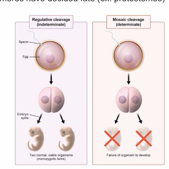
Regulative (Indeterminate) Fate of Cells
blastomeres are totipotent, don’t have pre-determined fate (ex. deuterostomes)
Mosaic (Determinate) Fate of Cells
blastomeres have a decided fate (ex. protostomes)
Evenness of Embryo Division
Holoblastic Cleavage: even cleavage, little yolk
Meroblastic Cleavage: uneven cleavage, forms animal and vegetal pole
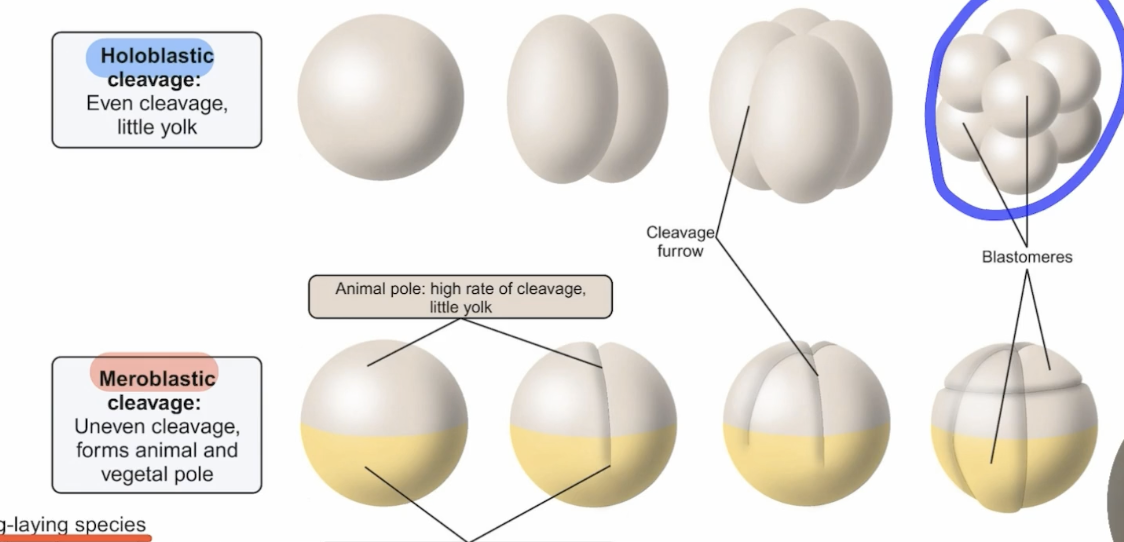
Meroblastic Cleavage
Performed by egg-laying species (except frogs, which have lots of yolk)
Uneven cleavage, forming an animal pole: high rate of cleavage and little yolk, and vegetal pole: low rate of cleavage, and lots of yolk, source of nutrients for the embryo
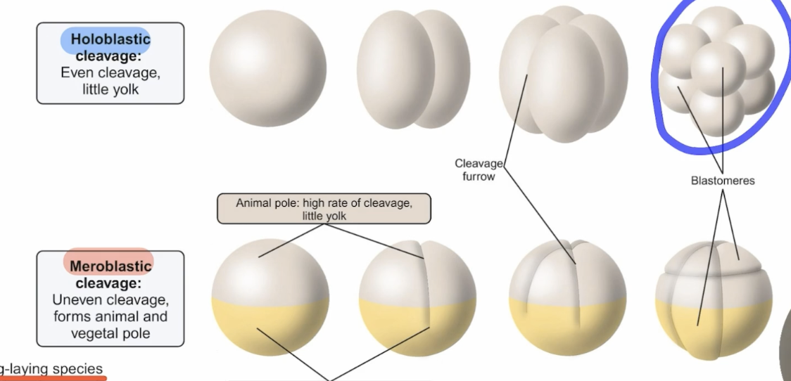
Holoblastic Cleavage
Cleavage happens very evenly, very little involvement from yolk, so it ends up dividing pretty evenly
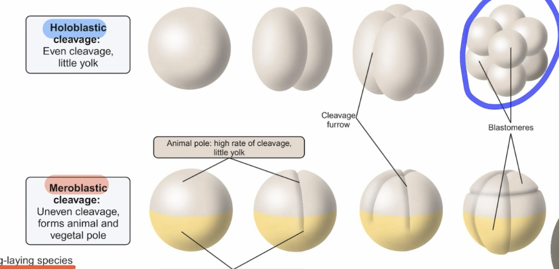
Morola
Solid ball of 16-32 blastomeres, dense, no fluid-filled cavity exists yet
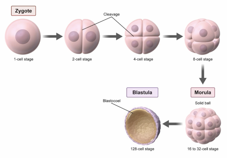
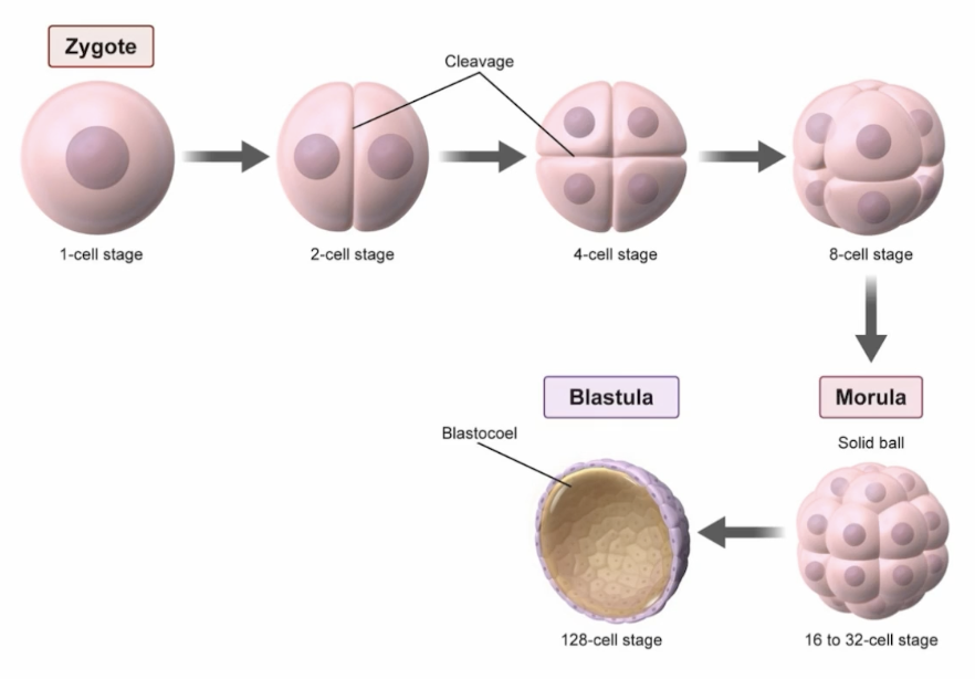
Blastula
Hollow sphere of cells surrounding a central fluid-filled cavity, the blastocoel, marking end of the cleavage stage and establishing the animal-vegetal axis, setting up spatial layout before gastrulation begins
Blastocyst
Mammalian, two cell lineages: inner cell mass (ICM) giving rise tot he embryo proper, and the trophoblast (forming the placenta, surrounding the blastocoel)
Hatches from the zona pellucida around day 5-6 to allow implanation into the uterine endometrium
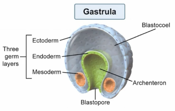
Gastrula
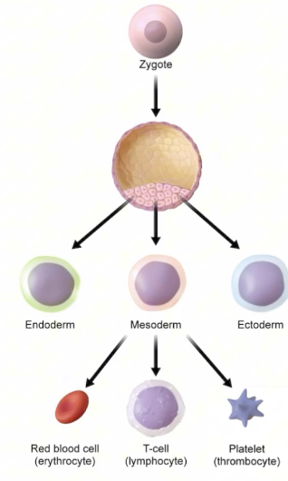
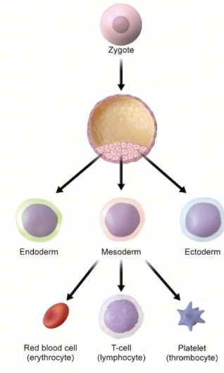
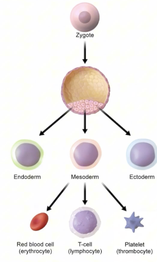
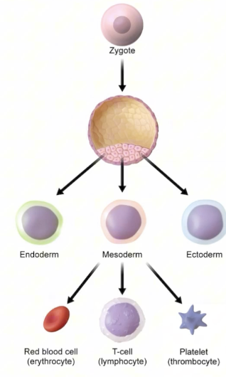
Triploblastic stage generated by the invagination and movement of cells, prpoducing the 3 primary germ layers: ectoderm, mesoderm, and endoderm.
For DAT, must memorize derivatives: ectoderm gives skin and nverous system, mesoderm gives muscle, bone, and blood, and endoderm gives the gut, lungs, and liver
Stem Cells
Undifferentiated cells with potential to develop in many different ways
Totipotent
Stem cell capable of developing into a complete embryo or differentiationg into any cell type
Pluripotent
Stem cell differentiating into any of the three germ layers
Multipotent
Stem cells differentiating into any cell type with a particular lineage
Differentiated
Differentiated cells are fully specialized and can only reproduce cells of their own type
Neurala
Term for embryo when it develops the nervous system
Stem Cells
Undiffferentiated cells with the potential to develop in many different ways
Notochord
Derived from mesoderm, stimulates ectoderm to thicken into a neural plate
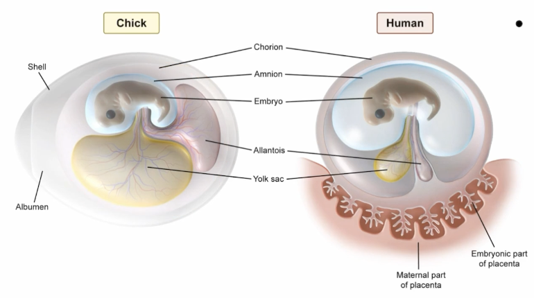
Extraembryonic Development
Structures and development outside of the embryo that provide protection and nourishment to the fetus—most embryos develop into embryotic organisms but some help to support it
Amnion
Innermost layer of the extraembryonic membrane, secreting amniotic fluid to cushion the embryo
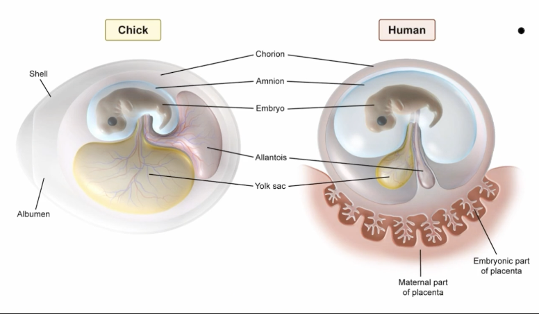
Amniotes
Have amnion (reptiles, mammals, birds)
Anamniotes
Lack amnion (amphibians, fish)
Chorion
Outermost layer of the extraembryonic membrane surrounding the developing embryo or fetus
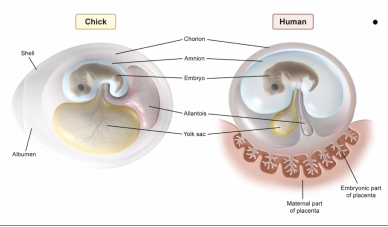
Chorion for Placental Mammals
Forms fetal half of placenta (nutrient exchange)
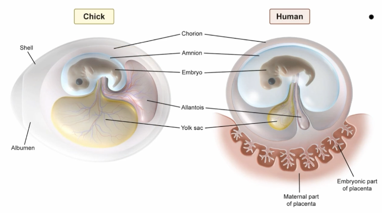
Chorion for Egg-Laying Mammals
Membrane for gas exchange is underneath the egg shell
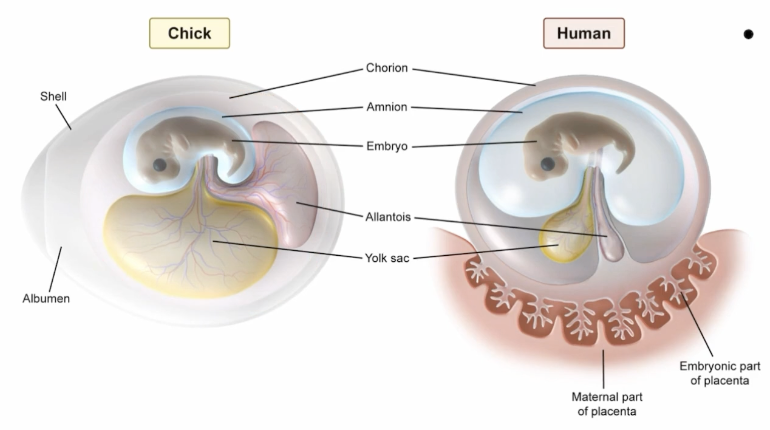
Allantois
Sac buds off archenteron, storing waste for disposal
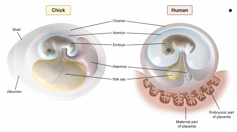
Allantois in Placental Mammals
Transports waste to placenta, forms umbilical cord and eventually urinary bladder in adults
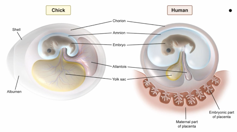
Allantois in Egg-Laying Mammals
Stores uric waste, later fuses with chorion to aid in gas exchange
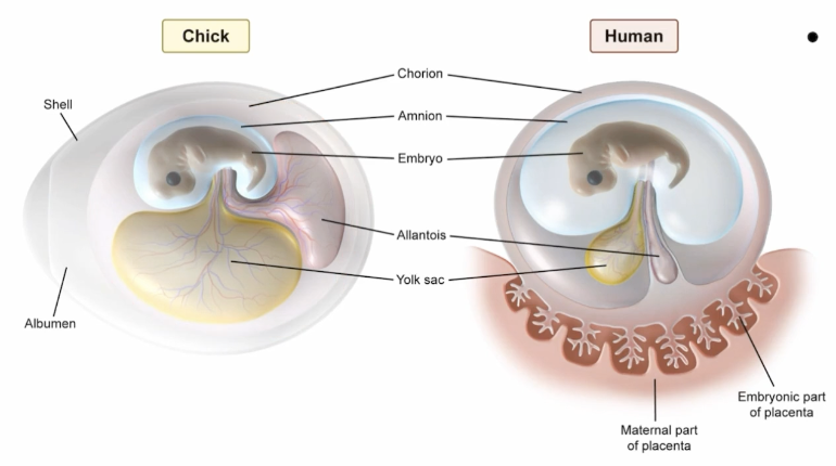
Yolk Sac in Placental Mammals
Doesn’t contain yolk. Functions temporarily until the placenta forms, providing early nutrients and serving as the first site of blood cell formation
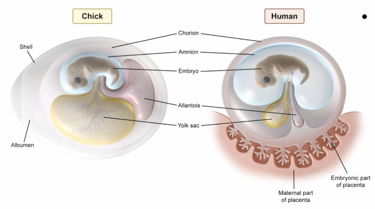
Yolk Sac in Egg-Laying Animals
Contains yolk, supplies all necessary nutrients to the developing of the embryo
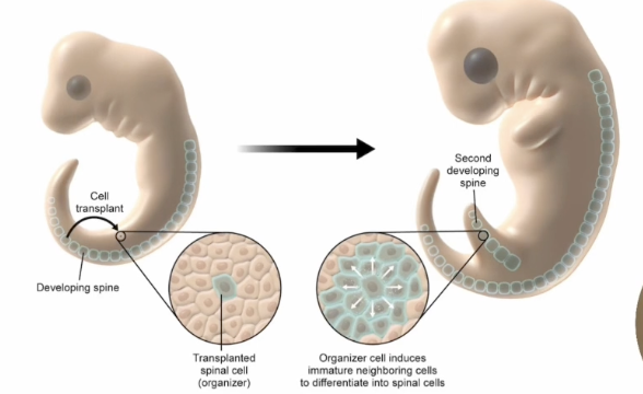
Embryonic Induction
Embryo contains cells known as organizer cells, and these organizer cells are going to secrete chemicals that instruct the cell around them on how to differentiate
Homeotic Genes
Determines what part of the embryo will develop into what structures
Master Controller
Turns certain gene expressions on/off
Homebox
A common DNA sequence homologous across different organisms that contain homeotic genes
HOX Genes
Subset of homeotic genes responsible for anterior-posterior (head-tail) organization
Apoptosis
Programmed cell death essential for normal fetal development, as well as health in adults (body modification)
Temperature-Dependent Sex Determination
Some reptiles determine sex by temperature rather than genetics
Pattern I: males in cold temp, females in warm temp (turtles)
Pattern II: females in low and high temp, males in intermediate temp (crocodiles)
Pattern I
Temperature-Dependent Sex Determination Pattern
Males in cold temps, females in warm temps (like turtles)
Pattern II
Temperature-Dependent Sex Determination Pattern
Females in low and high temperatures, males in intermediate temperatures (like crocodiles)
Oviparity
OFfspring develops in eggs, which hatch outside of the mother’s body
Example: chicken
Viviparity
Offspring develop inside the mother’s body, birth follows
Example: humans
Ovoviviparity
Hybrid between oviparity and viviparity, offspring develops in an egg and hatches within the mother’s body, birth follows
Example: sharks
HOX GEnes
Responsible for development of head to tail