Blood and Lymphatics
 Call Kai
 Learn
Learn Practice Test
 Spaced Repetition
 Match
Match Flashcards
 Knowt Play
1/37

There's no tags or description
Looks like no tags are added yet.
Name | Mastery | Learn | Test | Matching | Spaced | Call with Kai | Chat |
|---|
No analytics yet
Send a link to your students to track their progress
38 Terms
Agranulocyte
granules are NOT present in their cytoplasm
Basophil
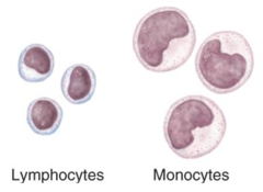
Bi-lobed dark purple nucleus with dark purple granules in cytoplasm.
Dark staining, grainy nucleus (lots of visible granules). Multilobed nucleus, can be difficult to see because of granules.
Granulocytes.
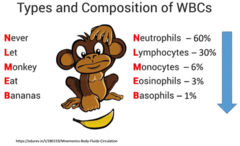
0-1% frequency in the blood
Eosinophil
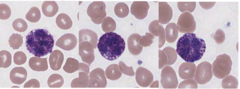
Bi-lobed dark purple nucleus with red-orange granules in cytoplasm.
Red staining, grainy cytoplasm (visible granules). Bilobed nucleus.
Granulocytes.
Erythrocyte / red blood cell
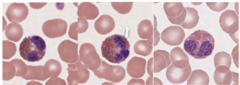
The most plentiful formed element, these cells appear pinkish-red on the outside and pale in the center. Functions to transport oxygen and carbon dioxide throughout the body. The normal count for people living at sea level is 4 - 6 million per cubic millimeter of blood.

Formed elements
red blood cells, white blood cells, platelets
Granulocyte
have granules in cytoplasm

Leukocyte / white blood cell
The five different types of cells are grouped by the presence or absence of granules in their cytoplasm. These cells are important in fighting infections and diseases. A normal count is 4,000 - 11,000 cells per cubic millimeter of blood.

Lymphocyte
The round dark purple nucleus takes up almost all of the cell. Only a slight "halo" of cytoplasm is visible.
Small 'crescent' of light cytoplasm. Large, round nucleus. About the same size as surrounding RBCs.
Agranulocytes.
20-45% frequency in the blood
Monocyte
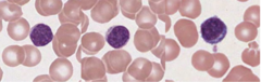
The largest of the white blood cells, this cell has a washed-out appearance with a large cytoplasm and kidney bean-shaped nucleus.
Large amount of light cytoplasm. Large irregular nucleus. MUCH larger than surrounding RBCs.
Agranulocytes.
4-8% frequency in the blood

Neutrophil
Multi-lobed dark purple nucleus with light purple granules in cytoplasm.
Light cytoplasm (granules don't stain well). 3-5 lobed nucleus.
Granulocytes.
40-70% frequency in the blood

Plasma
The liquid portion of the blood. 90% water. Protein and electrolytes. Important for transport.
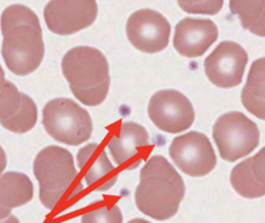
Thrombocyte / platelet
These fragments of megakaryocytes appear as small purple bits scattered among the red blood cells. They are important in the process of hemostasis (blood clotting). A normal count is 250,000 - 500,000 per cubic millimeter of blood.
Hematocrit
The percentage by volume of red blood cells in blood.
Hemopoiesis
the formation of blood cells and components
Hemostasis
blood clotting
Differential white blood cell count
Determines the percentage of each blood cell type in a person.
A blood smear is stained, and the percent of the different WBC types in a person's blood are determined. This is important in diagnosing clinical conditions where one type of white blood cell is elevated. For example, patients with an increased percentage of eosinophils may be having an allergic reaction to something.
Lymphatic system
Lymphoid organs
Consists of different organs, vessels, and tissues that make up a major portion of the immune system.

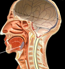
Lingual tonsil
A mass of lymphoid tissue located at the base of the tongue.
Lymph node [histology]
Lymph moves through these.
Acts as a filter to remove microorganisms and other foreign substances from the lymph.

capsule
connective tissue surrounding the lymph node
outer layer

cortex
outer region of lymph node

germinal center
central region of nodule, an area of rapid lymphocyte division

hilum
depressed, concave side of lymph node where efferent lymphatic vessels exit

Lymph nodule
(Follicles). Clusters of lymphocytes.

medulla
inner region of lymph node
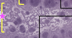
medullary cord
dark staining area in medulla, consists of lymphocytes and other cells
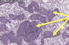
medullary sinus
major pathway through which lymph fluid flows through node
light staining portion of medulla

trabecula (pl. trabeculae)
inward extensions of capsule

Palatine tonsil
two masses of lymphoid tissue located at the back of the throat, one on each side

Pharyngeal tonsil
posterior wall of nasopharynx

Red bone marrow [histology]
The site of blood cell production.

Adipocyte
These cells are numerous as the bone marrow is a location for energy storage.
Fat storage cells in. In histology, they appear round and white.

Hemocytoblast
Stem cells from which all blood cells and cells fragments are made. Note: A stem cell is a cell capable of producing daughter cells that can differentiate into different cell types.
Small stem cells forming bone cells.

Megakaryocyte
Giant pink-stained cells that fragment to form many smaller platelets.
Large cell with cytoplasmic extensions (like the arms of an octopus) that stick into sinusoids. As blood flows through the sinusoids, bits break off. These are platelets.

Sinusoid
Small vessels through which blood cells enter the bloodstream.
Blood vessels in bone marrow where blood cells enter bloodstream. In histology, they appear as white spaces with a rust color, which is the red blood cells of the blood contained in them.

Spleen
A part of the lymphatic system and blood systems, is located just above the stomach, below the diaphragm, and inside the left rib cage. This small organ filters blood, removing cellular debris, microorganisms, and old or damaged blood cells. Stores blood cells and helps to fight infection by making lymphocytes. You can live without it, but you may be at a greater risk for certain types of blood infection. The degree of risk depends on your age and if you have other diseases.

Thymus gland
A small gland located behind the sternum and anterior and superior to the heart. Lymphocytes travel from the bone marrow to the here where they mature and become specialized T-cells. These T-cells are released from the here into the bloodstream after they have matured and are essential to fight infection and disease.

2 major components of blood
plasma and formed elements
