Cranial Nerve Examination - Abnormal findings
 Call Kai
 Learn
Learn Practice Test
 Spaced Repetition
 Match
Match Flashcards
 Knowt Play
1/50

There's no tags or description
Looks like no tags are added yet.
Name | Mastery | Learn | Test | Matching | Spaced | Call with Kai | Chat |
|---|
No analytics yet
Send a link to your students to track their progress
51 Terms
What abnormalities might you find when carrying out the general inspection?
Speech abnormalities
Facial asymmetry
Eyelid abnormalities
Pupillary abnormalities
Strabismus
Limbs (not specific CN lesions, but may be suggestive of neurological syndromes):
Spasticity
Weakness
Atrophy
Tremor
Fasciculation
If there was a problem with CN I (olfactory), what might you find on examination?
Anosmia or other changes to the patient’s sense of smell.
Anosmia
Partial or complete loss of the sense of smell.
What can cause anosmia?
Mucous blockage if the nose prevents odours from reaching CN I receptors.
Head trauma can shear CN I nerve fibres.
Genetics - congenital anosmia.
Parkinson’s disease (early feature)
COVID-19 - common feature (often transient)
What pupillary abnormalities might you see upon examination?
Abnormal pupil size
Dyscoria (abnormal pupil shape)
Anisocoria
Abnormal reflexes
What can cause abnormal pupil shapes (dyscoria)?
Congenital conditions
Posterior synechiae associated with uveitis
Eye trauma (peaked pupils)
Iritis
What is Posterior synechiae associated with uveitis?
Adhesion between the iris and lens / cornea due to inflammation in the uvea (rare). If the condition is very severe, then there can be associated glaucoma and permanent vision loss.

Peaked pupils
Irregularly stretched, teardrop shaped, or distorted pupils where a segment of the iris is pulled or stretched towards a point. In the context of trauma, it will likely be caused by a globe injury (eyeball injury).

Anisocoria
Asymmetry in pupil size. Causes include:
Congenital anisocoria
Migraines
Surgery side effects
Eye drops and other medicines
Trauma to eye, head, or arteries that supply blood to them (e.g. brain aneurysm, stroke, brain tumours etc.)
Oculomotor nerve palsy (causes large pupil)
Horner’s syndrome (causes small reactive pupil)

Bilateral miosis
Abnormal constriction of both pupils (normal in infancy!!). Causes include:
Senile miosis
Medications (e.g. opioids, barbiturates, pilocarpine etc.)
Brain trauma / haemorrhage / tumours / strokes
Poisoning
Untreated syphilis

Bilateral mydriasis
Abnormal dilation of both pupils. Causes include:
Eye examinations - use of some eyedrops
Medications
Brain / eye trauma
Microvascular cranial nerve palsy (blood flow to nerves is blocked)
Migraine / ocular migraine
Recreational drugs (cocaine, MDMA, LSD, methamphetamines, psychedelic mushrooms)
Sexual arousal
Adrenaline
If there was a CN II (optic) disorder, what might you find on examination?
Decreased visual acuity (eg. optic neuritis)
Abnormal pupillary light reflexes (shining a light on the affected eye will result in a reduced / absent direct and consensual response)
Abnormal colour vision (eg. optic neuritis)
Visual field defects
Abnormal blind spots (enlarged)
Abnormal fundoscopy
What are the different CN II disorders?
Glaucoma (fluid pressure inside eyes rises, damaging CN II)
Optic neuritis (inflammation of CN II)
Optic nerve atrophy (causes: trauma, toxins, ischaemia, disease)
Optic nerve head drusen (pockets of protein and calcium salts that build up in the optic nerve over time)
What types of visual field defects are there?
Bitemporal hemianopia
Homonymous field defects
Scotoma
Monocular vision loss
Bitemporal hemianopia
Loss of temporal visual field in both eyes resulting in central tunnel vision. It is typically caused by a tumour compressing the optic chiasm.

Homonymous field defects
Affect the same side of the visual field in each eye - can be hemianopias, or quadrantanopias. Typically caused by pathologies affecting visual pathways posterior to the optic chasm:
Stroke
Tumour
Abscess

Scotoma
A visual field defects where there is an area of absent or reduced vision surrounded by areas of normal vision. Causes include:
Optic neuritis
Demyelinating disease (e.g. MS)
Diabetic maculopathy
Retinal injuries
Cancer
Stroke
Age-related macular degeneration
Medications
Brain injuries
etc.

Monocular vision loss
Total loss of vision in one eye. Typical causes include:
Optic nerve pathology (e.g. anterior ischaemic optic neuropathy)
Ocular diseases (e.g. central retinal artery occlusion, total retinal detachment)
What causes an enlarged blind spot?
Papillitis - swollen optic nerve head (pictured). A dangerous subtype of this is Papilloedema - bilaterally swollen optic nerve head secondary to raised intracranial pressure (e.g. brain tumour, hypertensive crisis, intracranial haemorrhage).

What causes decreased visual acuity?
Decreased visual acuity has many potential causes including:
Refractive errors
Amblyopia
Ocular media opacities such as cataract or corneal scarring
Retinal diseases such as age-related macular degeneration
Optic nerve (CN II) pathology such as optic neuritis - will typically only be present in the affected eye
Lesions higher in the visual pathways
What is an abnormal pupillary light reflex?
No direct pupillary reflex present (no constriction in one pupil when light is shone on the ipsilateral pupil)
No consensual pupillary reflex present (no constriction in one pupil when light is shone on the contralateral pupil).
Relative afferent pupillary defect
Relative afferent pupillary defect
Normally light shone into either eye should constrict both pupils equally (due to the dual efferent pathways described above). When the afferent limb in one of the optic nerves is damaged, partially or completely, both pupils will constrict less when light is shone into the affected eye compared to the healthy eye. The pupils, therefore, appear to relatively dilate when swinging the torch from the healthy to the affected eye. This is termed a relative…. afferent… pupillary defect. This can be due to significant retinal damage in the affected eye secondary to central retinal artery or vein occlusion and large retinal detachment; or due to significant optic neuropathy such as optic neuritis, unilateral advanced glaucoma and compression secondary to tumour or abscess.
Abnormal accommodation reflex
Bilateral constriction and convergence is not observed.
What can cause colour vision deficiencies?
Congenital conditions (e.g. inherited red-green colour blindness)
Acquired:
Optic neuritis (typically red colour vision is reduced)
Vitamin A deficiency
Chronic solvent exposure
What CN II pathologies are visible via ophthalmoscopy?
Papilloedema (bilateral optic disc swelling)
Papillitis (only visible when it affects the nerve head)
Anterior ischaemic optic neuropathy (pale)
Optic disc neuropathy (pallor)
Pathological optic cupping
Congenital and structural abnormalities
Visual neglect / inattention
A condition in which an individual develops a deficit in their awareness of one side (hemispace) of their visual field. It is not caused by optic nerve pathology, but typically caused by pathology affecting the cerebral hemisphere, such as a stroke damaging the parietal lobe.
If there was a CN III (oculomotor) pathology, what might you find on examination?
Ptosis
Strabismus - eye pulled inferolaterally (down and out appearance)
Mydriasis (abnormal dilation of pupil)
Limited range of movement: unable to move up, down, or inward towards the nose
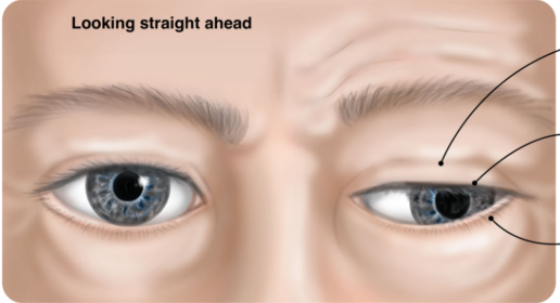
Ptosis
Drooping of the upper eyelid. Typically caused by:
Oculomotor nerve pathology
Horner’s syndrome
Neuromuscular pathology (e.g. Myasthenia Gravis)

What are some notable causes of CN III (oculomotor) palsy?
Intracranial aneurysm
Microvascular ischaemia
High impact trauma (low impact trauma should not cause this)
Recurrent painful ophthalmoplegic neuropathy
If there was a CN IV (trochlear) pathology, what might you find on examination?
Vertical diplopia when looking inferiorly (may notice patient compensating for this by tilting their heads forward and tucking their chin in)
Torsional diplopia (patient may compensate by tilting head go opposite side)
Limited range of movement: impaired ability to move eye downward and inward
What are some notable causes of CN IV (trochlear) palsy?
Congenital (most common)
Idiopathic (tends to be congenital, but presenting in adult life)
Moderate frontal head trauma (tends to be bilateral)
Hypertension
Diabetes
post-neurosurgery
Intracranial neoplasms
Herpes zoster virus (shingles)
etc.
If there was a CN VI (abducens) pathology, what might you find on examination?
Strabismus - convergent squint
Horizontal diplopia which is worsened when they attempt to look towards the affected side
Limited range of movement: impaired ability to abduct the eye (move outwards towards the side).

What are some notable causes of CN VI (abducens) palsy?
Idiopathic
Head injuries
Infections (e.g. meningitis, herpes zoster etc.)
Circulatory problems (stroke, microvascular ischaemia, brain haemorrhage etc.)
Systemic, inflammatory, and metabolic conditions (MS, T2 DM, hypertension etc.)
Blood disorders, cancers and tumours
Other neurological conditions (migraines etc.)
Medical procedures (LP, surgery etc.)
Strabismus
A condition in which the eyes do not properly align with each other when looking at an object. Pathology affecting the oculomotor, trochlear or abducens nerves can cause strabismus. Certain tests can be used to assess this (corneal light reflex test, cover test).

If the patient has pathology affecting CN V (trigeminal), what might you observe upon examination?
Loss of sensation in one or multiple of these areas:
Forehead (ophthalmic)
Cheeks (maxillary)
Lower jaw (mandibular)
Atrophy of the temporalis and/or masseter muscles.
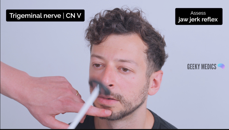
An inability to open the jaw against resistance or deviation of the jaw (typically to the side of the lesion) may occur in trigeminal nerve palsy.
Absent / reduced jaw jerk reflex if there is damage to the nerve itself.
Exaggerated / hyperactive jaw jerk reflex if there is UMN pathology.
Absent corneal reflex
Interpretation of the jaw jerk reflex
Normal jaw jerk reflex - slight closure of the mouth in response to tapping the chin with the tendon hammer.
Absent / reduced jaw jerk reflex if there is damage to the nerve itself.
Exaggerated / hyperactive jaw jerk reflex if there is UMN pathology.
Interpretation of the corneal reflex
Normal - direct and consensual blinking in response to unilateral corneal stimulation.
Abnormal - absence of direct and consensual blinking suggests a problem with either CN V or CN VII
Afferent branches = CN V (absent direct and consensual blinking when affected eye is stimulated, but not in the unaffected eye)
Efferent branches = CN VII (absent direct blinking when affected eye is stimulated, and absent consensual response when the non-affected eye is stimulated)
What notable pathologies affect CN V (trigeminal)?
Head trauma
Pressure from nearby blood vessels, tumours or swelling
Inflammation from certain infections (e.g. herpes zoster virus)
Circulatory problems (microvascular disease)
Autoimmune conditions (e.g. scleroderma)
Surgery complication
Trigeminal neuralgia (extremely painful chronic pain disorder)
Trigeminal neuropathy
Cluster headache
Lateral medullary syndrome (post-stroke or blood clot)
If the patient has pathology affecting CN VII (facial), what might you observe upon examination?
Changes in sense of taste (responsible for anterior 2/3 of tongue conveyance of taste)
Changes in hearing (paralysis of the stapedius muscle can result in hyperacusis)
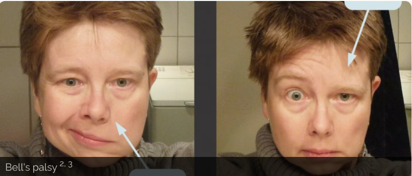
Asymmetry in the face at rest, particularly the forehead wrinkles, nasolabial folds and angles of the mouth.
Impaired ability to raise eyebrow(s) close the eye(s), blow out cheeks against resistance, smile or purse lips.
What are the two presentations of CN VII lesions?
UMN lesions
LMN lesions
Facial nerve palsy - UMN lesion
Presents with unilateral facial muscle weakness.
Upper facial muscles are partially spared because of bilateral cortical representation (frontalis function is therefore somewhat maintained)
Most common cause is a stroke
Facial nerve palsy - LMN lesion
Presents with unilateral facial muscle weakness - ALL ipsilateral muscles.
Most common cause is Bell’s palsy
What notable pathologies affect CN VII (facial)?
UMN:
Cortical stroke (typically MCA)
LMN:
Bell’s palsy
Pons pathology: stroke, demyelination (MS), tumour
Infections: meningitis, middle ear infections, tetanus etc.
Trauma
Tumours (meningioma, parotid gland, middle ear)
Surgery
Rare mononeuritis (diabetes, alcohol, HIV etc.)
If the patient has pathology affecting CN VII (facial), what might you observe upon examination?
Impaired hearing during the whisper test
Sensorineural hearing loss (Rinne’s + Weber’s tests)
Impaired balance (turning test, vestibulo-ocular reflex)
Sensorineural vs conductive hearing loss
Conductive hearing loss
Sound is unable to effectively transfer at any point between the outer ear, external auditory canal, tympanic membrane and middle ear (ossicles).
The patient will hear better when sound is conducted through bone rather than air.
Causes: excessive ear wax, otitis externa / media, perforated tympanic membrane and otosclerosis.
Sensorineural hearing loss
Dysfunction of the cochlea and/or vestibulocochlear nerve.
The patient will not hear well when sound is conducted through bone or air.
Causes: ageing,, excessive noise exposure, genetic mutations, viral infections, and ototoxic agents (e.g. gentamicin).
What does the Hallpike test assess?
If the test is positive, it indicates that the patient has benign paroxysmal positional vertigo (BPPV). This is not a problem with CN VIII (it is due to loose calcium carbonate crystals).
What notable pathologies affect CN VIII (vestibulocochlear)?
Idiopathic Sudden Sensorineural Hearing Loss (ISSHL - most common)
Excessive noise
Meniere’s syndrome
Ototoxic drugs
Labrynthitis
Accoustic neuroma
Small vessel disease
Brainstem lesions
Presbyacusis (age-related)
Congenital infections
If the patient has pathology affecting CN IX (glossopharyngeal), what might you observe upon examination?
Changes to taste (conveys taste from the posterior 1/3 of the tongue)
Difficulty swallowing
Difficulty with speech (also could be CN X pathology)
Absent gag reflex (also could be CN X pathology)
If the patient has pathology affecting CN X (V), what might you observe upon examination?
Difficulty with swallowing (also could be CN IX pathology)
Difficulty with speech (also could be CN IX pathology)
Weak, non-explosive sounding bovine cough caused by an inability to close the glottis.
Absent gag reflex (also could be CN IX pathology)
Deviation of uvula towards one side and asymmetrical elevation of palate.
What notable pathologies affect CN IX (glossopharyngeal)?
Trauma (e.g. styloid fracture)
Eagle syndrome (elongated styloid process)
Glossopharyngeal neuralgia
Iatrogenic
Tonsillar carcinoma
Stroke
What notable pathologies affect CN X (vagus)?