Comparative Anatomy Lecture Final ?
describe what morphology is
the use of morphological characteristics to compare evolutionary relationships between organisms
Jean Bapsitste de Lamark
believed in progressive change of characteristics dictated by use and disuse
believed evolution happened each generation
1/111
Earn XP
Description and Tags
#tibula
Name | Mastery | Learn | Test | Matching | Spaced |
|---|
No study sessions yet.
112 Terms
describe what morphology is
the use of morphological characteristics to compare evolutionary relationships between organisms
Jean Bapsitste de Lamark
believed in progressive change of characteristics dictated by use and disuse
believed evolution happened each generation
Carolus Linnaeus
used morphology to group organisms
taxonomic classification founder
Charles Darwin
survival of the fittest
tree of life
George Curvier
believed organisms exhibit ideal forms
father of comparative anatomy
Richard Owen
ancestral forms and homology
did not believe in evolution
named dinosaurs
ontogeny
an animals individual development from fertilization to death
gives us clues as to why form and function have changed in particular ways
invagination
one example of ontogeny: “how do we solve the surface area to volume ratio”
the respiratory and digestive system in surface are in relation to our mass → think villi
synapomorphy
shared derived characteristic
autoapomorphy
unique characteristics
pleisomorphy
ancestral characteristics
monophyly
common ancestor and all descendants
paraphyly
common ancestor and some descendants united by some characteristic
chordata characteristics
dorsal hollow nerve
endostyle
notocord
post-anal tail
pharyngeal gill slits
compare and contrast homology VS homoplasy
similarities: similar structures in different animals
differences: homology is when structures have morphological resemblance to each each other due to a shared common ancestor (forelimbs of mammals). homoplasy is when structures resemble one another but develop independently potentially due to the environment (wings of birds, bats, and reptiles).
vertebrata characteristics
backbone
inner ear specialization
radial fin muscles
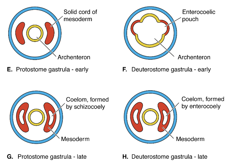
compared and contrast Protostome VS Deuterostome
similarities: they are both the first major division between organisms
differences: in protostomes the blastula develops a mouth first, has spiral cleavage and develops the coelom via schizocoely AKA the mesoderm splits to form the coelom. ex) crab
in deuterostomes the blastula develops a mouth second, has radial cleavage, and the coelom develops via enterocoely AKA the coelom buds off of the archenteron via enterocoelic pouches ex) fish
hemichordate characteristics
tripartite body (proboscis, collar, trunk)
link between invertebrates and vertebrates
have a stomochord
cephalochordate characteristics
myomeres (segmented muscles)
nervous system innervates muscles
“head” enclosed brain
locomotion!
craniate characteristics
“head” with tripartite brain
paired sense organs
neural crest cells (form when dorsal hollow nerve cord is developed)
neurogenic places (sense organ precursors)
muscularized hypomeres (contract the gills)
chordates VS craniates
chordates are filter feeders, craniates are pharynx feeders
Paedomorphism
vertebrate origins hypothesis: retention in adults of the larval form - child form
keep the overall larval form and specialize it
locomotion and different rate of development in reproductive organs drives this hypothesis
ex) axolotl
“new head” hypothesis
vertebrate origins hypothesis: Organisms centralized many structures in their head region (brain, eyes, ears) as well as changing feeding (muscular pharynx) means which makes you a better predator.
Gnathostome characterisitcs
vertebrates with jaws
paired nostrils
fill gill slits + visceral gill arches
chrondrichthyes characteristics
cartilaginous fish
cartilage skeleton strengthened by calcification
NOT BONES
Osteichthyes characteristics
bony fish aka teleosts
calcified bones
swim bladders
Sarcoptyergian characteristics
fleshy fish
fins are monobasic (have a humerus + femur)
tetrapod characteristics
limbs have digits
develop articulations (shoulder + hips bear weight now)
specialized sacral vertebra and sacral ribs
lose connection between skull and pectoral girdle
embryogenesis
make an embryo
morphogenesis
making of the morphology (how you make an organ)
two types of development
indirect = metamorphosis phase
(tadpole → frog)
direct = no intermediate stage
stages of development
fertilization
when sperm meets egg
cleavage
cell division without growth
gastrulation
formation of the three germ layers
three germ layers
ectoderm = outer skin
mesoderm = middle skin
endoderm = inner skin
ectoderm turns into….
epidermis
endoderm turns into…..
liver
mesoderm turns into….
notochord
neurulation
creation of the dorsal hollow nerve cord and neural crest cells
mesoderm differentiation
Created from enterocoelic pouches that bud off of the archenteron (gut) and form mesodermal segments called somites
1. Paraxial mesoderm:
segmented somites lateral to neural tube
2. Lateral plate mesoderm:
broad, unsegmented somite that lies ventral between archenteron and ectoderm
3. Intermediate mesoderm:
lies between the other two mesoderm layers
Paraxial mesoderm regions
sclerotome
forms vertebrate + occipital region of skull
myotome
forms voluntary + skeletal muscle
dermatome
forms the dermis
what does the meckel’s cartilage become
articular
what does the palatoquadrate become
quadrate
what are the two regions of the mandibular arch
palatoquadrate
mandible (meckel’s cartilage)
branchiomere
term used for embryonic development
refers to the segmentation of the gill region
evolution of chordates feeding style
filter feeds → pharyngeal feeders
region between each branchiomere (arch)
pharyngeal pouch
how many branchiomeres
7 total arches
branchiomere - arch I
mandibular
innervated by the trigeminal nerve (IV)
branchiomere - arch II
hyoid
innervated by byfacial (VII)
branchiomere - arch III
glossopharyngeal
innervated by glossopharyngeal (IX)
branchiomere - arches IV-VII
vagal 1, 2, 3, 4
innervated by vagus (X)
characteristics of the skeletal system
support / protection
attachment of muscles, tendons, and ligaments (locomotion)
framework of overall body shape
protection of internal organs
hemopoeitic
produce blood cells in the bone marrow
dynamic system
bimechanical strain, regulation of blood calcium levels, growth
why study bones?
easy to study since they preserve well (fossilization)
provides lots of information
can see where soft tissue attaches
predict locomotion
information about sense organs
diet of organisms (teeth shape)
3 classifications as bones
dermal VS endoskeleton
somatic VS visceral
cranial VS post-cranial
dermal VS endoskeleton
dermal = more superficial and develops as a membranous origin, consists of bony scales or large bony plates
endoskeleton = deeper and composed of cartilage then replaced by bones, ossification
2 types of ossification
intramembranous
endochondral
intramembranous ossification
formation of flat bones with mesenchymal cells that invade fibrous connective tissue, no cartilage model is present, flat bones are produced this way, this is the way bones heal after they are broken
endochondral ossification
bones are created through ossification of cartilage model, long bones are formed this way
somatic VS visceral
somatic = skeleton is associated with outer tube (mesoderm and ectoderm), most of the skeleton we think of
visceral = skeleton is associated with inner tube (endoderm), cartilage in the pharynx or gills
cranial VS post-cranial
head VS rest of the body
cranial skeleton regions
chondrocranium
protection of brain, neural crest cells derived
splanchnocranium
visceral skeleton, neural crest cells derived
dermatocranium
roofing bones, dermal bones, both mesoderm and neural crest cells derived
post-cranial skeletal regions
axial
notochord, vertebral column, ribs, fins, sternum
appendicular
appendages and associated girdles
cranial skeleton jobs
protect soft tissue (brain + sense organs)
involved in food gathering
passage for respiratory flow of water and air
major evolutionary changes occurred in the skull
chondrocranium jobs
protects brain and sense organs
most conserved evolutionarily
any change is done by fusion
splanchnocranium jobs
jaws and gill arches
primarily feeding and respiration
contains the mandibular arch
splanchnocranium evolutionary trends
loss of arches (associated with air breathing)
incorporation of bones into the head
jaw articulation changes
dermatocranium jobs
primarily superficial dermal bones that cover the other two regions
roofing bones
palatal series (roof of mouth)
encase mandibular cartilage
opercular series
ventral gills
evolution of jaw suspension and jaw articulation is driven by …..
predation
three places jaws attach to on the palatoquadrate
ethmoid process
basiethmoid
otic process
three ways jaws are suspended
Amphistylic
Hyostylic
Autostylic
Amphistylic
palatoquadrate anchored to chondrocranium and hyomandibular extends from otic capsule
EX] bony fish
![<ul><li><p>palatoquadrate anchored to chondrocranium and hyomandibular extends from otic capsule</p></li><li><p>EX] bony fish </p></li></ul><p></p>](https://knowt-user-attachments.s3.amazonaws.com/87a0a489-9330-4f51-97d3-d4cffbad86f6.png)
Hyostylic
palatoquadrate is stabalized only the Hyomandibular
EX] great white
![<ul><li><p>palatoquadrate is stabalized only the Hyomandibular</p></li><li><p>EX] great white </p></li></ul><p></p>](https://knowt-user-attachments.s3.amazonaws.com/b13c4aa2-96f3-470d-9caa-7a130b1814e6.png)
Autostylic
palatoquadrate fused to chondrocranium, frees up hyomandibular
EX] cat
![<ul><li><p>palatoquadrate fused to chondrocranium, frees up hyomandibular</p></li><li><p>EX] cat</p></li></ul><p></p>](https://knowt-user-attachments.s3.amazonaws.com/1fdda3c8-b340-42ce-8498-eb9ba7ee43e3.png)
adaptation VS exaptation
adaptation = trait which makes an animal better suited for their environment
exapataion = adaptation with a change in function (palatoquadrate + meckel’s cartilage articulation)
what does the hyomandibula become in amphibians
columella
transmits sound
connects the tympanic membranes in frogs
what does the quadrate-articular become in mammals
denture-squamosal articulation
increase in bite force
what does the quadrate become in mammals
incus
what does the articular become in mammals
malleus
what does they hyomadibular become in mammals
stapes
axial skeleton
portion of the skeleton that lies in the longitudinal axis of body
cranial skeleton, notochord, vertebral column, medial finds, ribs and sternum
post cranial skeleton
axial skeleton and appendicular without skull
give stability, rigidity, connection point for girdles
vertebrae common components
neural arches
hermal arches
neural and hemal spines
vertebral foramina
centrum
which craniates do not have vertebrae
Hagfish only have a notocord
lamprey have arcualia
Different types of vertebral columns
Amphicoelous
Procoelous
Opisthocoelous
Acoelous
Heterocoelus
amphicoelous vertebrae
both side of centra are concave, intervertebral pads present (derived from notochord), fishes
procoelous vertebrae
concave on cranial side, reduced chance of dislocation, intervertebral pads of ossified notochord, amphibians
opishocoelous vertebrae
convince caudal side, amphibians
acoelous vertebrae
flat surface, intervertebral disks present, mammals
heterocoelus vertebrae
saddle shaped centra, high mobility, bird necks
list the rib articulations
Basapothesis
Parapothesis
Diapothesis
Basapothesis rib articulation
vertebral rib attachment
Parapothesis rib articulation
small process for head of rib
Diapothesis rib articulation
transverse process for rib tuberculum
types of skeletogenous septum
dorsal
lateral
ventral
horizontal
tetrapod vertebrae evolution
centrum have become larger and well developed
increase in articulation sites
evolved ‘neck’ region
atlas + axis
regionalization of vertebrae
type of apotheoses: Zygapotheses
extend from neural arch and caudal
type of apotheoses: zygopothesis
from one vertebrae overlap the cranial (rostal)
type of apotheoses: zygapothesis
another type that helps with stability
what are the 2 major challenges which shape the axial skeleton over the years?
the type of environment the organism lives in
the type of locomotion the organism exhibits
hagfish vertebrae evolution
no true vertebrae
lamprey vertebrae evolution
have arcualia
not q true vertebrae but gives stability to notochord