MedSurg2 Unit 1
 Call Kai
 Learn
Learn Practice Test
 Spaced Repetition
 Match
Match Flashcards
 Knowt Play
1/329

There's no tags or description
Looks like no tags are added yet.
Name | Mastery | Learn | Test | Matching | Spaced | Call with Kai | Chat |
|---|
No analytics yet
Send a link to your students to track their progress
330 Terms
Approximate adult blood volume
5 L
Cardiac Output
Stroke Volume (amount of blood heart can pump with each beat) X Heart Rate
-If one is impacted, the other tries to compensate
Preload
(Rubber band / Stretch)
-Volume of blood in ventricles at end of diastole
(blood in chamber before heart contracts)
Afterload
(Pressure)
-Resistance L ventricle must overcome to circulate blood
(resistance heart faces as it contracts)
Contractility
Force of contraction
-Heart squeezing to empty
Cardiac Physical Assessment
-Inspection
Skin color & temp
Cap refill
Edema
JVD
Cardiac Physical Assessment
-Palpation
-PMI (ONLY at 5th ICS, Left MCL)
-Check for thrill (abnormal)
Feeling apical pulse in 2 or more locations could indicate
L ventricular enlargement
Feeling a thrill from a murmur could indicate
Valvular disorder
Cardiac arrest
Thrill over blood vessel could indicate
Partial obstruction (turbulent blood flow)
When is a thrill or bruit expected?
ONLY with dialysis
Cardiac Physical Assessment
-Auscultation
-BP
-HR, rhythm, S1, S2
-Murmurs (never expected)
S3 beat
Heart Failure
Normal in children/young adults
S4 beat
MI
HTN
CAD
Stenosis
Labs used for cardiac diagnostic assessment
CBC
BMP/CMP
PT, INR, aPTT, anti-Xa
CK-MB
Troponin I
BNP
Cholesterol
Cardiac Diagnostic Labs:
-CK-MB
Measures direct muscle damage to heart
-Increased reading = more damage
CK-MB lab values
0-3
Cardiac Diagnostic Labs:
-Troponin I
Measures heart damage
Troponin I lab values
< 0.5 ng/mL
Cardiac Diagnostic Labs:
-BNP
Measures cardiac fluid volume
(increased = worse HF)
< 100 pg/mL
BNP lab values
<100
WBC lab values
4,500-11,000
RBC lab values
M: 4.6-6.2
F: 4.2-5.4
Hgb lab values
M: 13-18
F: 12-16
Hct lab values
M: 42-52%
F: 35-47%
Platelets lab values
150,000-450,000
Sodium (Na+) lab values
135-145
Potassium (K+) lab values
3.5-5.0
Calcium (Ca+) lab values
8.2-10.2
Chloride (Cl-) lab values
98-107
Glucose lab values
70-99
Magnesium (Mg+) lab values
1.3-2.1
Phosphate (PO4) lab values
2.5-4.5
Serum Osmolality lab values
275-300
BUN lab values
10-20
Creatinine lab values
0.7-1.4
Total Cholesterol lab values
150-200
HDL lab values
M: 35-70
F: 35-85
(>70 desirable)
LDL lab values
<160
Triglycerides lab values
100-200
Cardiac Diagnostic Tests:
-ECG/EKG
Measures *electrical* impulses of the heart
Cardiac Diagnostic Tests:
-Stress Test
-Treadmill or chemical & hooked to ECG
-Pain or cardiac changes = positive test
Cardiac Diagnostic Tests:
-Traditional Echocardiography
US to check heart function
-No prep needed
Cardiac Diagnostic Tests:
-Transesophageal Echocardiography (TEE) *
-Probe down esophagus to get direct view of heart (invasive)
-Used to check for clots & rule out arrhythmias
TEE Pre Op
NPO
Throat numb
Consent
Meds
TEE Post Op *
Monitor gag reflex & check before giving food/water/meds
Cardiac Diagnostic Tests:
-Myocardial Perfusion Imaging (SPECT)
Radioactive US
-Checks for degree of muscle damage
-Radioactive isotopes given, they won't enter damaged area
Cardiac Diagnostic Tests:
-Cardiac Catheterization
Can be with or without angiography (dye)
-Cath through artery into heart
-Used to view arteries and/or place stent
Cardiac Catheterization
-Pre Op
-NPO 6 hrs
-Educate pt
-Check kidney function (BUN, Creatinine)
Cardiac Catheterization
-Pre Op Education
-Will be lying on hard table for 1 hr during procedure
-Meds given for comfort
-May have palpitations when cath is advanced
-Dye may cause heat, flushing, inc HR, feeling of needing to void (will last
Cardiac Catheterization
-Why check kidney function?
If it is poor, cannot use dye
Cardiac Catheterization
-Post Op * (7)
-Assess site frequently for bleeding ** (PRIORITY)
-Bed rest per protocol
-ECG & VS
-IV & PO fluids (flush out dye)
-Monitor UO (kidneys)
-Monitor pulse distal to puncture site
-Instruct pt to report chest pain, bleeding (wetness), or discomfort
Cardiac Catheterization
-Patient Education *
-Don't bend at waist, strain, or lift heavy objects for 24 hrs
-Avoid baths
-Call HCP if: bleeding, swelling, new bruising, pain at site, or temp ≥ 101.5
Hemodynamic Monitoring
Invasive system used in critical care to provide information about:
-Cardiac Effectiveness
-Blood Volume
-Tissue Perfusion
Hemodynamic Monitoring:
-Central Venous Pressure (CVP) Monitor
Measures blood pressure above the R atrium
CVP Normal Values **
*2-8*
-
Hemodynamic Monitoring:
-Intra-arterial BP Monitoring
IV cath into artery for direct BP reading
-Most accurate reading, fluctuates with every pulse
Hemodynamic Monitoring:
-Pulmonary Pressure Monitor
Invasive line into pulmonary artery with balloon on end
-Inc readings = LHF, high pressure in lungs and blood not moving forward
Electrical Impulses
-Pathway
SA node
AV node
Bundle of His
(If one doesn't work, will start in the next)
SA Node
Sinus Rhythm (normal)
-P wave for every QRS
-BPM: 60-100
AV Node
Backup if SA node stops working, can send impulses but triggers atrium at slower rate
-P wave messed up somehow (abnormal)
-BPM: 40-60
Bundle of His
If impulses start here, the SA and AV node have failed, heart starts dying
-Wide QRS complex
-BPM: 30-40 bpm
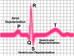
Electrocardiogram (ECG)
-Normal Reading
-P wave: SA node
-QRS: ventricular response
-T wave: vulnerable period (CANNOT shock here)
-ST Segment: check for MI here (elevated with MI)
Determining rate based on ECG
(All strips 6 seconds on test)
-Count R waves & times by 10

Determine regular vs irregular rhythm on ECG
Regular:
-All complexes match (P, QRS, T same on each one)
-P:R intervals all the same distance
When looking at an ECG, it's always important to do what?
Make sure what you SEE matches what you FEEL **
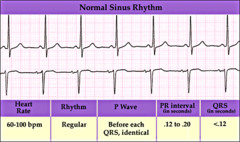
Normal Sinus Rhythm
-P wave for every QRS (SA node- Sinus)
-Regular (R waves all same distance)
-Rate 60-100
Types of Arrhythmias (Dysrhythmias)
-Sinus (P wave for every QRS)
-Atrial (irregular P wave but narrow QRS)
-Ventricular (wide QRS)
Stable Arrhythmias *
*NO S/S*
Have time to treat
Unstable Arrhythmias
*HAVE S/S* - need immediate intervention
-Altered mental status
-SOB
-Dizziness
-Chest pain
-Hypotension
-Shock
-Diaphoresis
-Cyanotic
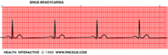
Sinus Bradycardia *
-P for every QRS, could be INVERTED
-Narrow QRS
-Regular
-Rate
Asymptomatic (stable) Sinus Bradycardia
-Treatment
No treatment
-Determine cause
Symptomatic (unstable) Sinus Bradycardia
-Treatment
1. Atropine (PRIORITY- pulls HR back up)
2. Transcutaneous pacing
3. Determine cause
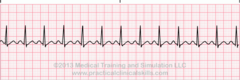
Sinus Tachycardia *
-*P for every QRS*
-QRS narrow
-Regular
-*Rate >100 bpm*
Sinus Tachycardia
-Treatment
1. Identify cause
2. Treat cause
-Ex: Acute blood loss- IVF & blood transfusion
Types of Pacemakers
-Transcutaneous (external)
-Implanted (internal)
Considerations for implanted pacemakers
Keep cell phone 6-12 inch away from device
-No cell phones in front shirt pockets
Pacemakers
-Potential Complications
-Failure to pace (shocks but nothing happens)
-Failure to capture (HR not where it needs to be)
Interventions if pacemaker fails to capture
Inc output
Change batteries
Reposition
Atrial Arrhythmias
Premature Atrial Complex (PAC)
Atrial Flutter
Atrial Fibrillation
Supraventricular Tachycardia
Premature Atrial Complex (PAC) *
-Extra beat before P wave
-Pause before next complex after occurs
-Irregular
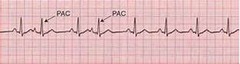
Premature Atrial Complex (PAC)
-Common causes
Caffeine
Stress
Anxiety
Hypokalemia
Premature Atrial Complex (PAC)
-Treatment
-Treat cause
-No meds unless frequent (>6 per min) ** (see general treatment)
Atrial Flutter *
-More than 1 P wave for every QRS
-Saw tooth P wave
-ALWAYS regular

Atrial Flutter S/S
-Could be asymptomatic (stable)
-Fatigue/lightheadedness
-Palpitations/chest pain
-SOB
-Hypotension
Atrial Flutter has *small* risk of?
Pulmonary or systemic emboli
Atrial Flutter
-Treatment
Determine stable vs unstable (see general treatment)
-Unstable = cardioversion immediately
Atrial Fibrillation *
-No P wave
-ALWAYS irregular
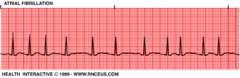
Types of atrial fibrillation **
-Controlled (stable): HR <100
-Uncontrolled (unstable): HR >100
When a pt has atrial fibrillation, they are at HIGH risk for?
Pulmonary or systemic emboli
Atrial Fibrillation S/S
-Could be asymptomatic (stable)
-Fatigue/lightheadedness
-Palpitations/chest pain
-SOB
-Hypotension
Atrial Fibrillation
-Treatment
Determine stable vs unstable (see general treatment)
Supraventricular Tachycardia (SVT) *
Atrial Arrhythmia
-High rate (>140)
-Regular
-Narrow QRS
-Can have sudden onset (be in sinus rhythm & then convert), be intermittent, & terminate suddenly **

Difference between sinus tachycardia and superventricular tachycardia
SVT has higher rate (>140)
SVT S/S
-Could be asymptomatic (stable)
-Fatigue/lightheadedness
-Palpitations/chest pain
-SOB
-Hypotension
Medication used to treat SVT
Adenosine
-Chemical cardio converter
-Stops heart & resets the electrical impulses (will have long pauses before flips back)
-Educate pt that they will feel heart stop

Adenosine
-Indication *
Treats SVT (converts back to NSR)
Adenosine
-Route *
IVP (rapid)
-6mg and may repeat with 12mg x2
Adenosine
-Side Effects
Arrhythmias
Hypotension
Dizziness
Dyspnea
Adenosine
-Know *
-May have asystole (heart stops before flipping back into NSR)
-Have emergency equipment nearby (incase doesn't start back)