Wk5 L11,12 - Lower Limb Bones, Joints and Muscles
1/80
There's no tags or description
Looks like no tags are added yet.
Name | Mastery | Learn | Test | Matching | Spaced |
|---|
No study sessions yet.
81 Terms
What are the main categories of lower limb muscles based on their actions?
Muscles that move the hip, Muscles of the thigh, Muscles of the leg, Intrinsic muscles of the foot.
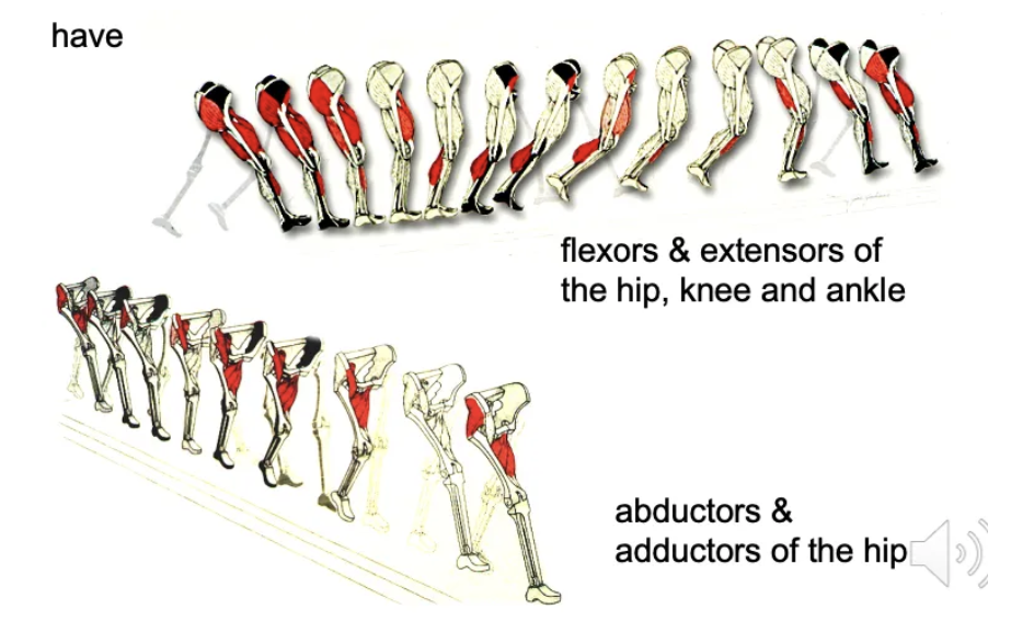
Which muscle groups are critical in the gait cycle?
Flexors and extensors of the hip, knee, and ankle; Abductors and adductors of the hip.
How are muscles in the lower limb organized?
They are organized into compartments, with muscles in the same compartment having similar functions and nerve supplies.
What are the five key groups of muscles that cross the hip joint?
Gluteals (posterior & lateral), External Rotators (deep to gluteals), Hamstrings (posterior, cross knee), Hip Flexors (anterior), Hip Adductors (medial).
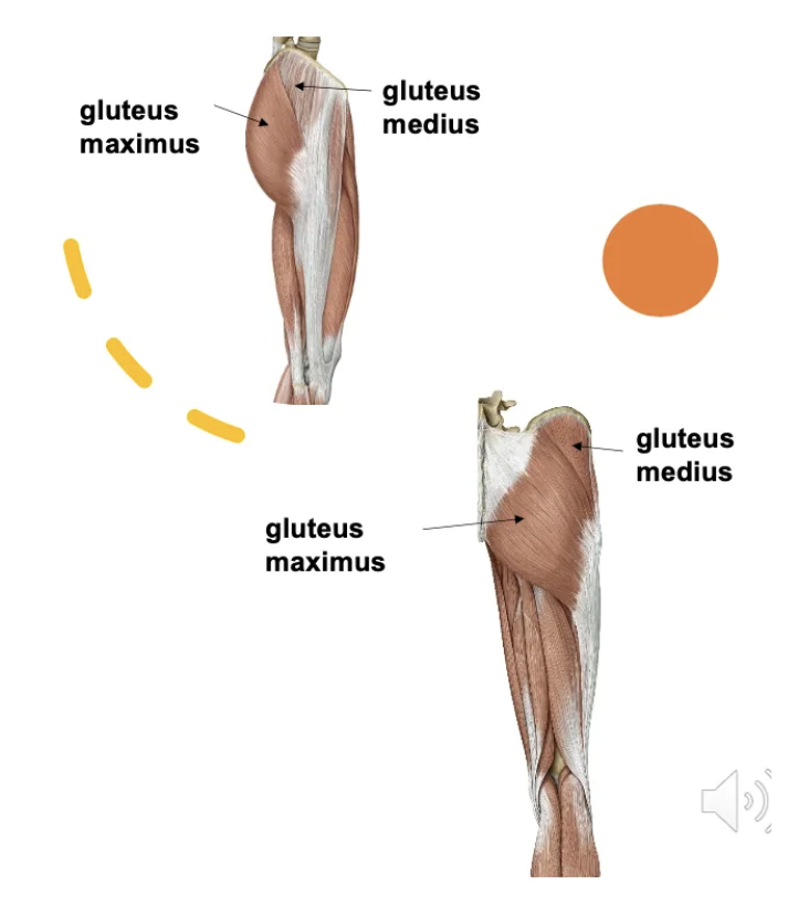
What is the primary function of the gluteus maximus?
Powerful hip extensor and lateral rotator. Also stabilizes the knee via the iliotibial band.
When is the gluteus maximus activated the most?
Engaged in climbing, running, and explosive hip extension (e.g., sprinting from a starting block).
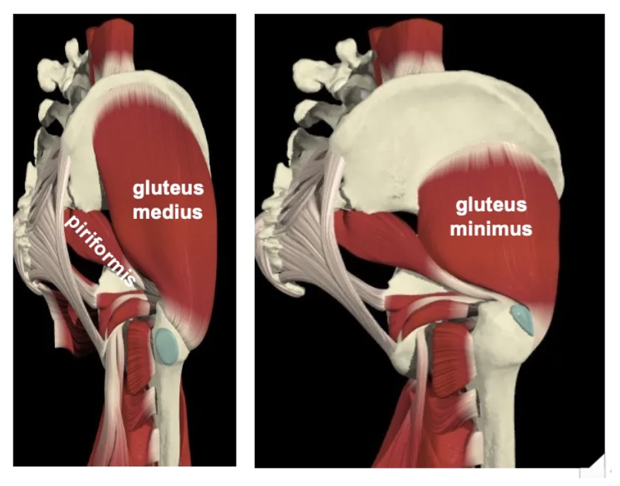
What are the functions of the gluteus medius and minimus?
Hip abduction and internal rotation. Work with the tensor fascia lata. Maintain pelvis level when standing on one leg.
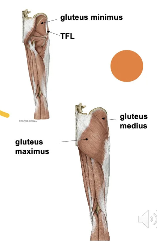
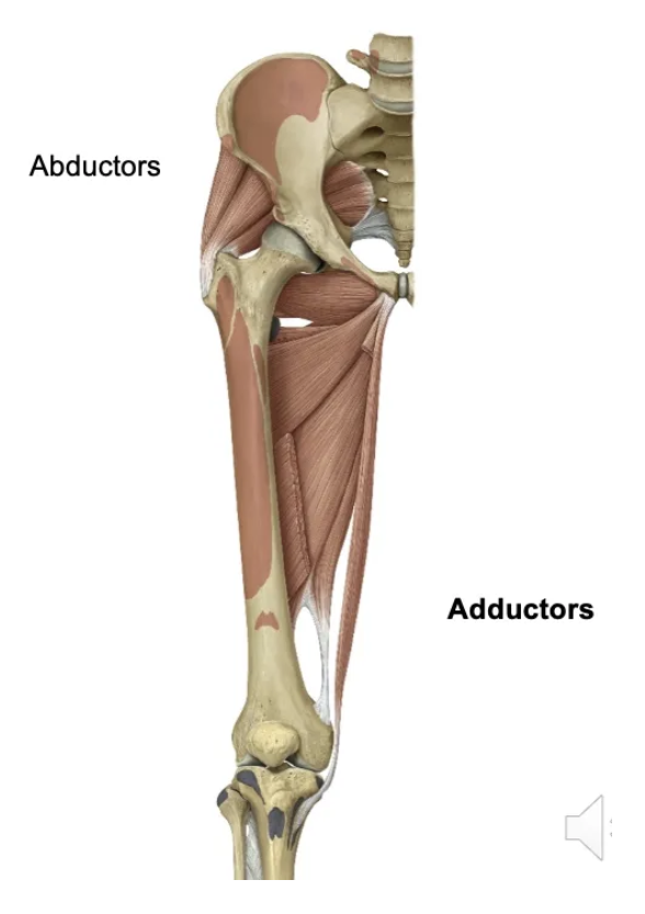
Which muscles act as hip abductors?
Gluteus medius, Gluteus minimus, Tensor fascia lata.
What is the function of hip abductors in locomotion?
Hold the pelvis level when standing on one leg. Essential for maintaining stability during walking.
Which muscles are the external rotators of the hip?
A group of 6 muscles that attach proximally to the pelvis and distally to the proximal femur.
What is the function of the piriformis muscle?
It is an external (lateral) rotator of the hip, passing posterior to the axis of rotation.
Which muscles function as medial rotators of the hip?
Gluteus medius, Gluteus minimus, Tensor fascia lata.
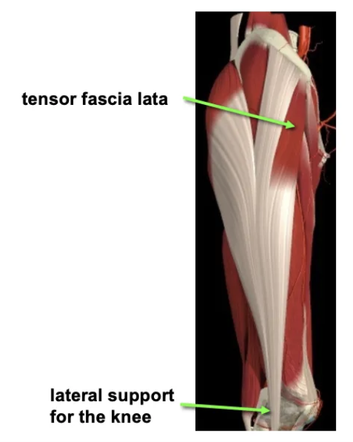
What are the attachments and functions of the tensor fascia lata?
Attachments: Ilium to tibia via the iliotibial band. Functions: Tenses the lateral fascia of the thigh, stabilizing the lateral knee.
What are the three muscles that make up the hamstrings?
Biceps femoris, Semitendinosus, Semimembranosus.
What are the actions of the hamstrings?
Hip extension, Knee flexion and rotation. Stretching occurs during hip flexion and knee extension.
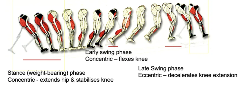
How do the hamstrings function during the walking cycle?
Early swing phase: Concentric knee flexion;
Stance phase: Concentric hip extension and knee stabilization;
Late swing phase: Eccentric deceleration of knee extension.
What are the hip adductors and their attachments?
Muscles: A group of 5 in the medial thigh compartment. Attachments: Pelvis to femur (4) and tibia (1).
What is the function of hip adductors?
Adduct the lower limb, assist in hip flexion or extension depending on hip position. Active in walking: return femur to midline in swing phase and stabilize pelvis in stance phase.
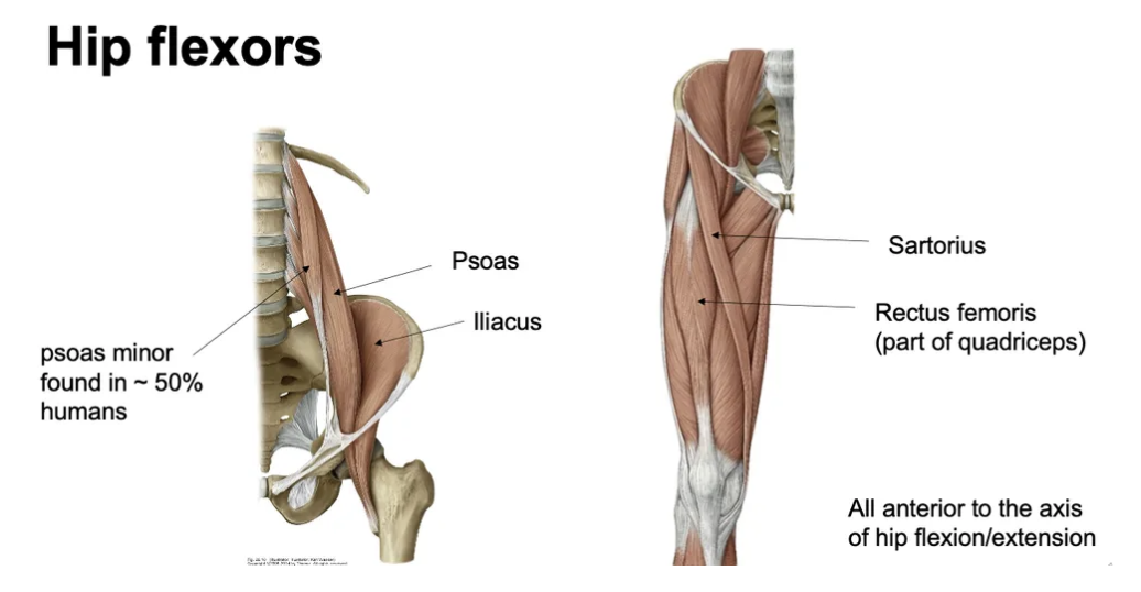
What are the main hip flexors?
Sartorius, Psoas, Iliacus, Rectus femoris (quadriceps), Psoas minor (present in ~50% of humans).
What is the primary function of the quadriceps?
Knee extension (powerful), Hip flexion (rectus femoris).
What are the four heads of the quadriceps?
Vastus lateralis, Vastus medialis, Vastus intermedius, Rectus femoris.
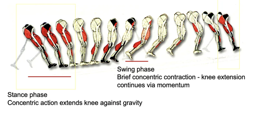
How do the quadriceps function during walking?
Swing Phase: Brief concentric contraction for knee extension.
Stance Phase: Concentric contraction to extend knee against gravity.
What role does the patella play in knee mechanics?
A large sesamoid bone embedded in the quadriceps tendon. Protects the knee joint. Aids in force transmission and knee extension.
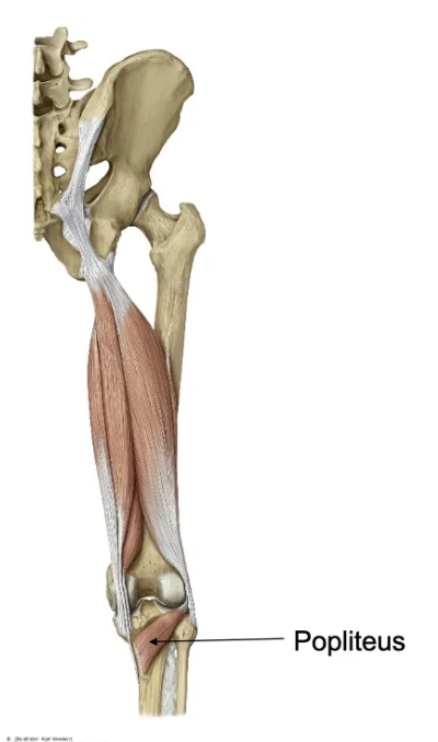
What is the function of the popliteus muscle?
It unlocks the knee by rotating it when the cruciate ligaments lock the knee in full extension, allowing muscles to relax while standing.
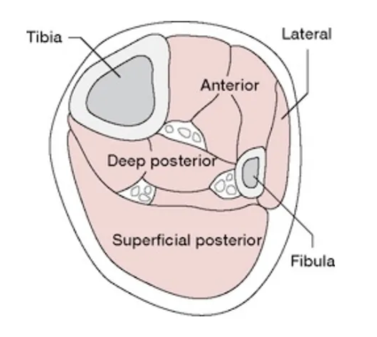
How are the muscles of the leg organised?
They are divided into four compartments:
- Posterior (Superficial): Gastrocnemius and Soleus – Plantar flexion
- Posterior (Deep): Toe flexion and inversion muscles
- Lateral: Eversion of the foot
- Anterior: Dorsiflexion, toe extension, and inversion
What are the main calf muscles, and what is their function?
The gastrocnemius and soleus attach to the calcaneus via the Achilles tendon and primarily perform ankle plantar flexion (pointing the toes).
How are the gastrocnemius and soleus positioned?
The gastrocnemius is superficial, while the soleus lies deep to it.
What functions do the calf muscles perform?
- Enable powerful ankle plantar flexion during walking, running, and jumping.
- Absorb impact forces when landing.
- The soleus acts as a postural muscle to maintain balance while standing.
What forces act on the ankle during landing?
- Achilles tendon: Handles up to 7× body weight
- Tibia: Takes up to 9× body weight
- Metatarsals: Endure 3× body weight
Muscles and connective tissues lengthen to absorb this force.
What happens during the stance phase of walking (Calf Action)?
The calf muscles contract strongly to push off the ground.
What are the muscles and function of the deep posterior leg compartment?
- Muscles: Toe flexors, Tibialis posterior
- Action: Inversion
What are the actions of the anterior leg compartment?
- Dorsiflexion (ankle)
- Inversion (subtalar/midtarsal joint)
- Toe extension (MTP, IP joints)
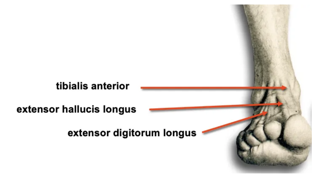
What muscles are found in the anterior leg compartment?
- Tibialis anterior
- Extensor hallucis longus
- Extensor digitorum longus
How does the tibialis anterior contribute to walking?
- Swing phase: Concentric contraction to clear the foot from the ground.
- Stance phase: Eccentric contraction to control weight transfer from heel to forefoot.
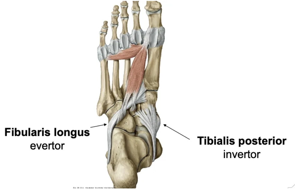
What muscles are found in the lateral compartment of the foot, and what is their function?
- Muscles: Fibularis longus, Fibularis brevis
- Action: Eversion of the foot
- Function: Supports foot arches
What are the types of foot arches?
Medial longitudinal
Lateral longitudinal
Transverse
What is the function of foot arches?
They distribute weight, absorb impact, and act as springs during movement.
What supports the foot arches?
Ligaments, tendons, intrinsic muscles, and plantar fascia.
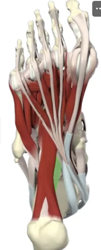
Which tendons support the foot arches?
- Tibialis posterior
- Flexor hallucis longus
- Flexor digitorum longus
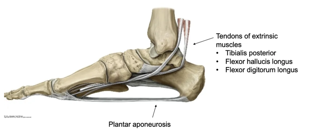
What is the function of the plantar aponeurosis?
It supports the arches and acts as a spring during gait, increasing walking efficiency.
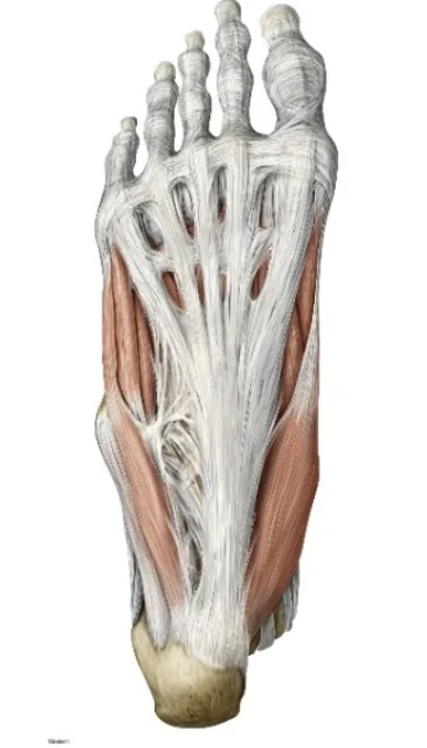
What is the function of the intrinsic plantar muscles of the foot?
- They flex, abduct, or adduct the toes.
- Help the foot adapt to different terrains.
How is toe abduction/adduction defined?
Movement away from (abduction) or toward (adduction) the 2nd toe.
What are the intrinsic muscles of the foot’s dorsum?
- Extensor hallucis brevis
- Extensor digitorum brevis
What is the function of the intrinsic muscles on the dorsum of the foot?
They extend the toes and lie deep to the extrinsic extensor tendons.
What are the three compartments of the thigh and their functions?
- Posterior: Hamstrings – Knee flexion and hip extension
- Medial: Hip adductors – Thigh adduction
- Anterior: Quadriceps & Sartorius – Knee extension and hip flexion
What are the specialisations of the lower limb?
The lower limb is specialised for locomotion and load-bearing.
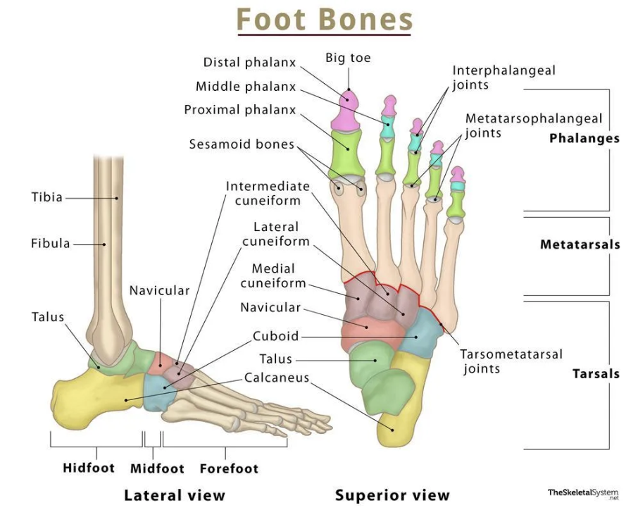
What are the bones of the lower limb?
Femur, patella, tibia, fibula, and bones of the foot.

What type of bone is the patella, and what is its function?
The patella is a sesamoid bone that provides leverage for knee extension.
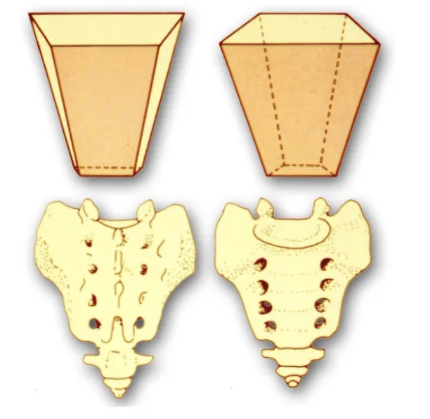
What bones form the pelvis?
Sacrum, two hip bones, and the coccyx.
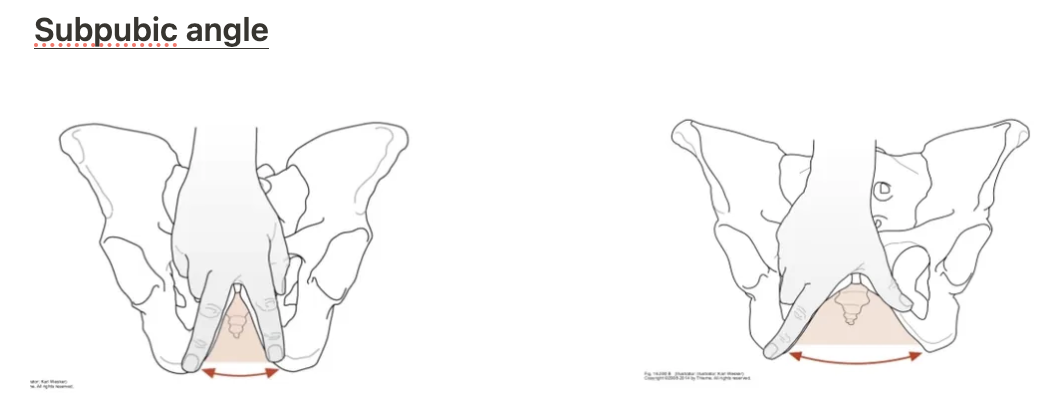
How does the female pelvis differ from the male pelvis?
The female pelvis has a larger subpubic angle, a broader pelvic inlet/outlet, and a greater distance between acetabula to facilitate childbirth.
What is the function of the sacrum?
It is wedge-shaped with five fused vertebrae and allows passage of spinal nerves through its anterior and posterior foramina.
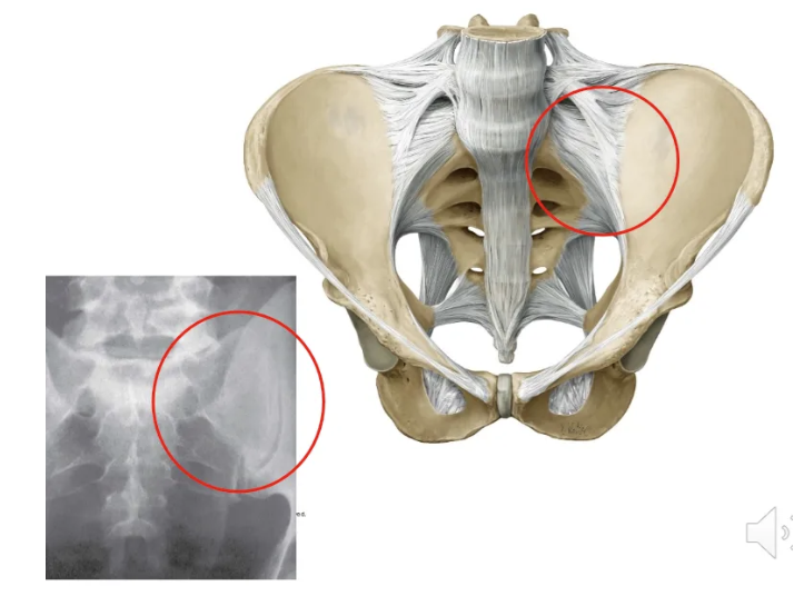
What is the function of the sacroiliac ligaments?
They attach the sacrum to the spine of the ischium and prevent anterior displacement of the sacrum.
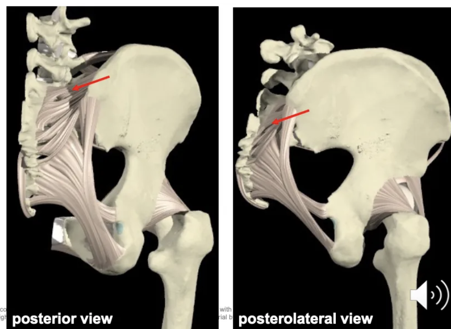
What is the function of the sacrotuberous ligament?
It attaches the sacrum to the tuberosity of the ischium and prevents anterior displacement of the sacrum.
What type of joint is the sacroiliac joint?
A fibrous and synovial joint with limited mobility.
Connects the sacrum to the ilium.
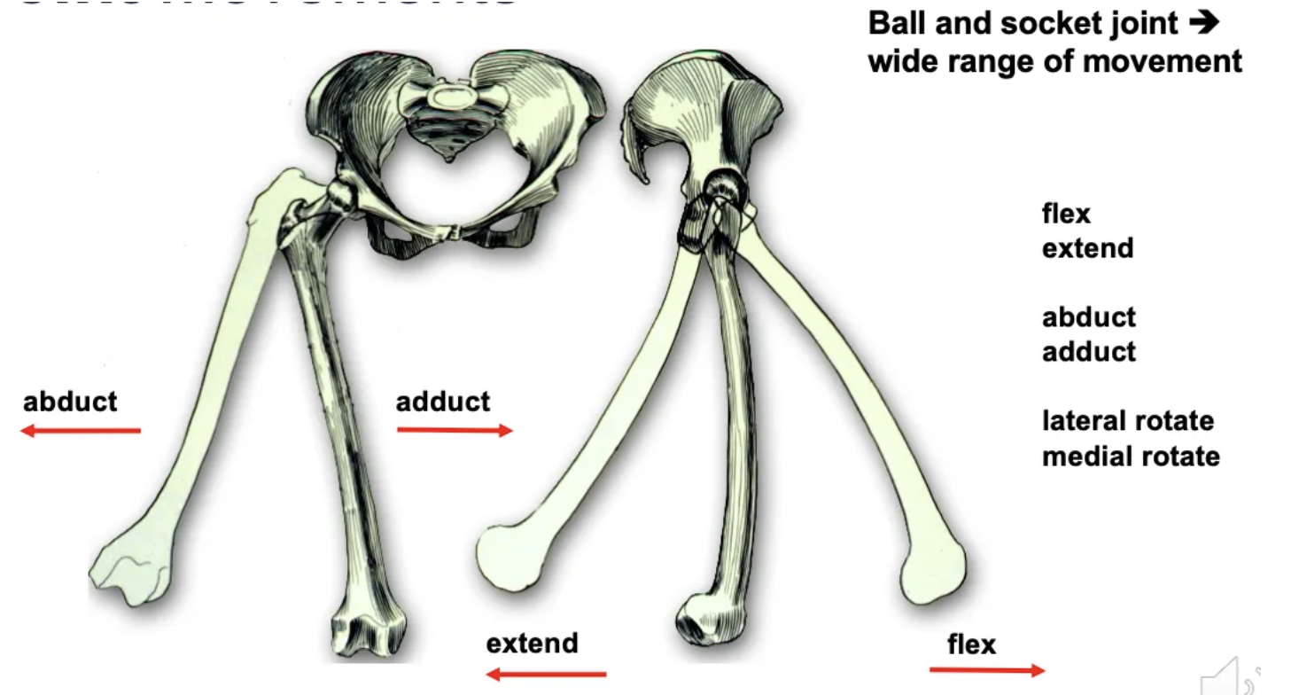
What type of joint is the hip joint, and what movements does it allow?
A synovial ball-and-socket joint allowing flexion, extension, abduction, adduction, lateral rotation, and medial rotation.
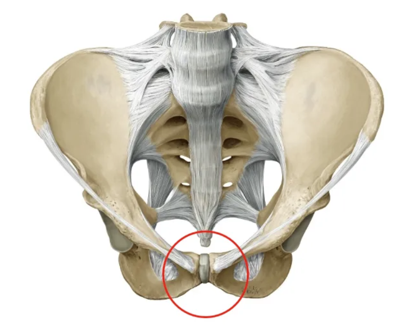
What type of joint is the pubic symphysis, and what happens to it during pregnancy?
A fibrocartilaginous joint with a cushioning disc; its mobility increases during pregnancy. Has limited mobility.
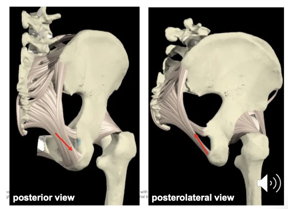
What are the major ligaments of the hip joint and their functions?
Pubo-femoral: Limits hip extension.
Ilio-femoral: Lateral, limits hip extension.
Ischio-femoral: Posterior, limits hip extension.
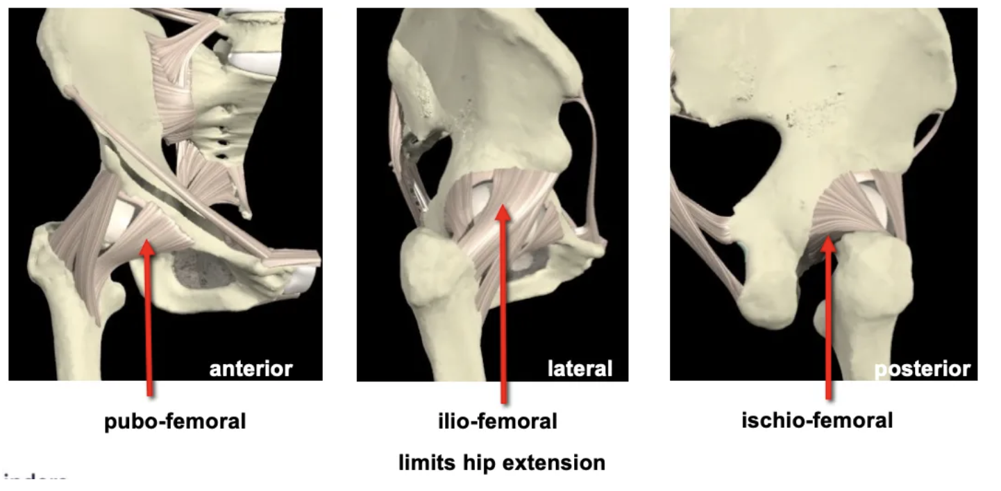
What happens to hip ligaments in vertical posture?
The hip joint is extended, causing the anterior ligaments to tighten.
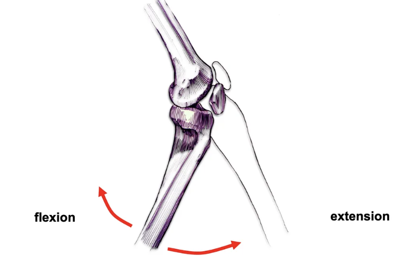
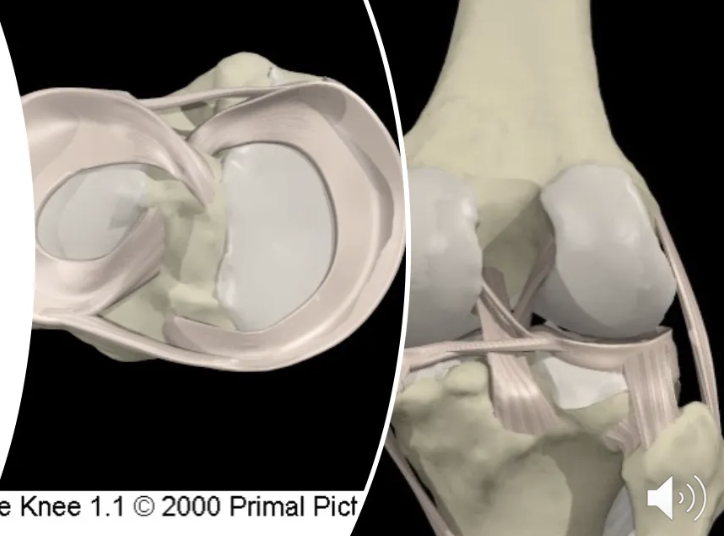
What type of joint is the knee?
A modified hinge joint (tibiofemoral joint) that allows flexion, extension, and limited rotation when flexed.
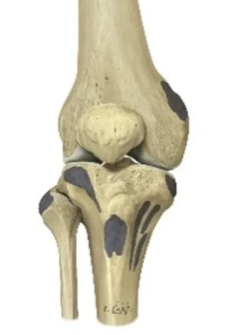
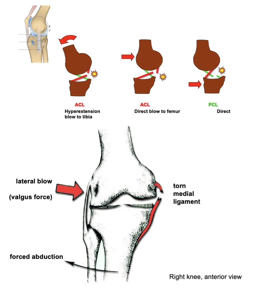
What are the key ligaments of the knee joint?
Anterior cruciate ligament (ACL): Prevents anterior displacement of the tibia.
Posterior cruciate ligament (PCL): Prevents posterior displacement of the tibia.
Medial collateral ligament (MCL): Stabilises the inner knee.
Lateral collateral ligament (LCL): Stabilises the outer knee.
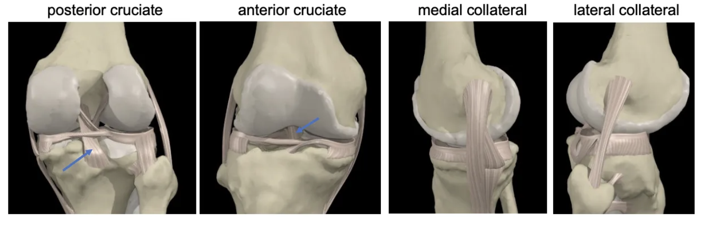
What is the function of the menisci in the knee?
The medial and lateral menisci act as shock absorbers and provide joint stability.
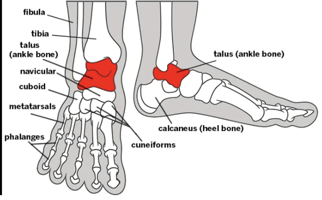
What bones make up the ankle joint?
The tibia, fibula, and talus.
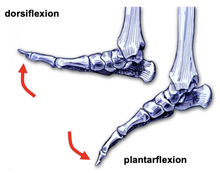
What type of joint is the ankle joint, and what movements does it allow?
A synovial hinge joint allowing dorsiflexion and plantarflexion.
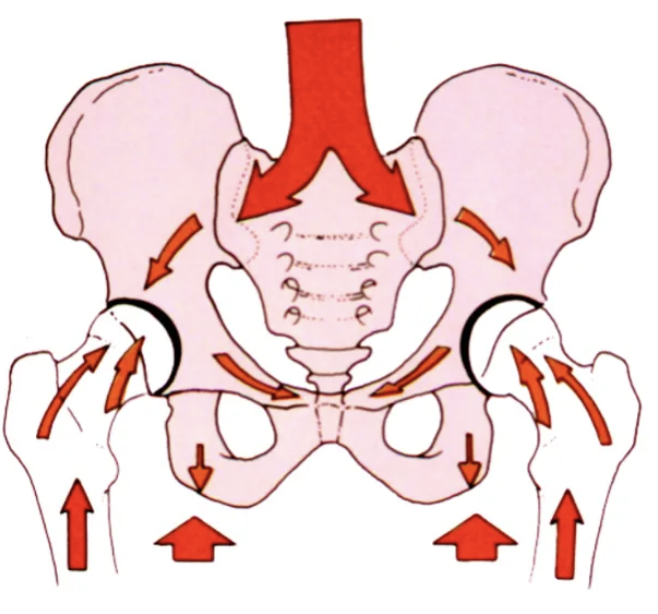
How does the pelvis bear loads?
Sacrum connects to hip bones, distributing force through pelvic arches to the femur. Ligaments, bones, and muscles help absorb impact.
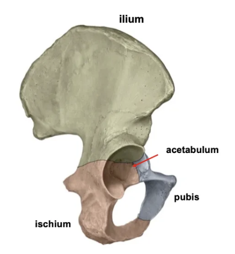
What bones form the hip joint?
The hip joint consists of the ilium, ischium, pubis, and the acetabulum.
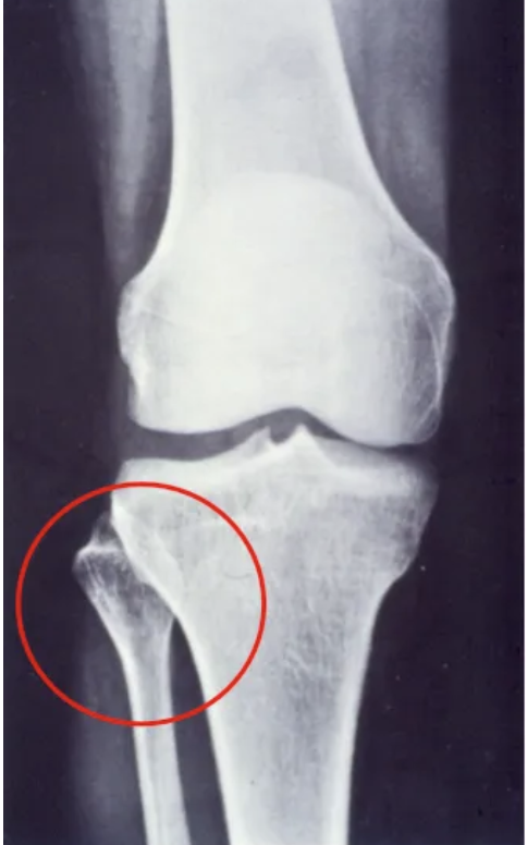
What type of joint is the superior tibiofibular joint (knee), and what movement does it allow?
It is a synovial, planar joint that allows slight gliding movement.
How do common knee ligament injuries occur?
ACL: Hyperextension or direct blow to the femur.
PCL: Direct blow to the tibia.
Medial Ligament: Lateral (valgus) blow or forced abduction.
What type of joint is the inferior tibiofibular joint?
A fibrous joint, more stable than a synovial joint.
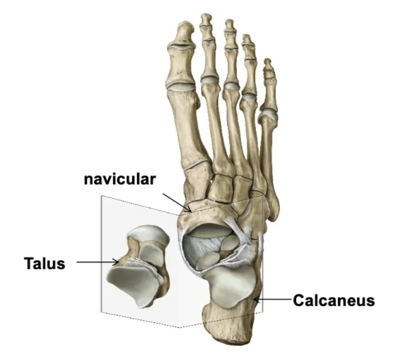
Where does the talus sit, and what is its role in gait?
The talus sits between the medial malleolus of the tibia and the lateral malleolus of the fibula.
It is wider anteriorly, providing increased stability during gait as it moves between the malleoli when the heel strikes the ground.
When is the ankle most stable?
The ankle is most stable in dorsiflexion and less stable in plantarflexion.
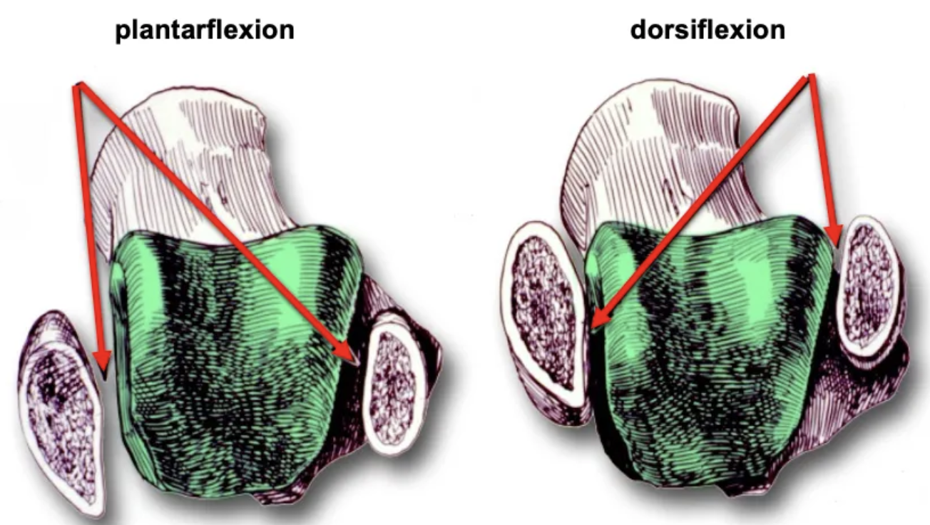
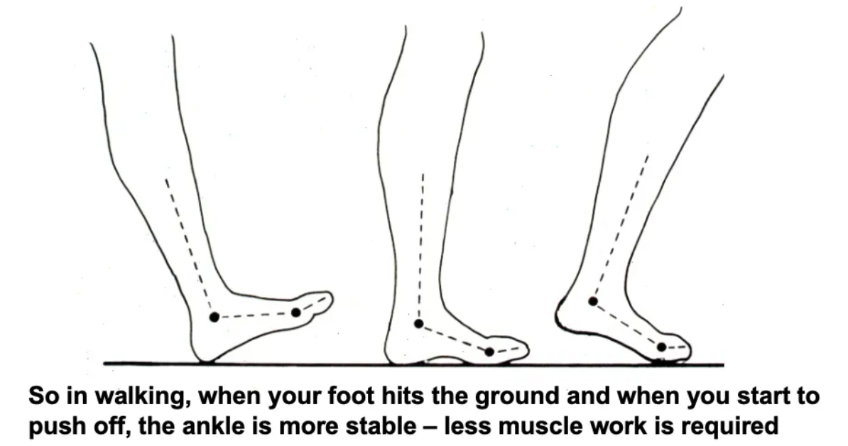
How does stability in walking change?
Stability is greater when the foot hits the ground or begins to push off, requiring less muscle activity.
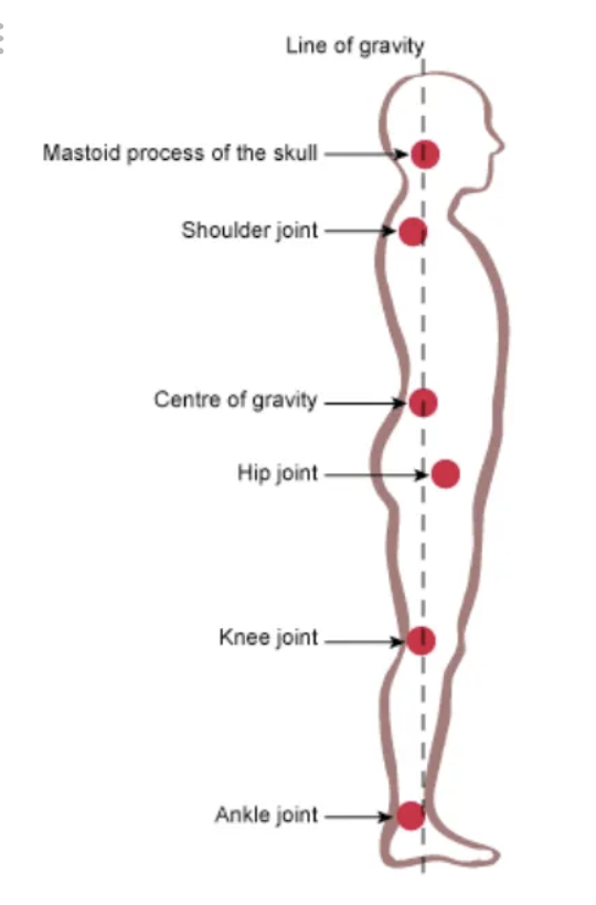
What does the line of gravity pass through in relaxed standing?
The line of gravity passes behind the hip (iliofemoral ligament bears the load), towards the front of the knees (cruciate ligaments bear the load), and in front of the ankle (ankle joint bears the load).
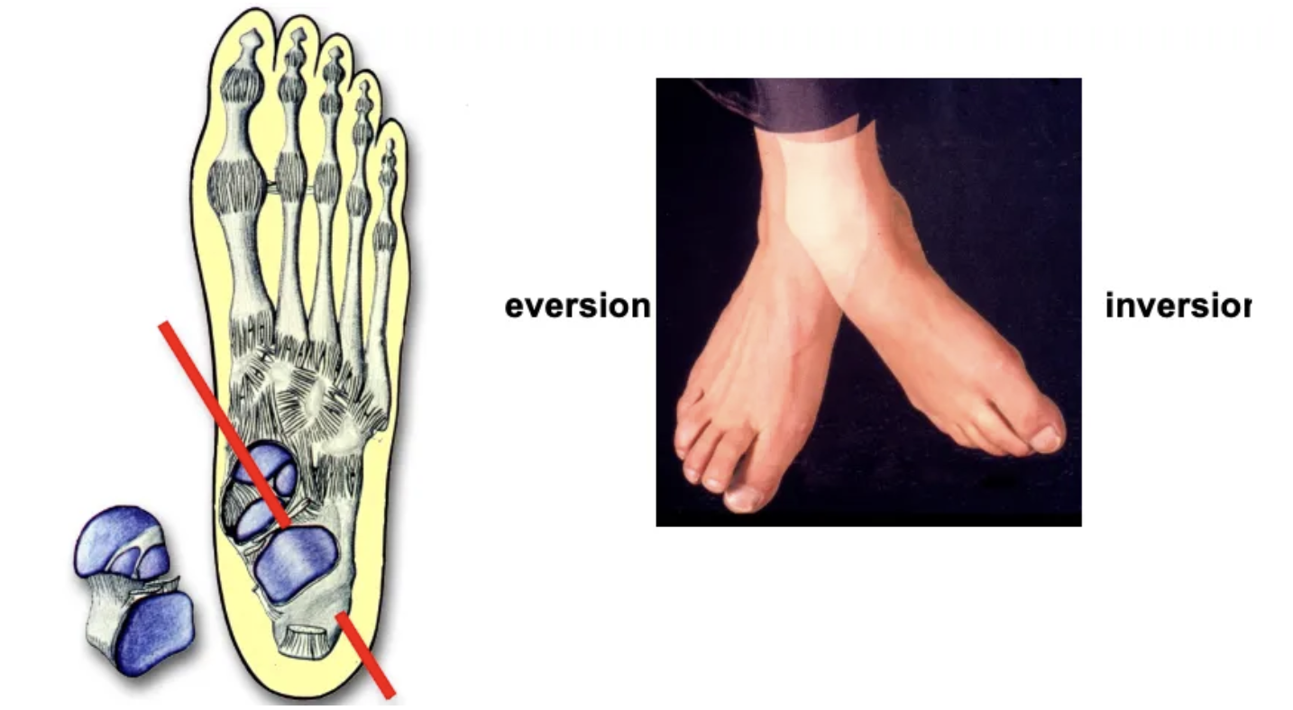
What is the function of the subtalar joint?
The subtalar joint is located between the talus and calcaneus
It allows inversion and eversion
It is a synovial joint with an atypical shape.
What is the function of the transverse tarsal/midtarsal joint?
The transverse tarsal joint allows the foot to adapt to uneven ground.
It is more mobile in plantarflexion and more stable in dorsiflexion, involving the navicular, cuboid, talus, and calcaneus.
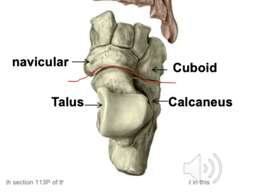
What type of joint is the talocalcaneonavicular joint?
The talocalcaneonavicular joint is a ball and socket joint, involving the navicular, talus, and calcaneus.
What types of joints are the metatarsophalangeal and interphalangeal joints?
The metatarsophalangeal joints are condyloid, and the interphalangeal joints are hinge joints.
What are the key ligaments of the ankle?
Deltoid ligament: Stabilizes the medial ankle.
Anterior talofibular ligament: Most commonly sprained ligament, stabilizes the lateral ankle.
What are the arches of the foot?
The foot has longitudinal and transverse arches, supported by bone structure, ligaments, muscles, and the plantar fascia.
What is the function of the plantar fascia?
The plantar fascia is a thick connective tissue that supports the arches, acts as a spring during gait, and increases efficiency.
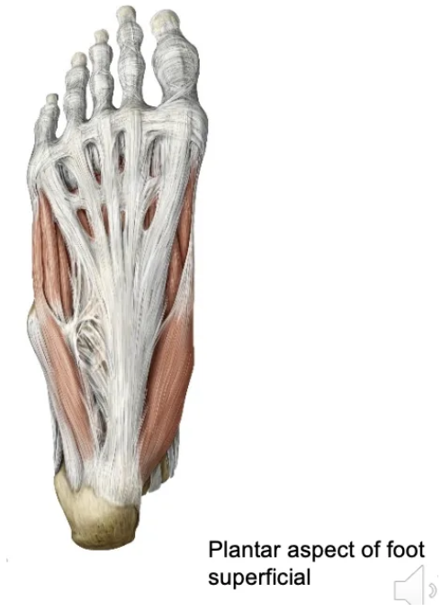
What role do extrinsic and intrinsic muscles play in the foot?
Tendons and muscles between the plantar fascia and ligaments provide dynamic support and enable movement.
What are the bones of the foot?
The foot consists of the tarsals (including navicular, talus, calcaneus), metatarsals, and phalanges.