Cell Biology Unit 1 Exam
 Call Kai
 Learn
Learn Practice Test
 Spaced Repetition
 Match
Match Flashcards
 Knowt Play
1/73

There's no tags or description
Looks like no tags are added yet.
Name | Mastery | Learn | Test | Matching | Spaced | Call with Kai | Chat |
|---|
No analytics yet
Send a link to your students to track their progress
74 Terms
Passive Transport
Movement of small uncharged molecules across membranes by simple diffusion → no energy or protein required, and moves down concentration gradient, must be hydrophobic
Facilitated Transport
Movement of ions, water, and hydrophilic molecules across cell membranes down concentration gradient via help of channel proteins → form a hydrophilic tube (uniporter)
Active Transport
Movement of ions, amino acids, and metabolites across cell membranes against concentration gradient via help of ATP powered pumps, symporters, and antiporters
Symporter
Transmembrane protein that catalyzes movement of one molecule against its concentration gradient with another ion traveling down an electrochemical gradient → both particles move in same direction
Antiporter
transmembrane protein that catalyzes movement of one molecule against its concentration gradient with another ion traveling down an electrochemical gradient → particles move in opposite directions
ATP Powered Pumps
Coupled chemical reaction transmembrane protein that facilitates the movement of a substrate against its concentration gradient with ATP hydrolysis (ATP → ADP + Pi)
GLUT-1
Glucose uniporter transport protein that undergoes a conformation change (triggered by glucose binding) to permit molecule passage FROM the blood TO inside the cell’s cytosol → provides a hydrophilic pathway, moves down concentration gradient, reversible
Vmax Glucose
Maximum uptake rate of glucose that GLUT-1 can mediate → higher glucose concentration means increased uptake
Km
The substrate concentration needed to reach half of Vmax and indicate enzyme-substrate affinity → a LOWER Km means HIGHER efficiency
GLUT-1 Rate Depends on:
Concentration of glucose and Km of the specific GLUT transporter
Aquaporin
Facilitated diffusion of water → parts of integral protein touching membrane are hydrophobic, but outer ends and the pore interior are hydrophilic
Osmotic Pressure
The minimum pressure required to prevent the inward flow of a solvent across a semipermeable membrane into a more concentrated solution → water flows from low solute to high solute concentration (consequently, it moves down its own gradient)
Frog Oocytes
Experiment with 2 cells. Top cells microinjected with aquaporin mRNA and cause lysis, bottom cells do not express them and remain impermeable to water → swelling rate of top cell used to quantify aquaporin water movement activity
Ca2+ ATP Pump
P-class pump located in the sarcoplasmic reticulum (SR) membrane that regulates muscle contraction and relaxation by storing, releasing, and reuptaking calcium ions → obtains an ATP binding site that initiates a conformation change and then binds 2 Ca2+. Also has a aspartyl-phosphate binding site that opens the protein and releases the Ca2+ (phosphate then kicked off) → pumped from cytosol to SR lumen
E1 vs E2 - Ca2+
Energy affinity states during Ca2+ ATP transport. High affinity in E1 for calcium in E1 and low affinity for calcium in E2
Na+/K+ ATP Pump
P-class pump located in the plasma membrane. ATP binds and 3 Na+ attach. ATP leaves as phosphate binds to different site and initiates a conformation change. 2 K+ attach. The 3 Na+ are pumped FROM the cytosol TO the outside of cell. Dephosphorylation leads to close and then pumps the 2 K+ FROM outside of cell TO the cytosol (Na+ is now high outside and K+ is now high inside)
E1 vs E2 - Na+/K+
E1: High affinity for Na+ and low affinity for K+
E2: High affinity for K+ and low affinity for Na+
V-class H+ ATP Pump
Symporter ATP pump that that moves H+ and Cl- from the cytosol to the lumen in order to create an acidic pH with HCl but eliminate all electrical potential
ABC Superfamily Pump
Ion and Molecule transporter that has two transmembrane domains, and two ATP binding sites → resistant cancer cells obtain way more of these, leading to excessive drugs pumped out and drug resistance
Na+/Ca+ Antiporter
High extracellular Na⁺ binds the transporter, Na⁺ enters the cell, the energy released drives Ca²⁺ out of the cell, the transporter resets and repeats → no ATP used. Na+ moves down its gradient, while Ca2+ moves up against its gradient
Congestive Heart Failure
A cell exhibiting no Na+/Ca2+ anti porter or Na+/K+ pump. Leads to exponential levels of Na+ and Ca2+ inside the cell and causes detrimental problems
Systemic Capillaries
Carry oxygenated blood from the left ventricle, through the arteries, to the capillaries in the bodily tissues. From the tissues the deoxygenated blood returns through a system of veins to the right atrium of the heart → high CO2 pressure and low O2 pressure
Cl-/HCO3- Antiporter in Systemic Capillaries
Water split into H+ and OH-. H+ binds to hemoglobin to release O2 while OH- binds to CO2 inside the cell and generates HCO3-. AE1 protein pumps HCO3- out and Cl- in to maintain neutral pH
Pulmonary Capillaries
Deoxygenated blood enters into the pulmonary arteries from the right side of the heart and is delivered here - the smallest blood vessels inside of the lungs and are attached to the walls of the alveoli → low CO2 pressure and high O2 pressure
Cl-/HCO3- Antiporter in Pulmonary Capillaries
Concentration of O2 is higher outside of the cell, so it can diffuse in. It binds to hemoglobin and releases H+. Additionally the AE1 protein pumps out Cl- and pumps in HCO3-. HCO3- broken down and sends CO2 out of cell. the remaining OH- and H+ come together and generate water.
Phagocytosis
Bulk flow across membrane in which bacteria is taken up inside a vesicle made from the membrane into the cell - endocytosis
Pinocytosis
Bulk transport of fluid droplets or small particles taken up inside a vesicle made from the membrane into the cell - endocytosis
Exocytosis
Secretion of these bulk transported particles (bacteria, fluid droplets, etc.)
In Vivo
Refers to inside the body
In Vitro
Refers to outside the body
Primary Cell Cultures
Samples taken directly from animals - obtain limited passages (50 to 100 times) . Often displays the differentiated properties of the organs from which they were isolated

Transformed Cells
Genetically modified cell samples that have infinite passages (immortalized) - usually cells that have undergone spontaneous genetic change, aneuploid (abnormal chromosome #), grow to higher densities, can differentiate into different cell types (ie. stem cell lines), solid surface often not required since they don’t have tight junctions

Cell Strain
Lineage of cells from one initial primary culture
Passage of Cells
Splitting of confluent cells into a new culture vessel - taking a portion of a growing cell culture and transferring it into fresh media and a new dish so the cells can continue to grow
Embryonic Stem Cell Growth Requirements
Inner cell mass separated from surrounding tissue and require matrigel to grow without presence of feeder cells
Induced Pluripotent Stem Cells (iPSCs)
Adult cells that scientists reprogram back into a somatic stem cell-like state with yamanaka factors. The produced hiPSCs behave like regular embryonic cells and can develop into any cell type but they come from adult tissues not embryos
Fluorescence Activated Cell Sorting
Cells in a culture a tagged with a fluorescent marker, they then pass through a machine that shines a laser, this machine detects which color the cell emits, the brightness of the fluorescence, the cell characteristics, and places either a positive or negative charge on it - separate cell populations can be sorted from there
Gating
Choosing what cells you want to isolate based on their fluorescent profile
A Light Microscopy
Exploring cell structure and visualizing proteins within cells
Magnification
Projection lens (10x) multiplied by objective lens (100x) = 1000x
Resolution
Ability to distinguish between 2 objects - the beam of any radiation cannot be used to probe structures smaller than its own wavelength (D = minimum “seeable” distance between 2 objects) → simple light microscopy can distinguish objects separated by approximately 0.2 um or more
D Calculation
(0.61/\) / Nsina → ( /\ is wavelength of radiation, N is refractive index of the air or fluid between the specimen and the objective lens ie. air, water, or oil) and a is the angular aperture)
Best Resolution
Shortest /\ (0.45 um), Immersion oil medium (N=1.5), Maximum a (sin(70) = 0.94) → TOTAL = 0.194 um
Brightfield Microscopy
Basic view of specimen with a refracted light source scaled with a condenser lens shining under the specimen
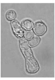
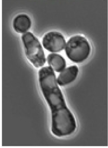
Phase Contrast Microscopy
Projection lens, phase plate in the objective, unobstructed light pointed down, objective sense, specimen, condenser lens, annular diaphragm, and refracted light source
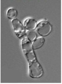
DIC Microscopy
Creates a 3D like image with a flat specimen by utilizing nomarski prisms to spit the light into 2 beams, travel along the cell, and rejoin into a singular beam to create specific contrasts and shadows
Sample Preparation - Fixation
Cross links macromolecules with free amino groups - takes the covalent or ionic bonds holding polymer molecules together and changes them into tough/rigid heat-resistant structures
Sample Preparation - Embedding
Using wax or resin on tissue samples to provide a solid and supportive structure for staining - thick samples only
Sectioning
Process of cutting the embedded sample into very thin slices using a microtome so it can be mounted and stained - thick samples only
Hematoxylin Staining
A blue-violet positively charged (cationic) stain that binds to negative charged nucleic acid - nuclear stain since it stains the nuclear blue/violet
Eosin Stain
An acidic negatively charged (anionic) stain that binds to basic amino acids like lysine and arginine. It stains the cytoplasm proteins in amino acids pink/red
Wright-Giemsa Stain
Histologic stain for blood and bone marrow cells - stains the nuclei blue/violet, stains the cytoplasm pale pink, and stains the erythrocytes pale pink. Differentiates white blood cell counts and studies red blood cell morphology (since red blood cells lack nuclei)
Benzidine Stain
Application causes a chemical reaction with red blood cells that detect hemoglobin deposits in histological sections

Fluorescence Microscopy
Absorbs light at one wavelength, excites it, and then emits light at another wavelength in order to visualize what cannot be seen in the bright field or phase contrast techniques → uses an excitation filter and a dichroic mirror
Immunoflourescence Microscopy
Adds antibodies to attach to a protein of interest. Then adds a second fluorescent antibody to highlight location through view under microscope
Confocal Laser Scanning Microscopy (CLSM)
Requires sectioning of thick specimens to focus on a chosen place while rejecting light that comes from out of focus areas (excludes light from other planes) → imaged volume must be thin

Forster Resonance Energy Transfer (FRET)
Determines if two proteins interact - when proteins are close enough together acceptor can absorb light from donor protein → different colors of light emitted

Fluorescence Recovery After Photobleaching
Membrane protein attached with fluorescent reagent. Those reagents are bleached with a laser. The rate at which the bleached areas recolorize measure how fast molecules move into the membrane or inside the cell → explains mobility and fluidity
Total Internal Reflection Fluorescence (TIRF)
Excites the interface between the cell and coverslip and identifies structures only growing on the coverslip such as microtubules and actin filaments

Two Photon Excitation Microscopy
E=hv OR E=hc/(/\) → reduces photobleaching
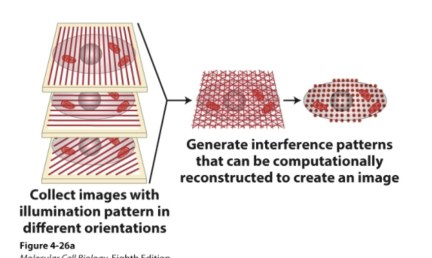
SIM Miscroscopy
Super resolution technique that uses patterned light and computational reconstruction to double resolution and create an image
STED Microscopy
Super resolution technique that uses a donut‑shaped laser to shrink the fluorescence spot and give higher resolution.

PALM Microscopy
Super resolution microscopy that turns fluorophores on/off one at a time and calculates their exact positions to be used for creating an image

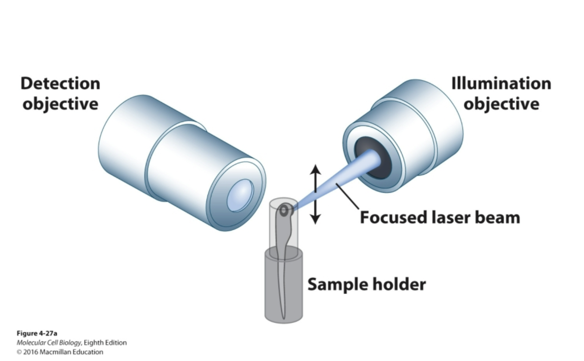
Light sheet Microscopy
Super resolution technique that uses a thin sheet of light to illuminate only one plane of a sample at a time and allow fast 3D imaging
TEM Microscopy
Uses transmitted electrons that are passing through the sample to create a 2D image. Obtains a resolution of 0.1-0.3nm

TEM Sample Preparation
Fixed with glutaraldehyde, embedded by dehydrating and utilizing solid plastic/epoxy block, sectioned to 50-100 nm thickness, and stained with heavy metal “negative” such as uranyl acetate → the sample is placed on a copper grid
Cryo-Electron Microscopy
A very powerful TEM that requires no sample preparation and is intended for live cells maintained at very low temperatures→ emits light on sample stationed on a rotating stage and records an image at each rotate stage

Scanning Electron Microscopy (SEM)
Creates a 3D sample image by detecting reflected or knocked off-electrons. Obtains a resolution of 0.5-4nm (higher D value, so therefore, lower resolution than TEM)

SEM Sample Preparation
Whole cells or unsectioned tissue specimens are fixed, dried, and coated with a heavy metal such as platinum
Sonication
Technique to isolate cell organelles by conducting ultrasonic vibrations to create homogenate or extract
Homogenate
Contains population of particles obtaining different densities, shapes, charges, and sizes (usually product of sonication)
Differential Velocity Centrifugation
Centrifuging homogenate for different increments of time to separate compounds based on their properties → organelle purification
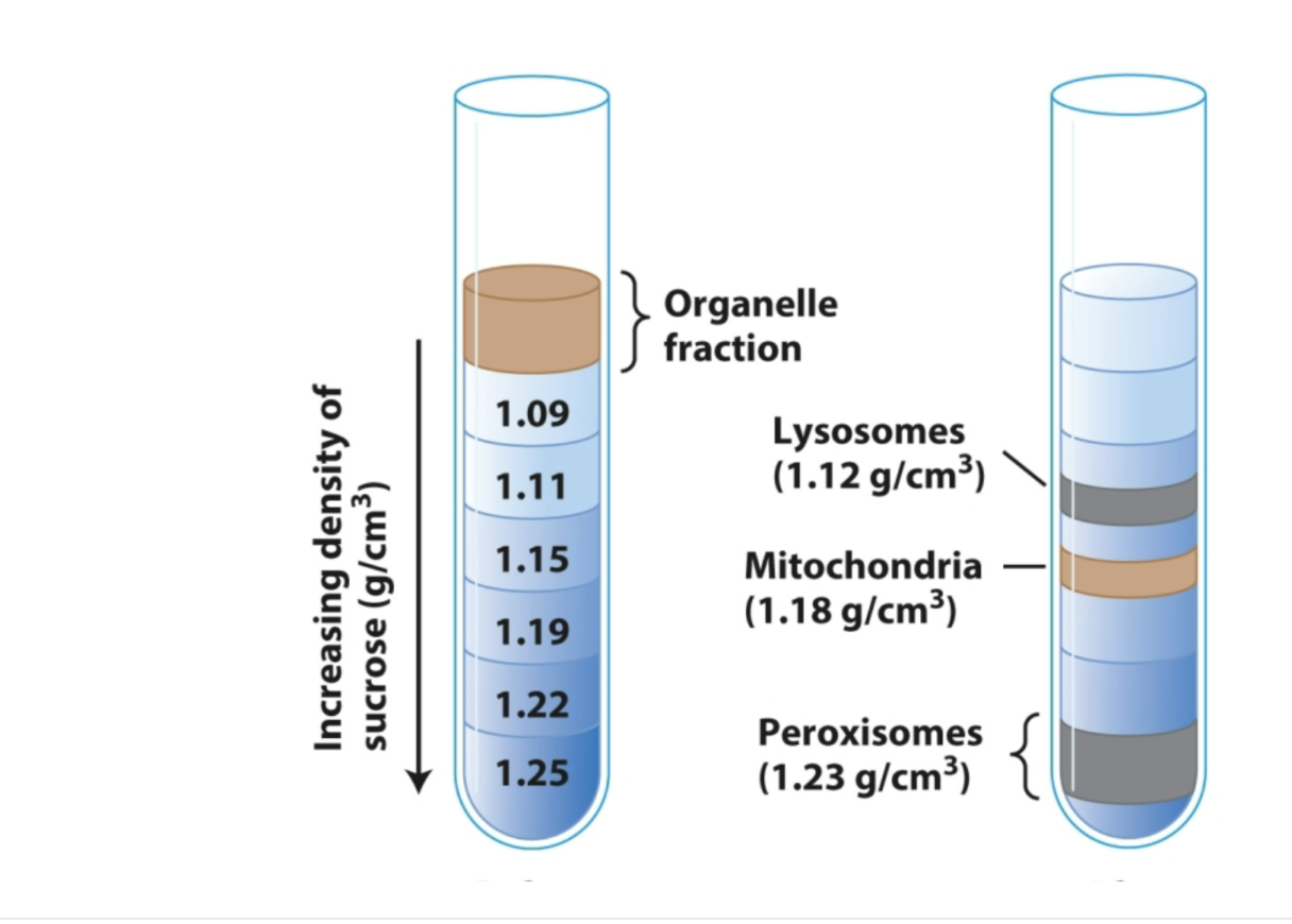
Equilibrium Density Gradient Centrifugation
Organelle purification method that separates molecules by spinning them in an increasing sucrose density gradient until each molecule reaches the position where its density equals the surrounding solution. Then it forms a distinct band that can be collected by puncturing the bottom of the tube (highest density at bottom leading to lowest density on top)
Anti-Clathrin
An antibody captured by protein A and aids in organelle purification → binds to Cathrin and confirms if the coated vesicles were successfully isolated