Unit 4: Plant and Animal Equilibrium
 Call Kai
 Learn
Learn Practice Test
 Spaced Repetition
 Match
Match Flashcards
 Knowt Play
1/87

Earn XP
Description and Tags
+ Neural Signalling
Name | Mastery | Learn | Test | Matching | Spaced | Call with Kai | Chat |
|---|
No analytics yet
Send a link to your students to track their progress
88 Terms
Gas Exchange as a vital function in all organisms
Intake of oxygen to metabolise energy
Removal of metabolic waste products i.e. CO2
Why do challenges become greater in larger organisms?
SA:Vol ratio decreases with increasing size
Increased distance from centre to exterior of organism
Properties of Gas Exchange Surfaces
Thin tissue layer
Permeable to respiratory gases
Moist (Gases are often dissolved; Exchange occurs in liquid medium)
Large SA
Maintenance of Concentration Gradients at Exchange Surfaces
Dense networks of blood vessels
Continuous blod flow
Continuous ventilation with air (lungs) / water (gills)
Adaptations of Mammalian Lungs for Gas Exchange
Alveolar lungs
Presence of surfactant (phospholipid and protein film) → Reduce surface tension of moist inner surface of alveolus → Prevents alveolus from collapsing when air is expired (Low air pressure)
Branched network of bronchioles + alveoli → Large SA of gas exchange tissue
Extensive capillary beds surrounding alveoli → Close access to capillaries → Easy access to / from bloodstream
High SA
Ventilation of Lungs (Mention Diaphragm, Intercostal muscles, Abdominal muscles, Ribcage)
Concentration gradient of O2 and CO2 is maintained by inhalation (intake of O2 from environment into lungs) and exhalation (expulsion of CO2 from lungs into environment)
Role of…
Diaphragm → Change volume of thoracic cavity
INHALATION: Contract → Flattening + Pushing down on abdomen
EXHALATION: Relax → Moves up
Intercostal muscles
INHALATION: Externals contract; Internals relax
EXHALATION: Internals contract; Externals relax
Abdominal muscles
INHALATION: Relax
EXHALATION: Contract → Pressure pushes diaphragm into dome shape
Rib cage
INHALATION: Moves upward and outward (due to contraction of external intercostals and abdominal muscles)
EXHALATION: Moves down and inward
Lungs & Thoracic Cavity
INHALATION: Expand → Lower internal pressure → Air flows in
Exhalation: Volume decrease → Raise internal pressure → Air flows out
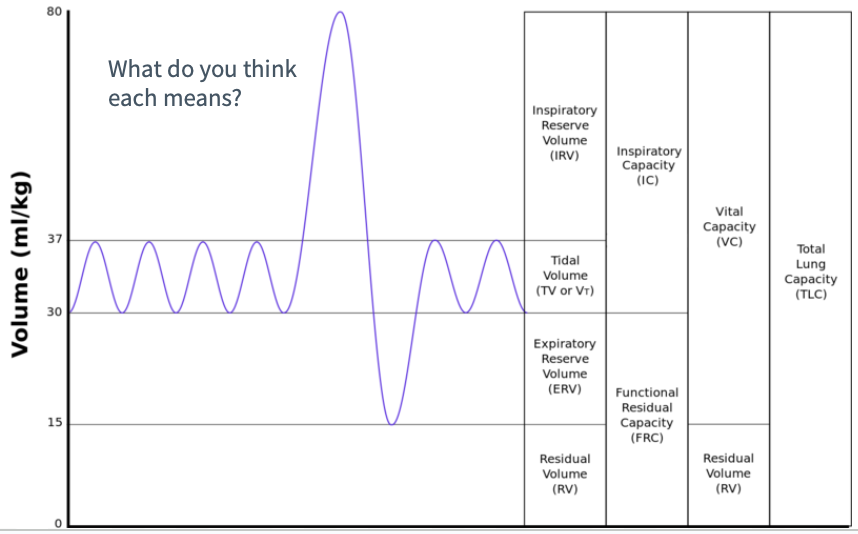
Measurement of Lung Volume (Spirometer graph)
Tidal Volume: Volume of air breathed in / out in a cycle at rest
Vital Capacity: Sum of inspiratory, expiratory reserves and tidal volume
Measure from maximum of inspiratory reserve to minimum of expiratory reserve
Inspiratory Reserve: Maximum volume of air that can be breathed in
Measured ONLY from maximum of tidal volume
Expiratory Reserve: Maximum volume of air that can be breathed out
Measured ONLY from minimum of tidal volume
Measurement of Lung Volume (How to use spirometer)
Ask subject to stand and breathe in and out using the device for ~15 seconds (Measure tidal volume)
Rest subject ≥1 minutes
Ask subject to breathe in maximum volume of air
Rest subject ≥1 minutes
Ask subject to breathe out maximum volume of air
Adaptations for Gas Exchange in Leaves
In leaf structure…
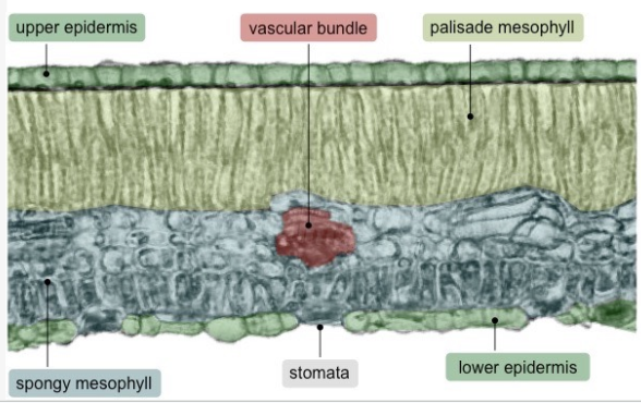
Cells on upper epidermis secrete waxy cuticle → Prevents water evaporation (Affects transpiration)
Air spaces in the spongy mesophyll (Main site of gas exchange) → Allow movement of gases + gas exchange
Stomata are on underside of leaf → Prevent obstruction → Maintain open channel for gas exchange
Stomatal Guard cells embedded on lower epidermis → Control opening and closing of stoma → Regulates amount of water lost; Responsible for gas exchange by diffusion → Controls transpiration rate
Turgid → Open // Flaccid → Closed
When plant wilts from water stress → Release of abscisic acid → Trigger efflux of potassium from guard cells → Decreased water pressure → Loss of turgor → Becomes flaccid and occlude opening
Veins are located centrally → Optimal access by all leaf cells
Draw and label a plant diagram
Transpiration & Gas Exchange + Factors
Transpiration = Consequence of gas exchange in leaf; Evaporation of water through open stomata
Factors include…
(Increased) Light → Increases rate
Stimulates guard cells to open stomata + Increase rate of photosynthesis → Requires increased diffusion of CO2 (in) and O2 (out)
(Increased) Temperature → Increases rate
Increase molecular movement → Increased water evaporation
(Increased) Wind speed → Increases rate
Removes water vapour at entrance of stomata → Increase water concentration gradient between inside and outside of leaf
(Increased) Humidity → Decreases rate
Reduce water concentration gradient between inside and outside of leaf
Stomatal Density
Varies between species due to long-term environmental factors
To calculate based on image…
Measure length and width of photograph
Using magnification formula, determine actual length and width
Count number of stomata
(At least half of stomata should be seen to be counted)
Express to nearest whole number as stomata per mm-2
Adaptation of Foetal and Adult Haemoglobin for Oxygen Transport
Capable of reversibly binding to both oxygen and carbon dioxide molecules
Saturated = Capacity is full
In HbF…
Quaternary structure – 2x alpha and 2x gamma polypeptide chains; Each contain haem group (Reversibly bind to oxygen)
Gamma polypeptides – Higher affinity for oxygen → Increased efficiency to obtain oxygen from mother’s blood across placenta
In HbA…
Quaternary structure – 2x alpha and 2x beta polypeptide chains
Cooperative binding of oxygen to haem groups
Binding of an oxygen molecule to a haemoglobin subunit alters its conformation → Increased affinity for oxygen; Easier for additional molecules to bind to remaining groups of haemoglobin
Allosteric binding of CO2
Forms carbaminohemoglobin
Induces conformational change → Reduced affinity for oxygen → Facilitates release of oxygen in regions with low oxygen partial pressure (E.g. respiring tissues)
Bohr Shift
Increase in CO2 → Increased dissociation of oxygen
Carbaminohemoglobin has decreased affinity for oxygen → Greater tendency to give up oxygen
Beneficial for actively respiring tissues
More CO2 → More O2 is released
Greater concentration of CO2 in muscles indicates that it is where oxygen is most needed for cellular respiration
Oxygen Dissociation Curve + Comparison between HbA and HbF
Represents affinity of haemoglobin for oxygen at different oxygen concentrations
Sigmoidal
As partial pressure of oxygen initially increase, rate of oxygen uptake by haemoglobin rises quickly due to cooperative binding (lil like chain reaction ykyk)
Eventually levels (Haemglobin is fully saturated with oxygen in regions of high oxygen partial pressure i.e. lungs)
Shifted left for HbF
Greater affinity for oxygen at almost every partial pressure of oxygen → More highly saturated with oxygen compared to mother (Due to gamma polypeptide chains)
Important in placenta where HbF and HbA must compete for oxygen in mother’s blood
Distinguishing Arteries and Veins in Micrographs
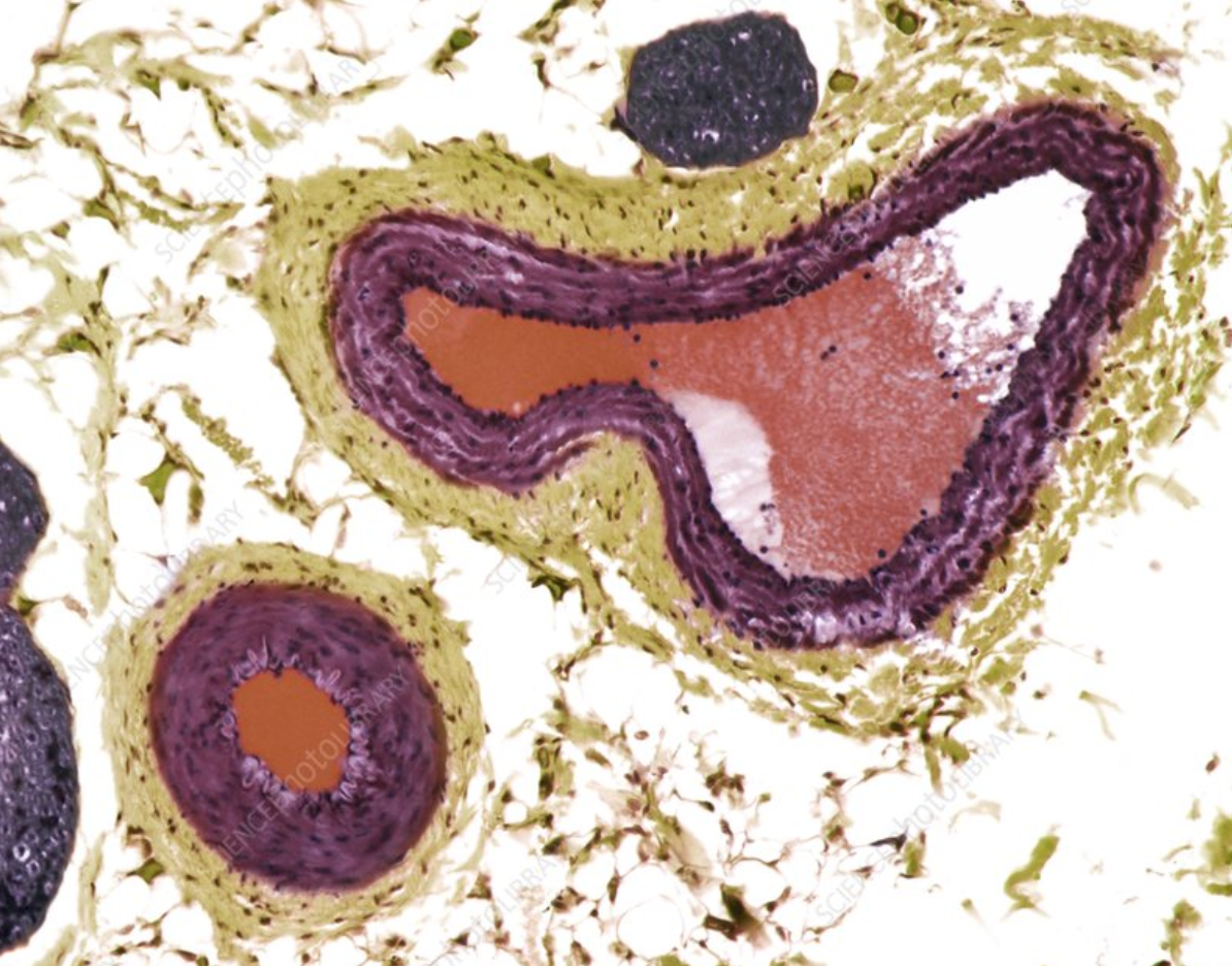
Structure of vessel wall + Thickness relative to diameter of lumen
Arteries: Thick smooth muscle layer; Regular circular shape
To transport blood at high pressures
Veins: Thin wall thickness; Wide lumens; Irregular shape
To transport blood at low pressures
Adaptations of Arteries for Transport of Blood Away From Heart
Layers of muscle and elastic tissue in walls of arteries
THREE LAYERS – Tunica adventitia, tunica media, tunica intima, endothelium
Muscle fibers help arteries stay rigid + maintain high pressure without bursting + contract → Increase pressure between pulses → Steady blood pressure
Smooth muscle layer (Controlled by ANS) changes lumen diameter → Regulate blood pressure
Elastin and collagen fibers allow arteries stretch / expand as blood flows + Elastic recoil helps push blood forward + Maintain pressure
Adaptations of Capillaries for Exchange of Materials Between Blood and Internal / External Environment
Blood from arterioles branch → Capillary beds → Venules
ONE LAYER – Only has tunica intima
Large SA + Narrow diameters due to branching
Ensures every cell is near a capillary and can receive oxygen
Slows down blood flow → More time for exchange (Only one cell is accommodated at a time)
Thin walls
Reduces diffusion distance
Fenestrations (pores) in capillaries where exchange needs to occur rapidly
Further enhances rapid exchange at intestines and kidneys
Adaptations of Veins for Return of Blood to Heart
THREE LAYERS – Tunica externa (elastin and collagen), tunica media (smooth muscle), tunica intimate (endothelium)
Presence of valves in areas where gravity is strong (e.g. legs)
Prevents back flow; Blood flows in one direction
Flexible wall; Have elastin
Allows to be compressed by muscle action (During activities like exercise where muscles contract) → Push blood towards heart
Coronary Heart Disease
Coronary arteries = Blood vessels at heart; Branched from aorta
Crucial in supplying oxygen to heart since very active and muscle-dense
Occlusion = Blockage caused by fatty plaque built up in inner lining of coronary arteries; May be caused by…
High blood cholesterol levels, smoking, hypertension, high blood sugar levels (Due to diabetes), genetic factors
Consequently…
Restricted blood flow to cardiac muscle → Chest pains + Deposition of minerals in plaque → Hard and rough plaque
Transport of Water during Transpiration + Properties of Water Involved
Loss of water by transpiration in leaf cells results in a negative water potential which draws water from xylem to the leaf. This generates a tension force known as a transpiration pull (a form of capillary action) which pulls water from roots, up the xylem, to the leaf.
Cohesion (attraction between water molecules due to hydrogen bonding) ensures that this column of water is continuous.
Adhesion (attraction to walls of xylem) allows water to counter gravity and travel upward by adhering to xylem walls
Adaptations for Xylem Vessels for Transport of Water
Lack of cell contents; Hollow dead cells + Incomplete / Absent end walls
Allows for unimpeded flow of water
Water movement is unidirectional (up)
Lignified walls
Provide strength → Withstand tensions during water transport
Prevent collapse under negative water potentials generated by transpiration
Pits (Microscopic holes in walls)
Enable entry and exit of water / water transfer
Transverse Section of Stem in Dicotyledonous Plant (HINT: 5 main features + 2-3 extra)
Vascular bundle
Function: Around edge of stem for structural support
Xylem
Function: Water and minerals transportation
Phloem
Function: Carbohydrate transportation
Cortex
Cylinder of tissue around stem; Has cells with secondary thickening in cell walls
Function: Provide support; Withstand bending and compression
Epidermis
Surface of stem made of cell layers; Embedded with stomata
Function: Waxy cuticle → Reduce water loss + Aid in gas exchange
Pith
Tissue of thin walled, parenchymal cells in centre of stem
Function: Storage of starch and oil + Secretion of resin
Cambium
Lateral meristem (cell growth region); Vertical cylinder in stem
Function: Produces secondary xylem and phloem through cell division
Fiber caps
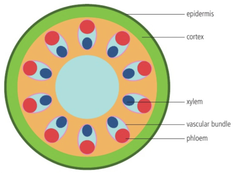
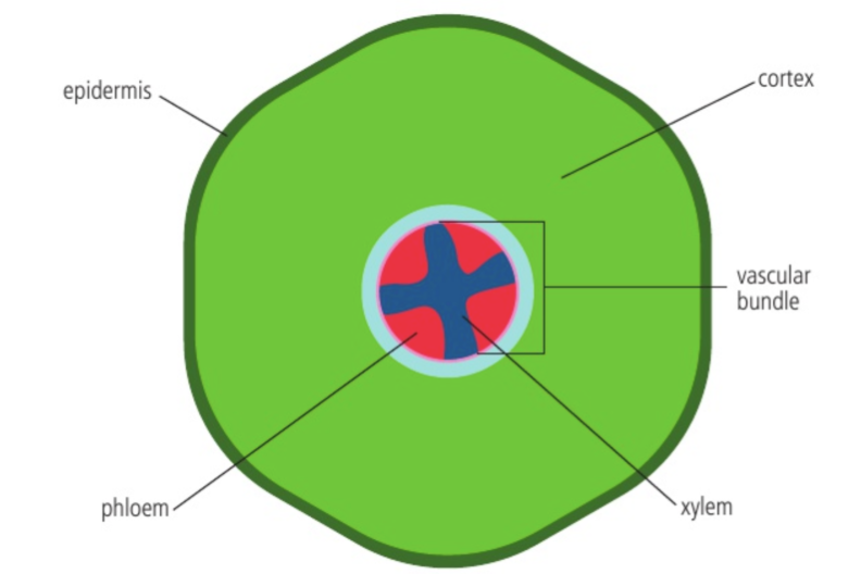
Transverse Section of Roots in Dicotyledonous Plant (HINT: 5 features)
Vascular bundle
Contains xylem (inside), phloem (outside), and cambium tissue
Function: Arranged in centre → Structural support; Withstand stretching roots
Xylem
Function: Water and minerals transportation
Phloem
Function: Carbohydrate transportation
Cortex
Cylinder of tissue around stem; Has cells with secondary thickening in cell walls
Function: Storage of reserve food
Epidermis
Surface of stem made of cell layers; Embedded with stomata
Function: has root hairs → Increased SA for substance exchange
Movement of Tissue Fluid in Capillaries
Tissue fluid = Aqueous portion of plasma including dissolved substrates; Formed by pressure filtration of plasma in capillaries
Leak out through fenestrations
Promoted by higher pressure of blood from arterioles
Lower pressure in venules allow tissue fluid to drain back into capillaries
Composition of Plasma and Tissue Fluid (HINT: 7 elements)
Cells
Plasma: Red + White blood cells, platelets
Tissue fluid: Phagocytes
Proteins
Plasma: Higher concentration; Large proteins and hormones
Tissue fluid: Lower concentration; Small proteins
Glucose
Plasma: Higher concentration; ~70-100 mg per 100 mL
Tissue fluid: Lower concentration; Used by cells for respiration
Fats
Plasma: Lipoproteins
Tissue fluid: N/A
Amino acids
Plasma: Higher concentration
Tissue fluid: Lower concentration
Oxygen
Plasma: Arterioles - High concentration; Venules - Low concentration
Tissue fluid: Lower concentration; Used by cells for respiration
Carbon dioxide
Plasma: Arterioles - Low concentration; Venules - High concentration
Tissue fluid: Higher concentration; Released by cells from respiration
Drainage of Excess Tissue Fluid ~ Lymphatic system
Presence of valves + Thin walls with gaps in lymph ducts
Lymphatic capillaries (small, gapped tubes with thin walls) facilitate easy movement of water and solute
Collection of lymph in lymphatic capillaries prevent build up and swelling around body cells
Valves assist with movement of lymph + Prevent back flow
Return of lymph to blood circulation
Through veins near heart
Facilitated by lymph nodes (Contain phagocytes to filter cellular debris & micro-organisms + immune cells)
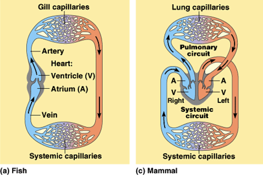
Single VS Double Circulation (Circuit diagrams)
FISH (& some invertebrates):
Heart → Artery → Gill capillaries → Systematic capillaries → Vein
Blood flows once in one circuit
One pathway
Two heart chambers
Low blood pressure → Low efficiency
MAMMALS (& birds):
Heart (Right atrium and ventricle) → (Pulmonary circuit) Lung capillaries → Heart (Left atrium and ventricle) → Artery → (Systemic circuit) Systemic capillaries → Vein
Blood flows once in two circuits
Two pathways; Pulmonary and systemic
Four heart chambers
High blood pressure → High efficiency
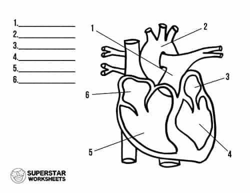
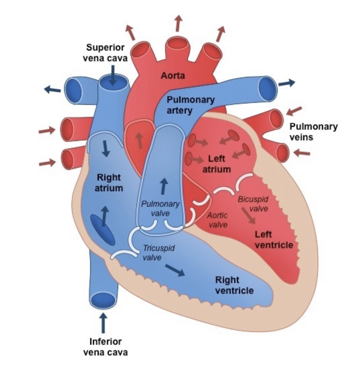
Adaptations of Mammalian Heart for Delivery of Pressurised Blood to Arteries (HINT: 8 features; Indicate direction of blood + Label)
Cardiac Muscle
Striated, branched, interconnected by intercalated discs
Allow for coordinated contraction and relaxation of heart
Myogenic (Originate from muscle, not nerve impulses)
Pacemaker
Specialised cells in right atrium
Generate electrical impulses; Regulate heart rhythm + Initiate heartbeat
Atria
Thin walls of cardiac muscles; At top chambers
Receive blood returning to heart + Help transfer to ventricles
Ventricles
Thicker walls; At bottom chambers
Forcefully contract to pump blood out of heart to lung (right) / body (left)
Atrioventricular valves (Tricuspid & Mitral valves)
Between atria and ventricles
Prevent back flow of blood from ventricles back to atria
Semilunar valves (Pulmonary & Aortic valves)
Located at exits of ventricles
Ensure blood flows one-way from ventricles to arteries
Septum
Thick muscular walls separating left and right parts of heart
Prevent mixing of oxygenated and deoxygenated blood
Coronary arteries
Small arteries and veins
Supply heart with oxygen and nutrients to fulfil high metabolic demands
Stages in Cardiac Cycle
NOTE: Electrocardiogram (ECG)
Atrial systole: Blood moves from atrium to ventricle through atrioventricular valves (tricuspid and mitral valves)
P-wave: Depolarisation of atria; Electrical impulse spreads from sinoatrial node to walls of atria causing muscle contraction
hey these are my flashcards so imma add whatever i want here hehe apparently the “sino-” prefix relates to “sinus” which in latin means hollow or curve so um thats coolPR-interval: Delay to reach atrioventricular node (causes ventricular systole); Time for impulse to travel is ~0.1 seconds
Atrial pressure: Increases → Falls ; Rise → Decline on graph
Fall when atria stops contracting
Aortic pressure: High; Flat on graph
Ventricular volume: Increase; Bump on graph
Ventricular pressure: Slight increase as blood flows in
(Beginning of ventricular systole) Isovolumetric contraction: Interval between closing of AV valves and opening of SV (Both AV valves and semilunar valves are closed); Amount of blood in ventricle remains same
AV valves closes when ventricular pressure > atrial pressure
QRS Complex: Polarisation of ventricles
Bundle branches and purkinje fibre carries signals from atrioventricular node to heart apex throughout ventricles
ok bio is weird idk why it's called the apex but its the very bottom tip of the heart
Atrial pressure: Increases; Bump on graph – c-wave
Blood in ventricles bulges against closed AV valves
Aortic pressure: High; Flat on graph
Ventricular volume: No difference; Flat line on graph
Ventricular pressure: Significant increase; Sharp gradient on graph
Ejection: Opening of semilunar valves (aortic and pulmonary valves) due to high ventricular pressure overcoming aortic pressure; Blood flows from ventricle into aorta and enters systemic / pulmonary circuit
T-wave: Repolarisation of ventricles; Ventricles relax
Atrial pressure: Decrease → Gradual increases; Decline → Slow rise on graph
Blood in ventricles are now exiting through SV → No longer pushing against AV
Since SV is closed and atria are relaxed, blood gradually flows back in → Atria becomes filled
Aortic pressure: Increase → Decrease; Rise → Fall on graph
Ventricular volume: Significant decrease; Decline on graph
Ventricular pressure: Initially increase (Higher than aortic pressure) → Decrease; Parabola / Rise → Decline on graph
Decrease due to repolarization
Isovolumetric relaxation: SV valves close, ventricles relax
End of T-wave: Ventricular repolarisation → Relax
Atrial pressure: Increases; Slight rise on graph
Aortic pressure: Small increase; Bump in graph
Blood in aorta briefly flows backwards and hit closed SV
Ventricular volume: No difference; Flat line on graph
Ventricular pressure: Decrease significantly (Below atrial pressure); Steep drop on graph
Ventricular filling: Since ventricular pressure is below atrial pressure, AV valves reopen, blood partially flows from atria to ventricles via passive filling
Flat line
Atrial pressure: No difference; Flat line on graph
Aortic pressure: Decreasing; Gradual decline in graph
Ventricular volume: Gradual increase; Rise on graph
Ventricular pressure: No difference; Flat line on graph
NOTE: Ventricular systole is from Q to T
On phonocardiogram…
“Lub” = AV closing
“Dub” = SV closing
Other Mechanisms of Water Transport by Generation of Root Pressure
When transpiration pull is insufficient for transport in xylem, e.g.
During high humidity which prevents transpiration
In spring, before leaves on deciduous plants have opened
Root pressure generates positive pressure potential
Active transport of minerals and ions from soil to root cells creates accumulation of solutes in cells
Lowers water potential inside cells → Causes water to move into cells by osmosis → Increases pressure inside cells → Positive pressure helps push water up the xylem
Phloem Translocation of Sap + Adaptations
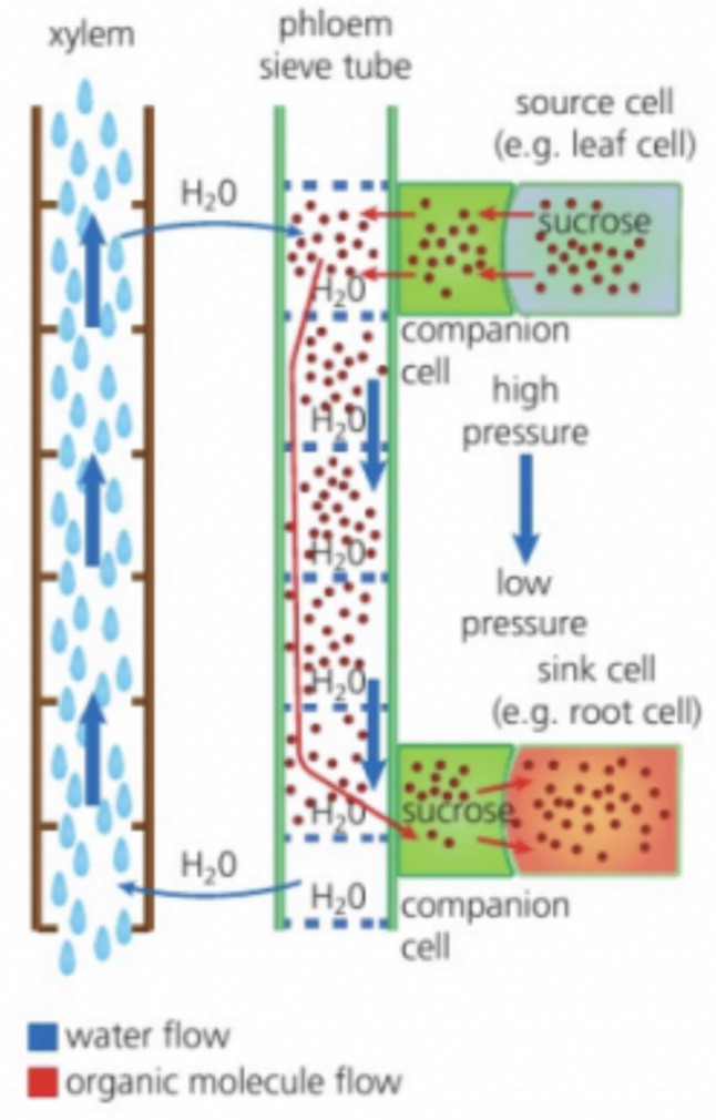
Active loading of sucrose into sieve tubes at source creates positive pressure potential + Water pressure created by entry of water by osmosis at source (Due to high solute concentration) → Translocation; Mass flow of phloem sap forward towards sink
Living tissue → Active transport + Response to environmental changes
Sieve tube elements
Sieve plates with pores → Allow sap to pass from one cell to another
Reduced cytoplasm and organelles + No nucleus → Ease flow of sap which travels within the tube like area of elements (Translocation)
Companion cells
Presence of many mitochondria → Actively load sucrose into phloem
Plasmodesmata between them → Allown cytoplasm of tube cell to be shared + Origin of proteins and ATP needed by sieve tube elements
Ease flow of sap
Enhance loading of carbon compounds into phloem sieve tubes at sources (leaves) and unloading of them at sinks (roots)
Importance of POSITIVE Pressure Potentials in Xylem
Aids in water uptake, counteracts transpiration pull, and structural support?
System Integration
Necessary process in living systems
Coordination = Needed for component parts of a system to collectively perform an overall function
Hierarchy of Body Systems
Cells → Tissues → Organs → Organ systems
Integrated in a multicellular living organism
System Integration & Emergent Properties + Example
System Integration = Responsible for emergent properties
Emergent properties – Properties that result from various interactions in system + Do not exist in individual components of the system
For example, for the cheetah…
Cellular level
Cheetah DNA → Controls body shape + Pigmentation of the fur → Creates spots for camouflage → Advantage in hunting and hiding
Tissue level
Loose hips and shoulder joints + Flexible spine → Flexes to store potential energy & springs back to release → Running
Smaller teeth → Larger nasal passage → Quick air intake
Organ level
Enlarged heart → Effective delivery of glucose & oxygen to muscles → Rapid physical response
Position of eyes → Maximum binocular vision → Assess distances to prey accurately
Organ system
Breathing system → Rapid delivery of oxygen to muscles + Circulatory system → Delivers blood to muscles + body ⇒ Run at high speeds
Organism level
Narrow paws → Minimal contact with ground + Blunt claws → Increase traction + Long tail → Counterbalance ⇒ Run at high speeds & Fast acceleration for chasing prey
Integration of Organs in Animals (Role of Endocrine)
Regulate homeostasis within body; Widespread response
Slow but long-lasting
Controls longevity, growth, and homeostasis (Involuntary factors)
Hormones = Chemical messengers; Manufactured by glands
Relies on circulatory system to be transported
From endocrine cell → Bloodstream → Target cell
Integration of Organs in Animals (Role of Nervous)
Receive information from various sensory inputs; Localised effects
Integrate information and trigger electrical impulses to send out signals
Neurons transmit and receive impulses
May be result from hormones released by glands in endocrine system
Quick but short-lived → For survival
Integration of Organs in Animals (Role of Circulatory System)
Helps transport…
Glucose and oxygen to the nerves and endocrine glands for energy
More generally, transport oxygen from lungs → legs
Hormones throughout body (endocrine signalling)
Waste products like CO2 and ammonia out of body
Urea produced by liver as waste from protein metabolism → Kidneys for filtration → Urine
Integration of Organs in Animals (Example)
Epinephrine
Sensory organs transmit information
Nervous system to trigger fight-or-flight response → Signal release
Adrenaline is released from adrenal glands → Body responses
Organisation of Nervous System
Central nervous system
Brain
Spinal chord
Peripheral nervous system
Somatic
Autonomic
Sympathetic
Parasympathetic
Role of Brain in Nervous System (HINT: 6 Parts)
Brain = Central information integration organ
Processes information combined from several inputs
Learning & memory
Three main areas:
Cerebrum
Divided into cerebral hemispheres consisting of four lobes
MAIN ONES TO KNOW:
Frontal Lobe: Learning new information
Reasoning, motor control, emotion, language
Temporal Lobe: Storing and retrieving memories
Managing emotions
Processing information from sense; Understanding language
Occipital Lobe: Memory formation
Visuospatial processing; Distance & depth perception
Color determination
Object and face recognition
Cerebellum
Coordinates voluntary movements; Balance and equilibrium
Brainstem
Connects cerebrum, spinal chord and cerebellum
Controls involuntary impulses + ANS
Sensory Neural Pathway (Neural Pathway Pt. 1)
Input to spinal cord and cerebral hemispheres through sensory neurones
Sensory receptor is stimulated
(Transduction) Physical signal is converted into electrical impulse down axon to spinal cord and brain
Sensory neurons convey messages from receptor cells to CNS
Electrical signal causes release of neurotransmitter at a synapse between sensory neuron and interneurone
Neurotransmitter stimulates interneuron to form action potential which travels along its axon
Action potential is transmitted until activates interneurones in higher parts of brain → Perception; Conscious awareness of touching happens
Neural Motor Pathway (Neural Pathway Pt. 2)
Output from cerebral hemispheres to muscles through motor neurons
Stimulus in brain → Forms action potential in upper motor neurone
Action potential stimulates release of neurotransmitter in synapse between upper and lower motor neurons
Forms action potential in lower motor neurons which stimulates release of neurotransmitter at neuromuscular junctions
Connected to skeletal muscle fibers
Neurotransmitters stimulate muscle fibers → Form muscle action potentials → Muscle contraction (movement)
Role of Cerebellum in Skeletal Muscle Coordination
Receive feedback impulses from moving area of body & sensory organs → Sends out impulses to motor neurons → Coordinate & Fine-tuning movement; Smooth and balanced muscular activity
Note initiation of muscle contractions is from motor cortex of cerebrum
Cerebellum does NOT initiate muscle contractions
Coordinates posture, balance, etc.
Overall control of movements of body including skeletal muscle contraction and balance
Nerve Bundles + Transverse section of Nerve
In cross section diagram of brain…
Gray matter = Neuronal cell bodies
White matter = Myelinated axons
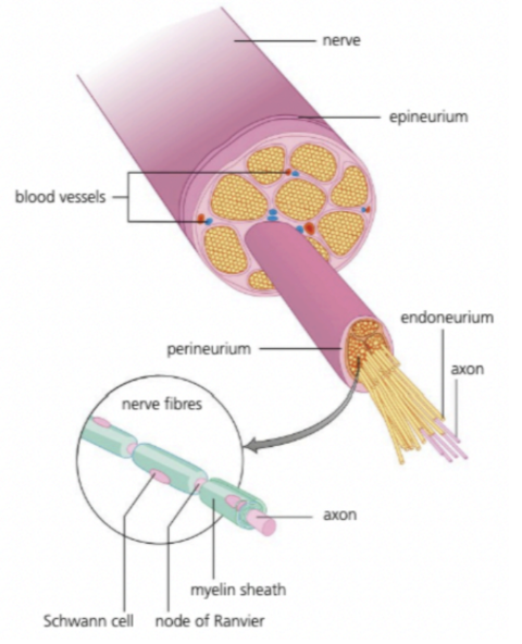
Nerves = Bundles of sensory and motor neurones; Covered by layers of protective sheath (Made of connective tissue)
Endoreneurium = Tissue surrounding each individual nerve fiber
Perineurium = Smooth connective tissue surrounding each fiber bundle
Epineureum = Fibrous tissue surrounding many bundles of nerve fiber; Covers most large nerves
Neurones can be unmyelinated or myelinated…
Schwann cells wrapped around axon
Nodes of Ranvier = Intervening areas between Schwann cells
Action potentials can skip from one node to another → Faster transmission compared to unmyelinated axons
Spinal Cord and Unconscious Processes
Spinal cord = Integrating center for unconscious processes
Receptors in the body receive stimulation information and pass it to spinal cords and onto cerebral hemispheres
Receptors for unconscious processes:
Osmoreceptors
Change in osmotic pressure / Concentration of bodily fluidsBaroreceptors
Blood pressure and help with its regulationProprioceptors
In muscles, tendons, and joints; Body position and movementReceptors for conscious processes:
Proprioceptors
Photoreceptors
In eyes; Respond to light + Allow sightThermoreceptors
Changes in temperatureMechanoreceptors
Respond to mechanical stimuli (touch, pressure, vibration)Chemoreceptors
Detect chemical stimuli (taste, smell)
White matter = Carry information to / from CNS & body
Grey matter = Integration & processing of information
Conscious VS Unconscious processes
Conscious = Deliberate and voluntary
Unconscious = Automatic and involuntary
Example of Involuntary Response
Pain reflex arc = Involuntary response; Skeletal muscle as effector
Evoluntary advantage to limit damage to body tissue by generating quick reaction involving minimal (three) neurons
Free sensory neuron (Pain receptor i.e. nocireceptor in hand) is stimulated → Initiate afferent (sensory) action potentials → Travel through hand → Joins a spinal nerve
Afferent neuron forms a synapse with a short interneuron in grey matter of spinal cord
Interneuron synapses with a motor neuron → Forms action potential → Travel to effector (arm muscles) → Movement (pull away)
Sensation of pain must travel to cerebrum
Circadian Rhythm
Circadian rhythm = Controlled by biological clock; 24 hour cycle
Modulation of sleep patterns by melatonin secretion
Regulated by pineal gland, hypothalamus, and pituitary gland
Melatonin = Amine hormone
Diurnal pattern of melatonin secretion by pineal gland → Helps establish cycle of sleeping and waking
When awake, LOW melatonin, HIGH cortisol
When asleep, HIGH melatonin, LOW cortisol
Exposure to light in retina inhibits melatonin production → Wakefulness during day / light period
Epinephrine and Vigorous Activity
Medulla of adrenal glands (located at upper side of kidneys) secrete epinephrine to prepare body for vigorous activity
During times of stress; Fight-or-flight response
Epinephrine = Peptide hormone
Widespread effects of epinephrine in the body
Increased heart rate → Increased delivery of oxygen to tissues
Increased breathing rate
Stimulates glycogenolysis in liver cells → Increased blood glucose
Enables increased rates of aerobic respiration → Increased ATP production in muscle tissues → Facilitates intense muscle contraction
Hypothalamus, Pituitary Gland, and Endocrine System + Example
Hypothalamus and pituitary gland controls endocrine system
Hypothalamus = Has receptors that detect changes in blood + Receives signals from neurons
Signals stimulates neurosecretory / glandular cells to produce and secrete hormones that stimulate / inhibit hormone release in pituitary gland
Anterior and posterior lobes secrete their own hormones
For ADH…
Hypothalamus produces ADH and sends to posterior pituitary
Osmoreceptors in hypothalamus sense water content of blood
If necessary, transmits action potential to cells in posterior pituitary gland
Signals posterior pituitary gland to secrete hormone into bloodstream
ADH reaches target tissue (collecting tubules in nephrons) → Higher water reabsorption
Maintain homeostasis by negative feedback
Feedback Control of Heart Rate
Feedback control of heart rate following sensory input from baroreceptors and chemoreceptors
Baroreceptors = Monitor stretch in arteries → Indirect measurement of blood pressure + Volume of blood pumped by heart
Distension of arterial wall → Increased rate of action potentials sent to medulla
Carotid sinuses / Arch of aorta
Chemoreceptors = Detect oxygen, carbon dioxide and pH levels in capillaries
When cellular respiration is increase, O2 ↓ CO2 ↑ pH ↓ (CO2 combines with water in blood; forms carbonic acid) → Increased rate of action potentials to the medulla
Carotid sinuses / Arch of aorta
Role of medulla in controlling heart rate
Sends nerve impulses to SA node to…
Adjust heart rate
Regulate stroke volume
Feedback Control of Ventilation Rate
Feedback control of ventilation rate following sensory input from chemoreceptors
Changes in pH levels of blood are monitored by chemoreceptors in the brainstem; Caused by…
Increased physical activity → Increased respiration rates → Higher concentration of CO2 → Increased H+ ions → Increased action potentials to medulla
In blood, CO2 combines with water to form of carbonic acid which later becomes bicarbonate and hydrogen ions
Control of ventilation rate using signals to the diaphragm and intercostal muscles
Control of Peristalsis by Enteric Nervous System
Enteric nervous system = Controls digestive system
CNS + ENS → Control of peristalsis in digestive system
Initiation of swallowing of food
Food forms into boluses in alimentary canal
Peristalsis (Under involuntary control of ENS)
Contraction and relaxation of circular and longitudinal muscles
Keep food moving in one direction
(From oesophagus → intestines → rectum)
Egestion of faeces (Under voluntary control of CNS)
Peristaltic reflex works by…
Triggered by boluses (food formed in alimentary canal); Bolus causes distension in surrounding area → Activates stretch receptors in ENS
Receptors connect with relay neurons
Relay neurons connect with motor neurons
Contract smooth muscle behind bolus + Relaxes smooth muscle ahead → Push forward + Create space for food to pass through
ENS ensures passage of material through gut is coordinated
Phototropism
Positive phototropism = Directional growth response to lateral light in plant shoots
Phytohormones + Examples
Phytohormones = Signalling chemicals which control growth, development, and response to stimuli in plants
Examples include…
Auxin → Plant cell elongation
Cytokinin → Increased rate of cell division
Ethylene → Promote fruit ripening
Gibberellin → Control stem elongation, seed germination, flowering, and dormancy
Auxin & Auxin Efflux Carriers
Auxin = Phytohormone produced in growing regions of plants (Tips of shoots, roots, and growth buds)
In growing areas where evenly concentrated, cell elongation is uniform
When concentrated on certain areas → Differential growth
Can enter cells easily by diffusion but require efflux carriers to exit cell
Auxin efflux carriers = Maintaining concentration gradients of phytohormone
Can be positioned in a cell membrane on one side of cell
If all cells coordinate to concentrate efflux carriers on same side of cell, auxin can be actively transported from cell to cell through plants tissue → become concentrated in specific part of plant
Promotion of Cell Growth by Auxin
Cell elongation is facilitated by…
Auxin stimulates synthesis of proton pumps
Promotes secretion of hydrogen ions into apoplast
Apoplast = Area including cell wall and intercellular spaces
Acidfies cell wall and activate expansin
Loosen hydrogen bond cross links between cellulose molecules
Absorption of water generates high turgor internal pressure + Loosened cellulose fibers are allowed to slide past each other and reposition themselves
Forms new hydrogen bonds as elongated cell (walls)
Concentration gradients of auxin → Differences in growth rate needed for phototropism
Elongation of cell on shaded side for new growth and turn towards the sun
Auxin and Cytokinin in Regulating Growth
Root tips produce cytokinin (promotes cell division) → Transported to shoots
Transported via xylem fluid
Shoot tips produce auxin (promotes cell elongation) → Transported to roots
Transported via phloem sap
Interactions between them ensure root and shoot growth are integrated
HIGH auxin, LOW cytokinin → Rooting
LOW auxin, High cytokinin → Shooting
At certain concentrations, they works synergistically with each other
Feedback Control of Fruit Ripening
Positive feedback in fruit ripening and ethylene production
Ethylene = Stimulates changes in fruits for ripening which in turn increases production of ethylene
Benefits of positive feedback mechanism
Ensuring fruit ripening is rapid and synchronised
Due to its gaseous nature, also transmits to adjacent fruits
Rapid ripening in large volume → Effectively attracts herbivores to consume fruit and disperse seeds (propagation)
Homeostasis & Homeostatic Variables
Maintenance of internal environment of organism
Variables are kept within preset limits despite fluctuations in external environment
Homeostatic variables in humans may include
Body temperature, blood pH, blood glucose concentration, and blood osmotic concentration
Negative Feedback Loops in Homeostasis
Negative feedback loops are…
Used to return homeostatic variable from higher or lower values to a set point and keeps values within a narrow range
Positive feedback loops would amplify responses which is less applicable
Example of Role of Hormones in Homeostasis
Regulation of blood glucose by secretion of insulin and glucagon
By pancreatic endocrine cells
α-cells secrete glucagon
β-cells secrete insulin
Transported through blood stream
Insulin works by…
Opening protein channels in cell membranes → Allow facilitated diffusion for glucose uptake
Stimulate muscles to take in more glucose / store as glycogen → Reduce blood glucose levels
Glucagon works by…
Acts on stored glycogen in liver and muscles
Glycogen is hydrolysed to glucose → Enters bloodstream; Increased blood glucose level
Type 1 Diabetes
Physiological changes
Autoimmune condition; β-cells are destroyed → Insulin can’t be producedRisk factors
Family history, ageMethods of prevention
N/ATreatment
Insulin injections, blood glucose monitoring, diet control
Type 2 Diabetes
Physiological changes
Insulin resistance; Body receptors don’t respond properly to insulin and cells cannot take in glucoseRisk factors
Family history, obesity, lack of exerciseMethods of prevention
Good eating habits, regular exerciseTreatment
Diet control, exercise, and medication
Example of Negative Feedback Control
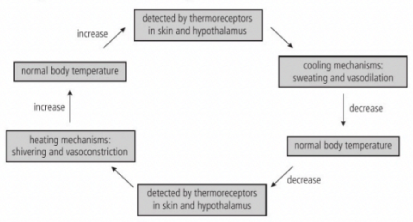
In thermoregulation…
Thermoreceptors in hypothalamus and skin (peripheral) sense temperature of blood
Peripheral thermoreceptors send impulses to the hypothalamus
Hypothalamus responds by initiating cooling or heating mechanisms through stimulating pituitary gland
HEATING MECHANISM: Hypothalamus stimulates the pituitary gland to release hormones to activate the thyroid gland → Thyroid gland releases thyroxin to increase metabolic rate of body → Generate heat
COOLING MECHANISM: Hypothalamus stimulates the pituitary gland to release hormones that inhibit the release of thyroxin → Decrease metabolic rate of body → Reduce heat production
Example of effectors of temperature change: Muscle and adipose tissue
Hypothalamus initiates shivering
Generate heat by muscle movement
Marine mammals also have layer of blubber (adipose tissue) for insulation
Helps retain warmth generated by internal metabolic activities
Thermoregulation Mechanisms
Ectotherms = Animals that don’t control their internal body temperatures
Endotherms = Maintain steady internal temperature
Birds and mammals maintain body temperature through behavioural and physiological means
Cerebrum initiates conscious behaviours
E.g. Move around more to generate muscle heat, dress warmer, stay inside / Seek shade, use air conditioning, wear light clothing, stop exercising
Thermoregulation in humans
HEATING MECHANISM
Vasoconstriction
Arterioles near skin become smaller → More blood is shunted to internal organs / Capillaries receive less blood → Preserve vital internal organs + Prevent heat loss to surroundings
Shivering
Muscle movement → Generate heat
Uncoupled respiration in brown adipose tissue (Lots of mitochondria)
Undergo cell respiration uncoupled from ATP production / Oxidise glucose → Generate heat
Hair erection
Release of epinephrine → Goosebumps on skin + Raised hairs → Create insulating layer of air between fur and skin → Protect body from cold air
COOLING MECHANISM
Vasodilation
Hypothalamus sends impulses to arterioles near skin → More blood travel through capillaries in skin → Release of more heat to surrounding air
Sweating
Hypothalamus initiate perspiration → Heat from body is transferred to water in sweat → Evaporation of sweat from skin → Evaporative cooling
Role of Kidneys in Osmoregulation and Excretion (HINT: 5 parts)
Excretion VS Osmoregulation
Osmoregulation = Regulation of osmotic concentration (osmoses per L)
Excretion = Production of urine for leaving body
Nephron = Filtering unit in kidney; ~1.25 million in each kidney
Thin tubules → Increased SA:V ratio
Capillaries wrapped around → Movement of substances to bloodstream
Within the nephron…
Glomerulus = Capillary bed
Ultrafiltration of substances from blood under hydrostatic pressure
Bowman’s capsule
Collect filtrate from glomerulus
Proximal / Distal convoluted tubule & Loop of Henle
Pathway for filtrate to travel; Adjust water and salt concentrations
(Peritubular) Capillary bed
Surrounds tubules for reabsorption into blood
Collecting duct
Collect final urine to send out of kidney
Role of Glomerulus, Bowman’s Capsule, and Proximal Convoluted Tubules in Excretion
Ultrafiltration (In bowman’s capsule) remove solutes from blood plasma under high pressure
Blood enters via afferent (wide) arteriole → Branches out to glomerulus → Exit through efferent arteriole (narrow diameter → high hydrostatic pressure)
Fenestration in walls of glomerulus → Substances move into Bowman’s capsule (Becomes part of filtrate)
Water, glucose, Na+, Cl-, amino acids, urea'
Useful substances (H2O, Na+, Cl- and glucose) are then reabsorbed into peritubular capillaries
Filtrate travels into proximal convoluted tubule for selective reabsorption (All glucose, amino acids, vitamins, hormones, water, and most mineral ions are reabsorbed)
Tubule has microvilli → Increased SA for reabsorption
Mineral ions and vitamins are actively transported by protein pumps and carrier proteins
Glucose and amino acids are cotransported via symporters across apical membrane with sodium
Water follows movement of mineral ions (passive diffusion via osmosis)
Toxins and unwanted solutes are left in filtrate → Excreted in urine
Role of Loop of Henle
Descending limb = Permeable to WATER / Impermeable to SODIUM IONS
High osmotic concentration in medulla creates osmotic gradient
Water leaves filtrate by osmosis
Solute concentration in filtrate increase as loop descends
Ascending limb = Permeable to SODIUM IONS / Impermeable to WATER
Active transport of sodium ions into medulla, followed by Cl- ions
Solute concentration in filtrate decreases as loop ascends
Maintains high osmotic concentrations in medulla
Facilitates water reabsorption in collecting ducts
Remaining liquid enters distal convoluted tubule
Output of urine is more dilute than input
Large amounts of salts are removed
Osmoregulation in Collecting Ducts (ADH)
Role of osmoreceptors in the hypothalamus
Monitors water content of blood → Control release of ADH
Changes to the rate of antidiuretic hormone secretion by the pituitary gland
ADH changes permeability of collecting duct
ADH switches location of aquaporins between cell membranes and intracellular vesicles in cells of collecting ducts
When ADH present, aquaporins (where water move OUT of collecting duct) are inserted into membranes to allow water reabsorption
Changes in Blood Supply in Response to Activity
Controlled by vasodilation and vasoconstriction
To…
Skeletal muscles
Can withstand large volume changes
Vasodilation in muscle arterioles to increase blood during exercise
Gut
Can withstand large volume changes
Vasoconstriction in gut arterioles to decrease blood during exercise
Brain
Maximum blood flow during REM sleep
Kidneys
Maximum blood flow during sleep and wakeful rest
Neurons + Structure
Cells within nervous system that carry electrical impulses along their fibers
Cell body = Cytoplasm and nucleus; Elongated nerve fibres project from it
Axon = Long, single fiber
Dendrites = Multiple shorter fibers
Nerve Signals / Impulses
Action potentials propagated along nerve fibers
Impulses = Electrical; Involves movement of positively charged potassium and sodium ions
Generation of Resting Potential by Sodium-Potassium Pumps (HINT: 3? Nah. 2? Ok)
BTW: Potential difference ~ Voltage
Pumping establish and maintains concentration gradient of potassium and sodium ions
Energy from ATP drives pumping of sodium and potassium ions AGAINST concentration gradient through sodium-potassium pumps across plasma membrane of neurones
Three Na+ OUT
Two K+ IN
Negative resting potential (-70 mV) due to imbalance of positive and negative charges across membrane
Stages of Nerve Impulse (Membrane Potential and Polarisation)
Membrane potential = Difference in charge across membrane of neurons; Allows creation of electrical signals
Resting potential = Difference in charge when neuron is NOT firing
Action potential = Difference in charge when neuron is FIRING; Rapid change resulting in nerve impulse
Stages:
Resting potential
Outside is relatively more positive; Na+ are concentrated OUTSIDE / K+ = INSIDE
Depolarisation = Change from resting to action potential
Opening of Na+ channels in response to signal initiated at dendrite (Inside becomes positive) → Passive influx of Na+ ions
Repolarisation = Restoration from action to resting potential
Opening of K+ channels (Inside becomes negative) → Passive efflux of K+ ions
Refractory period = Between action potentials
Since concentration of K+ is OUTSIDE / Na+ = INSIDE, sodium potassium pump restores concentrations to resting potential via anti porter action
Leakage channels allow for back flow of ions
Variation in Speed of Nerve Impulse
Myelinated VS Non-myelinated: Myelinated axons conduct faster
Skin temperature receptor axon: 5 μm; 20 ms-1
Schwann cells around the axons provide insulation
Electrical signals can jump between nodes of Ranvier and travel fast
Giant axons of Squid VS Non-myelinated: Axons with greater diameter conduct faster
Squid: 500 μm; 25 ms-1
Internal organ axon: 1 μm; 2 ms-1
(Chemical) Synapses
Junction between neurons / between neurons and receptor / effector cells
Where signals are transferred through neurotransmitters
A signal can only pass in ONE DIRECTION across a typical synapse
Presynaptic cell = Neuron transmitting a signal and releasing neurotransmitters into synaptic cleft
Postsynaptic cell = Neuron receiving the signal by detecting presence of neurotransmitters
Processes in Synapses (Release of Neurotransmitter from presynaptic membrane)
Uptake of calcium in response to depolarisation of presynaptic membrane
Action as signalling chemical inside neurone
Steps:
Action potential arrives
Depolarisation of presynpatic membrane → Uptake of calcium
Calcium ions = Signalling chemicals → Trigger movement of vesicles through cell
Vesicles fuses with plasma membrane
Neurotransmitter is release into synaptic cleft
Neurotransmitter binds to receptors on postsynaptic neuron
Processes in Synapses (Generation of Excitatory Postsynaptic Potential)
Diffusion of neurotransmitters across synaptic cleft
Binding to transmembrane receptors
Processes in Synapses (Depolarisation and Depolarisation During Action Potential)
Action of voltage-gated sodium and potassium channels (See before)
Stimulus causes their opening and subsequent diffusion of ions along concentration gradient
Need for threshold potential to be reached for sodium channels to open
Action potential must reach minimum threshold potential in order to be self-propagated
Propagation of Action Potential Along Nerve Fibre
Due to local currents
Diffusion of sodium ions inside and outside axon cause threshold potential to be reached
Depolarised area is initially localised
Na+ ions that have entered diffuse to nearby areas → Raise axon voltage high enough to overcome threshold potential
Saltatory Conduction in Myelinated Fibers
Saltatory conduction = “Jumping” of impulses between nodes (depolarised region) → Achieve faster impulses
Ion pumps and channels are clustered at nodes of Ranvier
Ion movement only occurs at nodes instead of whole axon → Less energy to fuel sodium-potassium pump + instantaneous response
Action potential is propagated from node to node
Effects of Exogenous Chemicals on Synaptic Transmission + Example
Exogenous chemicals = Chemical from outside cell / body
Neonicotinoids = Pesticide that blocks synaptic transmission
Binds to acetylcholine receptors
Cannot be broken down by acdtylcholinesterase (Irreversible binding) → Block cholinergic synapse
Cocaine = Drug that blocks reuptake of transmitter
Affects dopamine, serotonin, and norepinephrine
Binds to transporters that remove excess neurotransmitters from synaptic cleft
Prevent reabsorption of neurotransmitters → Increased concentration → Enhances effects on postsynaptic neurons + Increased production of receptors → Increased sensitivity → Addiction / Depression
Excitatory VS Inhibitory Neurotransmitters + Generation of Excitatory VS Inhibitory Postsynaptic Potentials
EXCITATORY –
Acetylcholine
Exists in many types of synapse including neuromuscular junctions
Excitatory within neuromuscular junction
Inhibitory within heart
Acetylinecholinesterase breaks it down for recycling → Prevent sustained activation
Generate action potential by increasing permeability of postsynaptic membrane to positive ions
Na+ ions in synaptic cleft diffuse into postsynaptic neuron
INHIBITORY –
GABA
Hyperpolarization inhibits action potentials
Binds to specific receptor causing Cl- to move into / K+ move out of postsynaptic neuron
Postsynaptic membrane becomes hyperpolarized (More negative than normal) → Hard to overcome threshold potential
Summation of Effects of Excitatory and Inhibitory Neurotransmitters in Postsynaptic Neuron
Multiple presynaptic neurons interact with all or nothing consequences in terms of postsynaptic depolarisation
EPSP → Depolarisation
IPSP → Hyperpolarisation
EPSPs combine via temporal or spatial summation → Threshold is reached → Action potential
Accumulation of EPSPs over time / in certain area
EPSP + IPSP → NO Action potential
Perception of Pain
By neurons with free nerve endings in skin
Nerve endings have channels for positively charged ions
Open in response to stimulus
E.g. High temperature, acid, or certain chemicals like capaiscin
Entry of positively charged ions causes threshold potential to be reached
Nerve impulses pass through neurons to brain → Perception of pain
Consciousness
Emergent property from interaction of individual neurons in brain