Gas Transport & Exchange
 Call Kai
 Learn
Learn Practice Test
 Spaced Repetition
 Match
Match Flashcards
 Knowt Play
1/75

There's no tags or description
Looks like no tags are added yet.
Name | Mastery | Learn | Test | Matching | Spaced | Call with Kai | Chat |
|---|
No analytics yet
Send a link to your students to track their progress
76 Terms
2 ways to transport oxygen
Dissolved in Plasma
Attached to haemoglobin
What oxygen exerts a partial pressure
Only oxygen dissolved in plasma
(oxygen in Plasma has a Partial Pressure - all the Ps)
Is oxygen soluble
Only very slightly

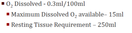
Why can’t we just rely on oxygen dissolved in plasma for our oxygen requirement
We need haemoglobin to supply the other 235ml O2 needed
For every 1g of haemoglobin (Hb) how many ml of O2 is it carrying
1.34 ml
How much Hb is in 100ml of blood
Hb 15 g Hb/100 ml
How much oxygen is in 100ml of blood
20.1mlO2
When CO is 5L how many ml of oxygen being delivered (ml/min)
1005ml/min
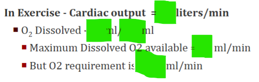
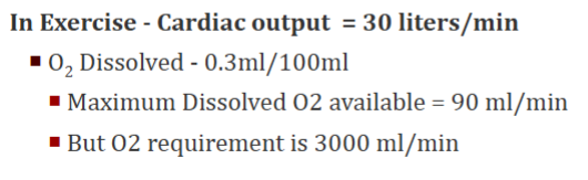
When CO is 30L how many ml of oxygen being delivered (ml/min)
6030 ml/min
How many oxygens does haemoglobin generally give up at rest
1
Generally 3/4 molecules are still attached
O2 capacity of blood formula



O2 saturation of Hb formula
Oxygen content in blood / (oxygen-carrying capacity of Hb (1.34 mL) x Hb conc. in blood)
How much O2 is Hb carrying / How much is possible for it to carry
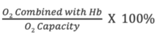
O2 saturation of Hb in mixed venous blood
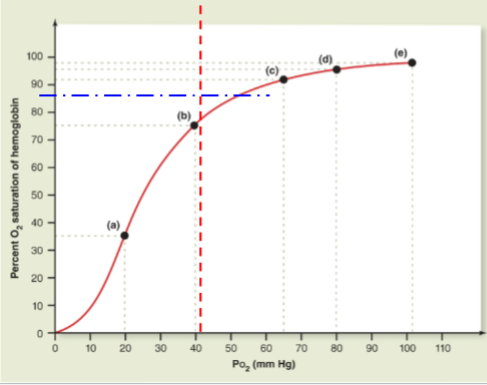
75% saturated (so up to 75% of O2 supply to tissues is in reserve
(this would make sense - Hb only usually gives out 1 of 4 O2s so 75% would be left)
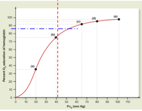
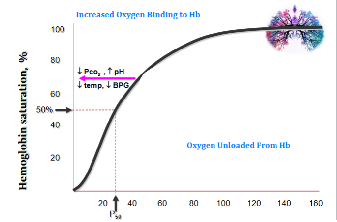
name this curve
Oxygen dissociation curve
What is an oxygen dissociation curve
For a given PO2 what percentage of haemoglobin is going to be saturated
(PaO₂ represents the amount of dissolved O₂ in plasma , not in RBCs. e.g. this would be high in the lungs)
(Think of PaO₂ as the pressure pushing oxygen into:
Hemoglobin (to bind it),
Tissues (to deliver it).)
Explain the shape of the graph
Flat upper portion: For a big drop in PO2 you only get a small drop in haemoglobin saturation
Much sharper decrease
The first oxygen is difficult to give up. The oxygens become easier to lose after that
if the oxygen dissociation curve moves right what does this mean
Any time we need more oxygen to be released from haemoglobin for use in cells the graph moves to the right.
E.g. during exercise we need haemoglobin to be less saturated as we need it to give up oxygen more easily to the muscle
The temperature goes up - it goes up during exercise
Space under the graph = oxygen bound to Hb. Space over the graph = oxygen given up to tissues (if we move the graph right we decrease the area under the graph (O2 bound to Hb) and increase area above the graph (O2 given to tissues) which is what we want during exercise!)
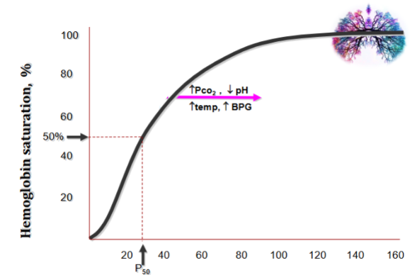
If the oxygen dissociation curve moves left what does this mean
e.g. foetal haemoglobin
(BPG binds to deoxygenated haemoglobin (Hb) and reduces its affinity for oxygen, which helps haemoglobin release oxygen to tissues)
Polycythaemia
Overproduction of red blood cells
(from cancer or high altitudes)
(Poly = many. Cyto = cell. Haemia = blood)
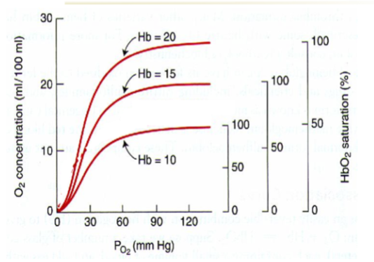
Effect of anaemia and polycythaemia on oxygen concentration curve
Doesn’t change the shape of the curve
People with anaemia (Hb = 10) have a lower oxygen concentration despite the same oxygen saturation as those with higher Hb levels.
People with polycythaemia (Hb = 20) have higher oxygen concentration despite the same oxygen saturation as those with lower Hb levels.
4 types of hypoxia & what are they
(Hypo- = low, -oxia = oxygen, 👉 So: Hypoxia = low oxygen (in tissues))
Hypoxic Hypoxia - Low Arterial PO2 (Pulmonary Disease)(not enough O2 entering the blood from the lungs)
Anaemic Hypoxia - Decreased ability to Carry O2 (Anaemia or CO Poisoning)(low iron → low Hb → low O2)
Circulatory Hypoxia (Stagnant Hypoxia) - Slow blood flow - pools in legs (Shock, Local Obstruction) (Good O2, Good Hb, Bad delivery - low blood volume / clot / heart failure)
Histotoxic Hypoxia - Toxic Substance Stops Tissue Using Available O2 (cyanide)(oxygen is there, delivery is fine, but cells can’t use it due to poisoning)
(4 places for problems:
Lungs → Blood
Blood Plasma → Hb
Hb → Cells
Cells use of O2)
How is CO2 transported
70% is transported as carbonic acid (H2CO3) - dissociates into H+ and bicarbonate (HCO3-)
23% is bound to amino globular proteins in Hb molecule forming carbaminohemoglobin
7% is transported as CO2 dissolved in plasma - CO2 has a higher solubility coefficient than O2
What happens when CO2 enters red blood cells from respiring tissues and combines with water
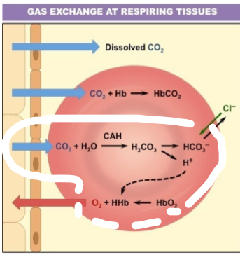
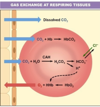
Role of chloride ion transport in & out
Keeps electrical neutrality
In respiratory physiology, work = (formula)
In respiratory physiology, work = pressure x volume.
Lungs opening easily isn’t always a good thing, why?
In emphysema are highly compliant - doesn’t mean person is healthy
(High compliance in emphysema = lungs too floppy → can't recoil → air trapping + poor gas exchange.)
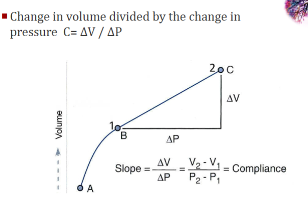
Compliance formula
Tidal volume in ml
approx. 500ml
Patient inhales 500 mL of air from a spirometer with a starting pressure of -5 cmH2O ending at -10 cmH2O. Calculate compliance

Compliance normal value
100ml/ cm of water
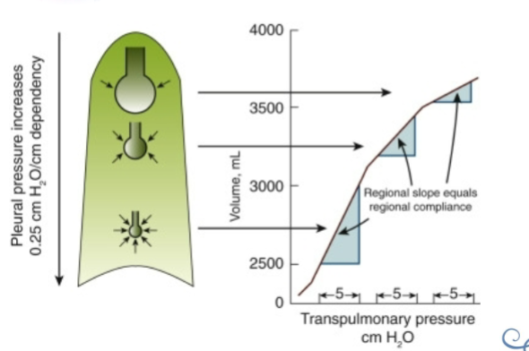
How does compliance change throughout the lungs
Alveoli at the bottom are easy to open - very small - easier to expand before they reach their maximum size
Alveoli at the top are hardest to open
What significance does compliance change throughout the lungs have
Pneumonia in the bottom of the lungs will be a bigger problem than something affecting the top of the lungs.
Factors That Influence Compliance
Posture
Age - Compliance often ↑
High standing Diaphragm - Pregnancy, Obesity, Scoliosis
Lung Disease
If lung is unventilated for long period (C↓)
↑ surface tension within alveoli (C↓)
Lung engorged with blood (↑pulmonary venous pressure) (C↓)
Pneumothorax - Trauma, diving, underlying lung pathologies
Effect of fibrosis on compliance
You need a huge increase in pressure to open lungs - very low compliance
Name 3 restrictive disorders that decrease compliance and therefore increase stiffness & work needed to inflate lungs
Pneumonia
Toxicity
Fibrosis
Name a disease that increases compliance, reducing stiffness and work needed to inflate lungs
COPD
(Emphysema & chronic bronchitis)
Compliance is dependant on what 3 things
Distensibility - ease to stretch/deflate – Elastin
Elasticity - Collagen (Without elasticity, lungs enlarge with every breath. Compliance would eventually become zero - Not compatible with life)
Surface Tension
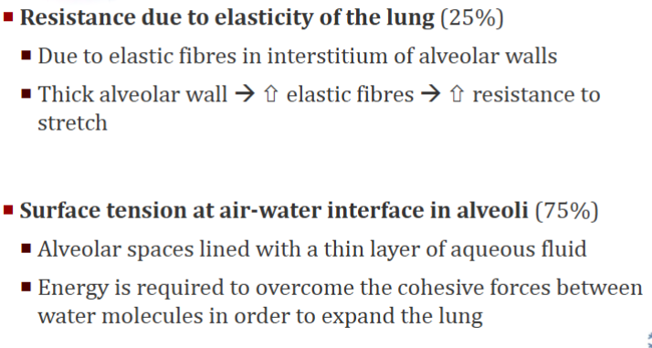
What are the 2 determinants of lung compliance at an alveolar level
What produces surfactant
Type II alveolar cells
What stimulates Type II alveolar cells to produce surfactant
Cortisol plays a key role in stimulating surfactant production in Type II alveolar cells.
What happens to infants without enough surfactant
Infant Respiratory Distress Syndrome (low compliance)
How is Infant Respiratory Distress Syndrome treated
Exogenous surfactant
Does surfactant increase/decrease surface tension
↓Surface Tension
Does surfactant increase/decrease lung compliance
↑ Lung compliance
Other roles of surfactant
↑ Stability of the alveoli
↓Small alveoli emptying into larger ones
Helps to keep alveoli dry because the “surface tension” of the alveolus liquid layer would tend to draw water into the alveolus
What does the Law of Laplace show
Smaller alveoli have higher pressure inside
(it is la place of pressure)
is surfactant hydrophillic/hydrophobic/amphipathic
amphipathic
Surfactants are composed of approximately ___% lipids and ___% proteins
approximately 90% lipids and 10% proteins
These Surfactant specific proteins are hydrophobic/phillic
SP-A
SP-B
SP-C
SP-D
SP-B and SP-C are two small hydrophobic proteins
SP-A and SP-D are large hydrophilic proteins
Most abundant neutral lipid in surfactant
Cholesterol
responsible for generating a near-zero surface tension at the air–liquid interface of the alveoli during compression
DPPC (lipid)
(REALLY long name!)
The effect of DPPC is stronger in higher/lower alveolar volumes
lower
Role of Surfactant Protein A (SP-A)
Immune defence
Role of Surfactant Protein B (SP-B)
Reduces Surface Tension (B chill - no tension)
Antibacterial (Ban Bacteria)
Which surfactant protein is only found in the lung
SB-C
(You only C it in the lung)
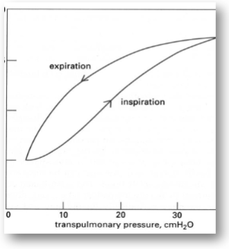
Explain the Pressure-Volume Curve
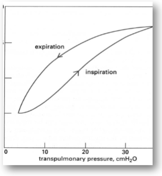
Early in inspiration there is a small change in volume for a large pressure change
At this time the slope (V/P) is small and compliance is low
As inspiration continues the slope is steeper indicating an increase in compliance
The V/P relationship during expiration is different than during inspiration - hysteresis
Volume at any pressure is greater for deflation than inflation
Most of the hysteresis and also most of the compliance of the lung appears due to the surface tension within of the alveoli
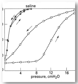
How would the addition of saline to the lung affect the lines on this graph
(Inflation of the lung with saline eliminates air-liquid interface and surface tension)
It is much easier to inflate lung with saline as elastic recoil of the lung is diminished due to lack of surface tension
Also not much difference between inflation and deflation – no hysteresis
Why does hysteresis happen
Changes in Surfactant activity: Surface tension greater in inspiration
Stress relaxation: Inherent property of elastic tissues (crinkled structure of collagen in the lung)
Redistribution of gas: fast and slow alveoli
Lung compliance formula
Change in lung vol / change in transpulmonary press
Chest wall compliance formula
Change in lung vol / change in transchestwall press
Total compliance within the respiratory system in ml / cmH2O
Approx. 100 ml / cmH2O
Flow formula
Flow = Pressure/Resistance
Factors That Effect Flow
1) Diameter(radius)
2) Velocity
3) Anatomy
4) Lung volume
5) Inspiration
6) Expiration
What does resistance depend on
Whether flow is laminar or turbulent
Airway dimension
Viscosity of the gas.
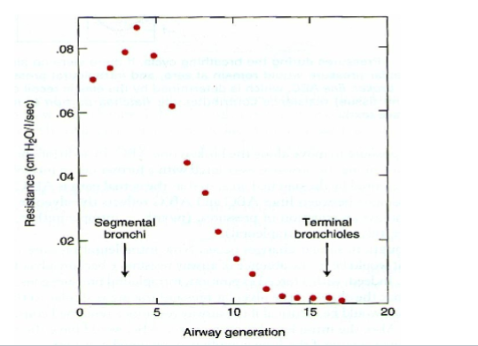
What is the Major Site of Airway Resistance
Most Resistance in Medium Sized Bronchi
What is a Site of low Airway Resistance
Terminal Bronchioles Offer Very Little Resistance
How does innervation cause a decrease in airway diameter
The vagus nerve innervates the smooth muscle of the airways.
It releases acetylcholine (ACh) which constricts airways
How does innervation cause an increase in airway diameter
Activation of Adrenergic Receptors
Adrenaline, Isoprenaline, β2-Adrenergic Agonists
Diseases in which airway narrowing occurs
COPD, asthma - increase airway resistance
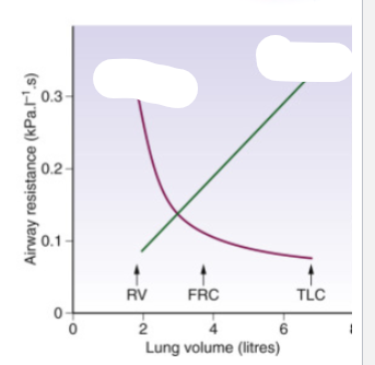
which is resistance & which is conductance
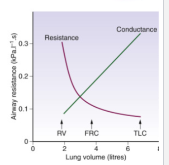
Does resistance increase/decrease above FRC
Decrease - increased airway diameter
Effect of Decrease Lung Volume below FRC to RV
Airway Resistance
Airway Diameter
Decrease Lung Volume below FRC to RV causes
• Increased Airway Resistance
• Decreased Airway Diameter
Small Airways (Base of Lung) May Close Completely
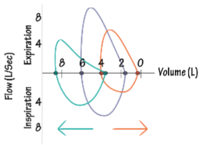
what is this called & what does it represent
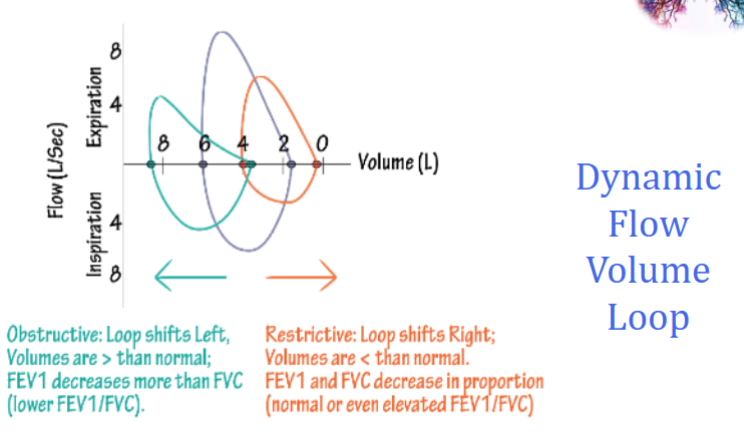
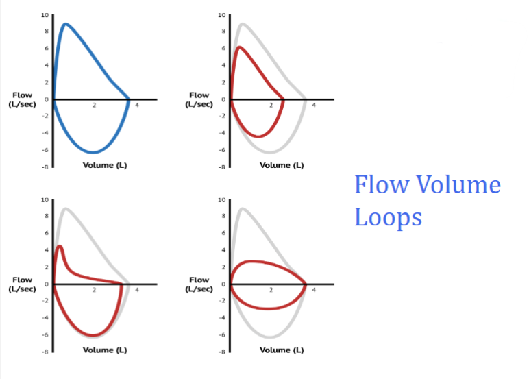
Which of these are normal & what is wrong with the rest