Lecture 2 gross anatomy
 Call Kai
 Learn
Learn Practice Test
 Spaced Repetition
 Match
Match Flashcards
 Knowt Play
1/143

There's no tags or description
Looks like no tags are added yet.
Name | Mastery | Learn | Test | Matching | Spaced | Call with Kai | Chat |
|---|
No analytics yet
Send a link to your students to track their progress
144 Terms
What is considered the thorax?
Region between the neck and abdomen
What makes up the thorax
thoracic wall, thoracic cavity, and the organs contained within it.
Boundaries of the thorax
Superior thoracic aperture, inferior thoracic aperture
Superior Thoracic aperture
passageway for structure between thorax and neck
inferior thoracic aperture
Passageway for structures between thorax and abdomen
enclosed by diaphragm
Bony elements of the thoracic wall
Ribs, vertebrae bodies (T1-12) sternum, costal cartilages
Rib types
True ribs (1-7) false ribs (8-10) Floating (11-12)
Costal cartilages localtion
Costal margin Rib 7-10
sternum components
manubrium, body, xiphoid process
Joints of the thoracic wall
costovertebral, intervertebral, costotransverse, costochondral, sternocostal
Intervertebral
2 Thoracic vertebrae plus intervertebral disc
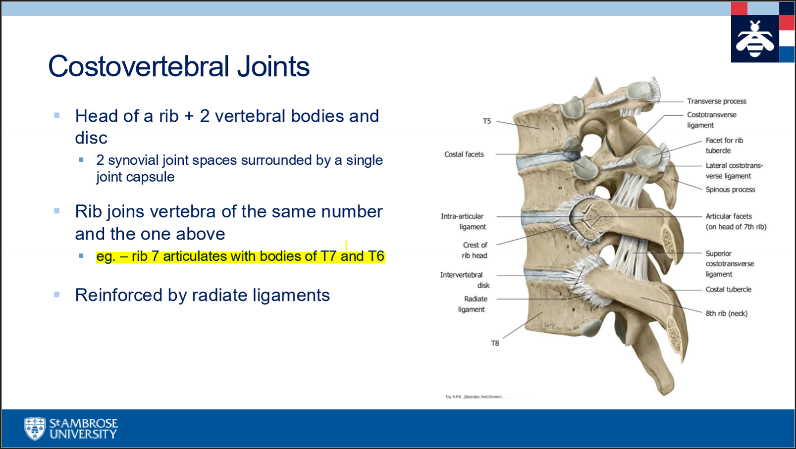
Costovertebral
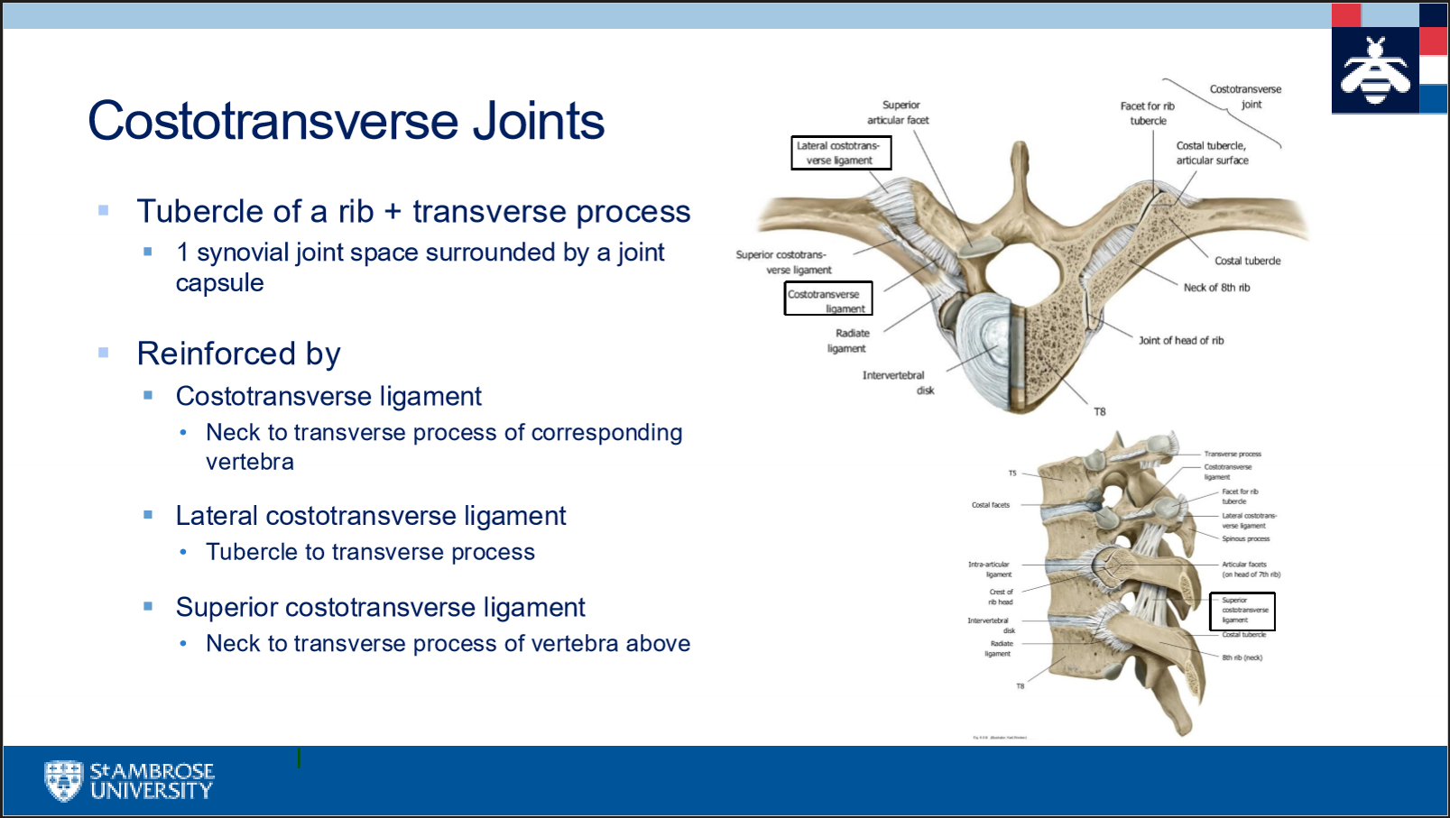
costo transverse
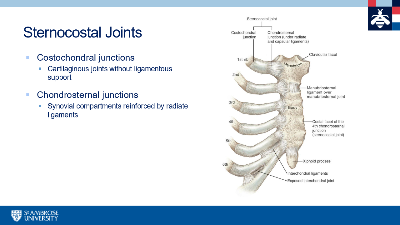
costochondral junction
end of rib to costal cartilage
sternocostal
sternum to costal cartilage
Rib motion during respiration
sternum moves forward and upward during inhalation
A-P dimension of the upper thorax increases ( pump handle)
M-L dimension of the lower thorax increases (bucket handle )
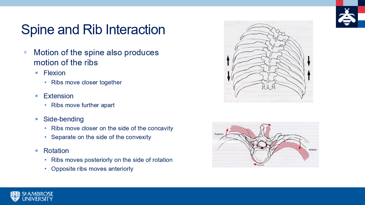
Spine and Rib interaction
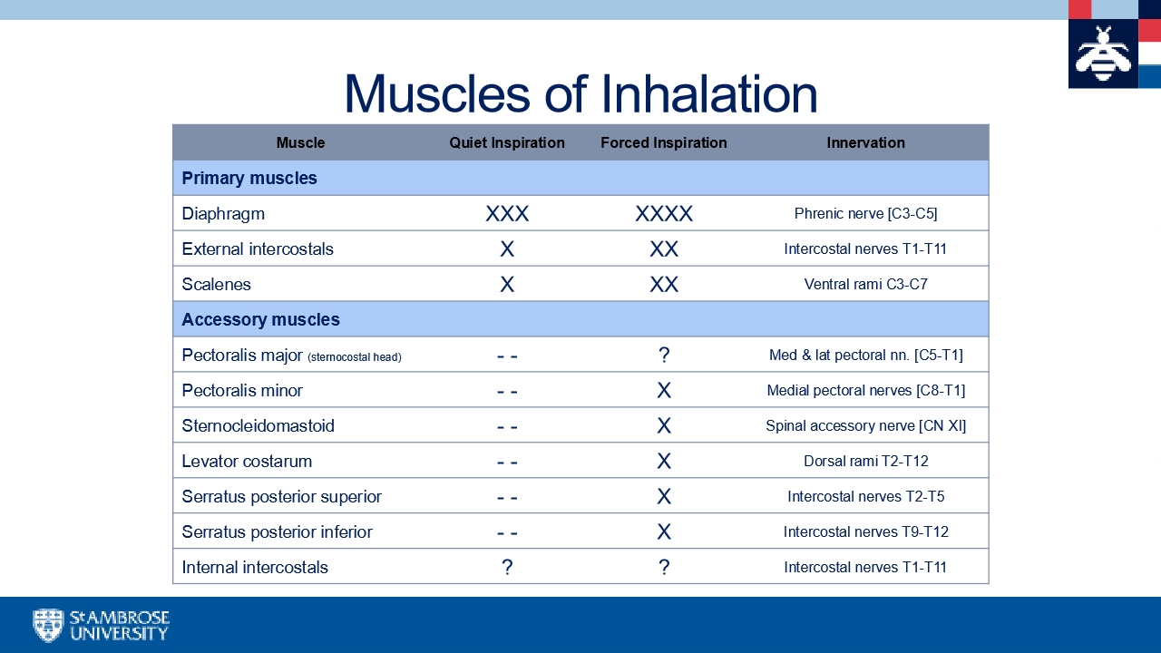
Muscles of inhalation
Diaphragm
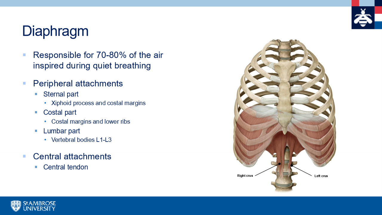
central tendon of the diaphragm

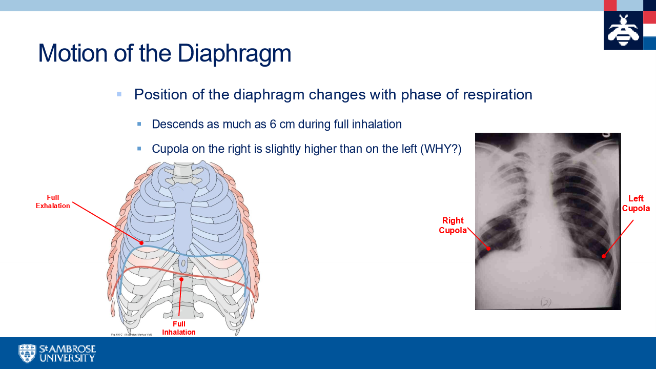
Motion of the diaphragm
Apertures of the diaphragm
opening s that allow passage of structures between thorax and abdomen
Aortic hiatus, esophageal hiatus, caval opening
Aortic hiatus
Aorta, thoracic duct, azygos vein
esophageal hiatus
esophagus, anterior and posterior vagal trunks
caval opening
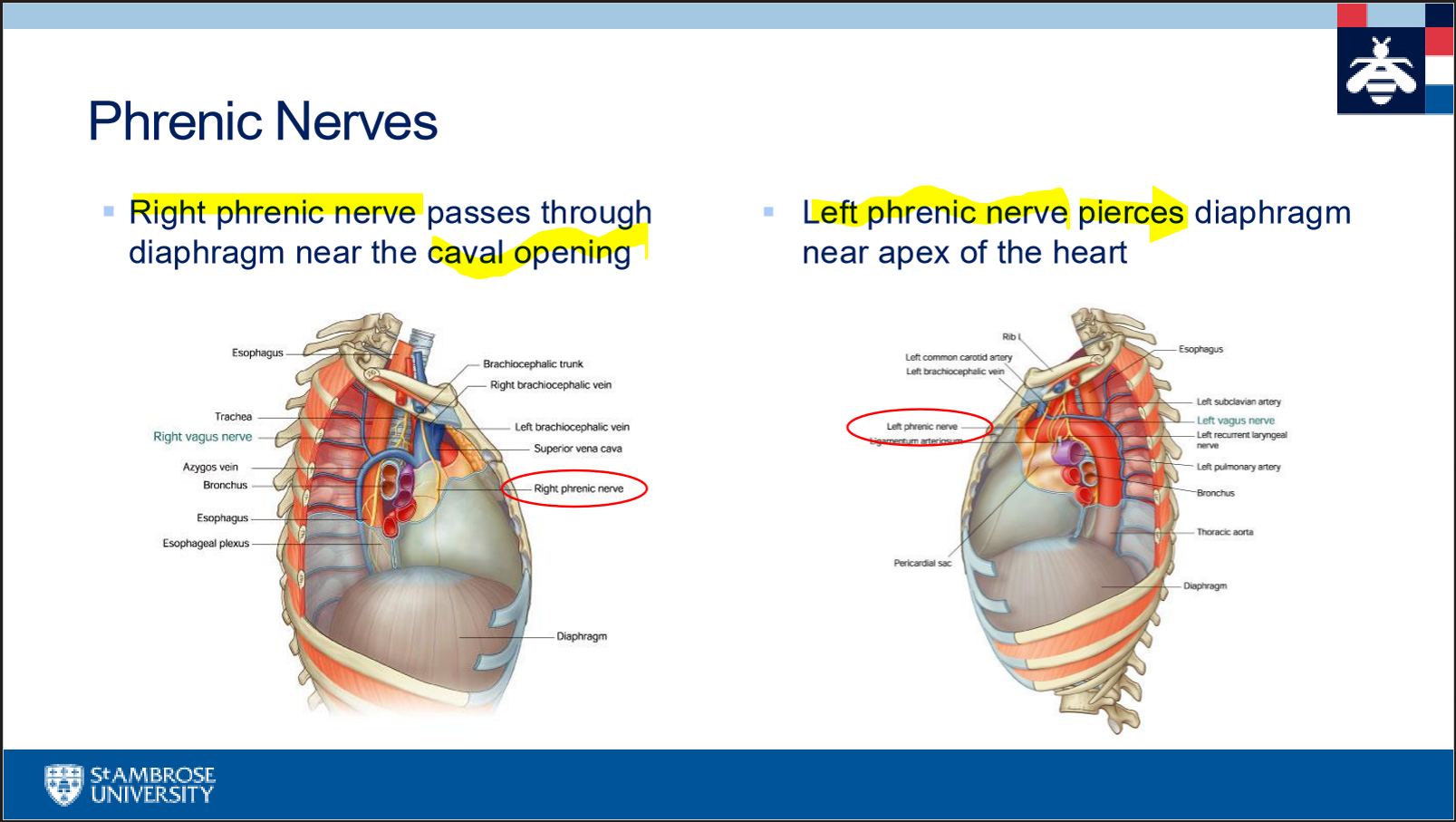
inferior vena cava and right phrenic nerve
innervation and blood supply of the diaphragm
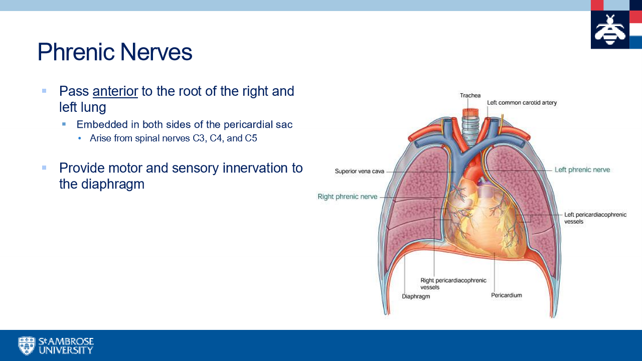
Phrenic nerves (C3-5) pierce the diaphragm and inervate it from its abdominal surface
inferior phrenic arteries first paired arteries that arise from the abdominal aorta
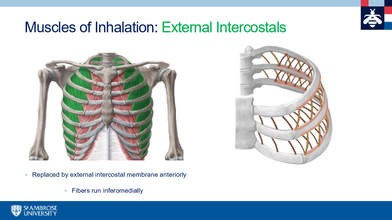
muscles of inhalation
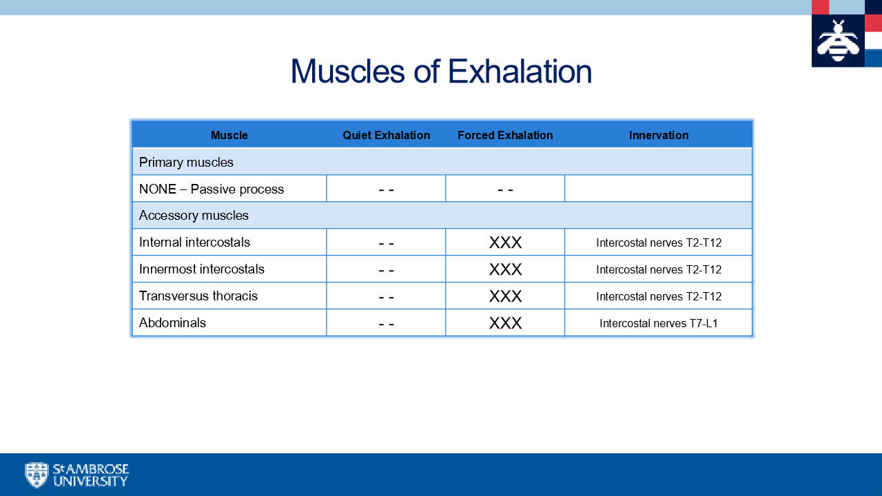
muscle of exhalation
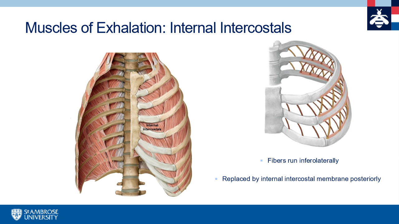
internal intercostals
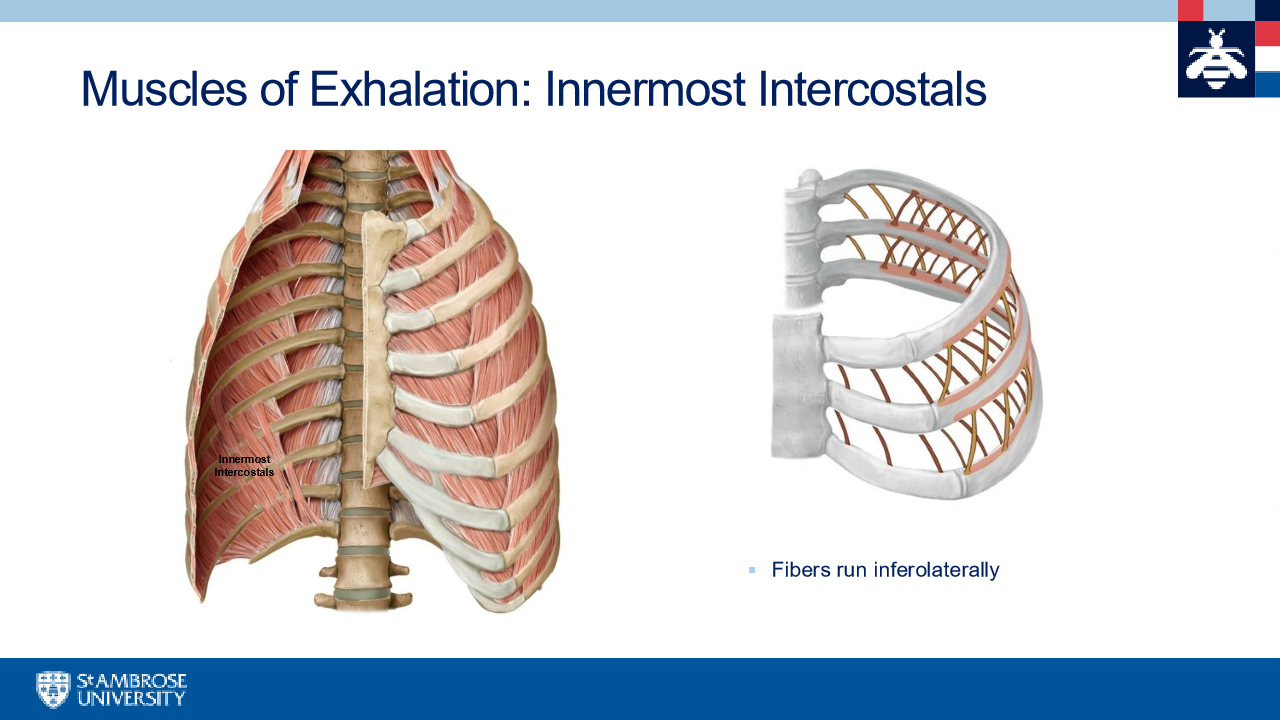
innermost intercostals
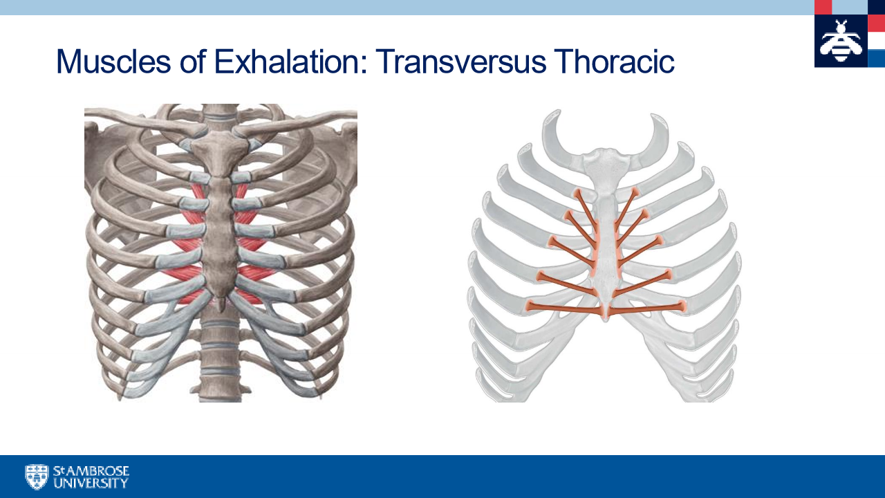
transversus thoracic
deep seen when flip ribcage over
muscles of pectoral region
pectoralis major, pectoralis minor, subclavius
pectoralis major
1 clavicular head and 2 sternocostal head
pectoralis minor
coracoid process and ribs 3-5
subclavius
rib 1 to anterior clavicle
nerves of the thoracic wall
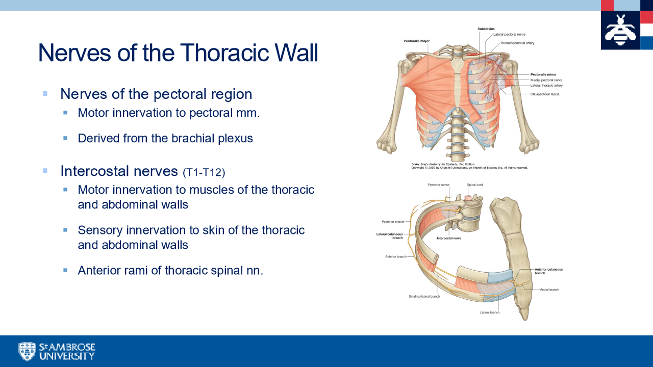
pectoral nerves
lateral and medial pectoral nerves
lateral pectoral nerve (C5-C7)
derived from lateral cord of the brachial plexus
emerges at upper medial border of pectoralis major along with pectoral artery
innervated pectoralis major
median pectoral n (C8-T1)
derived from medial cord of the brachial plexus
usually pierces through pec minor
Supplies pectoralis minor and major
intercostal nerves
all other spinal nerves correlrte to their vertebrae
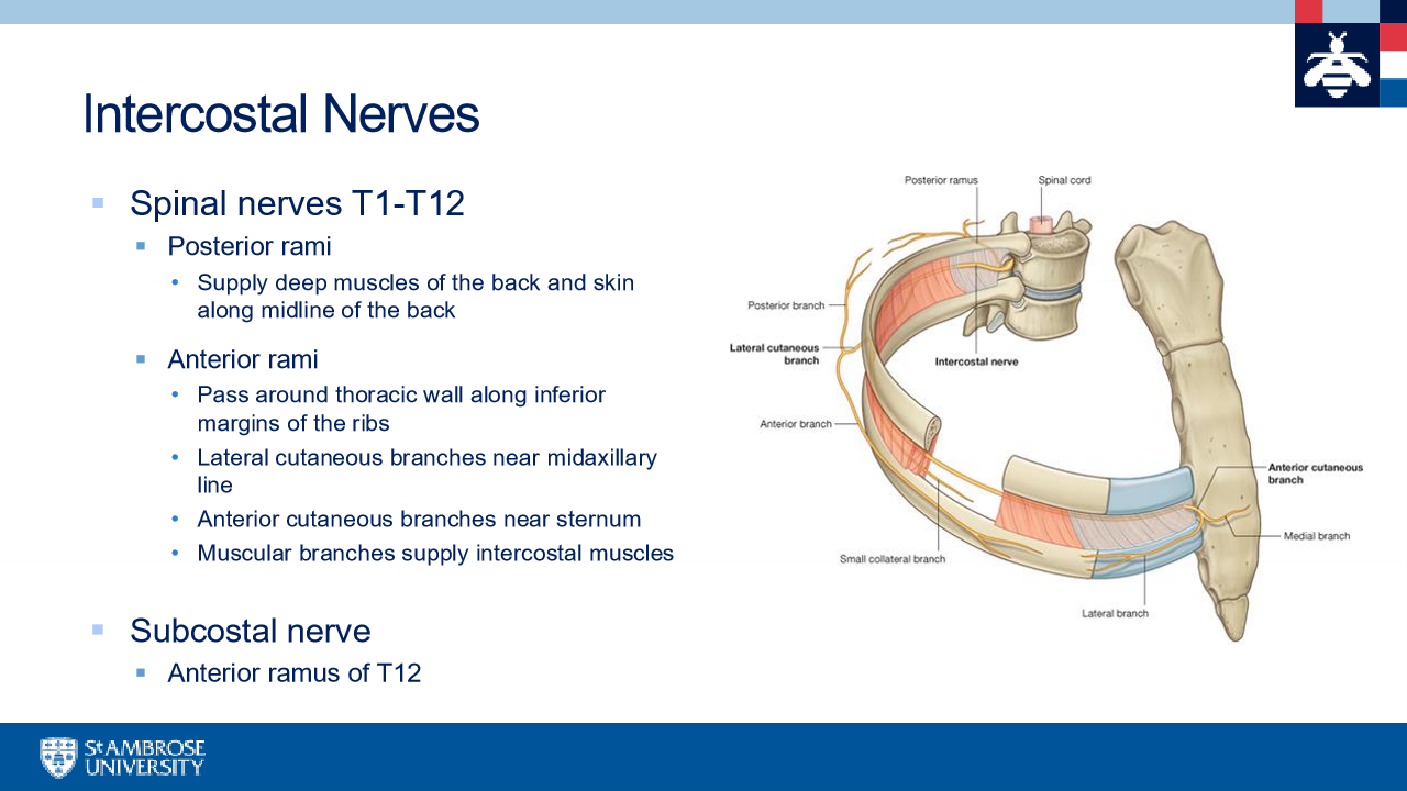
key dermatomes
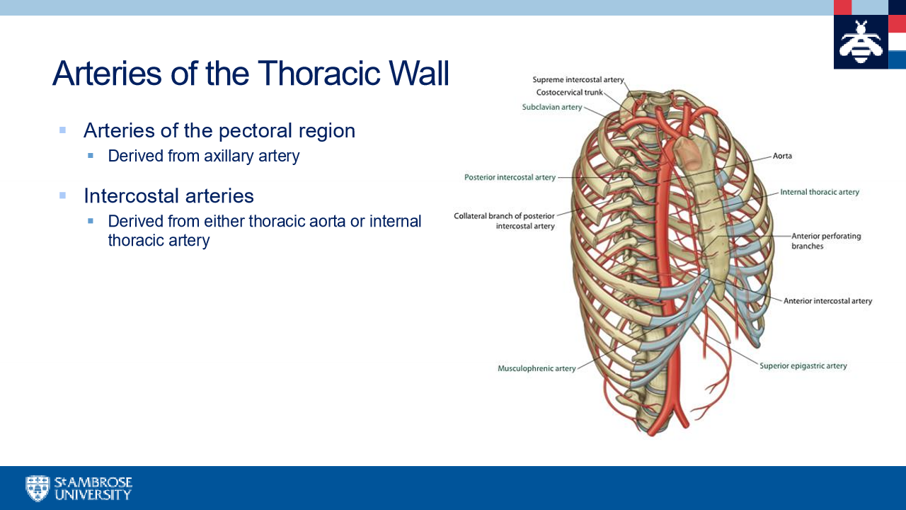
arteries of the thoracic wall
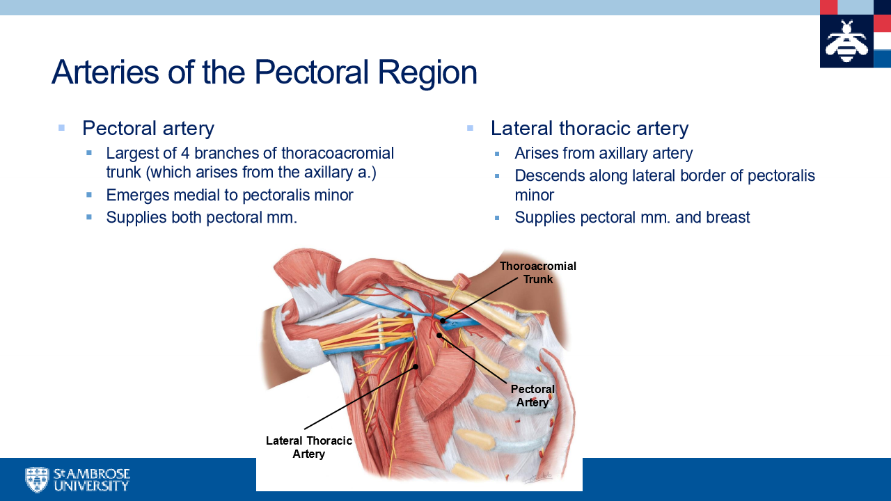
arteries of the pectoral region
pectoral artery runs with lateral pectoral nerve
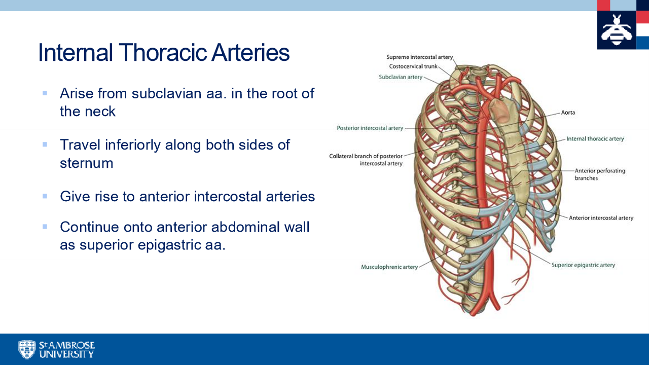
internal thoracic arteries
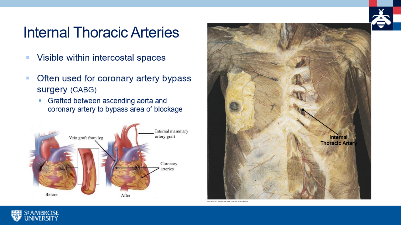
What is interanl thoracic arteries used for
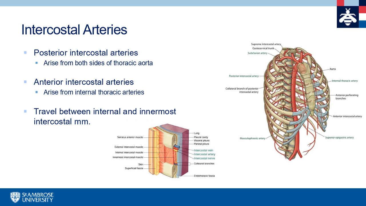
intercostal arteries
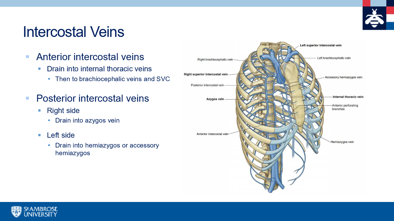
intercostal veins
Azygos system of veins
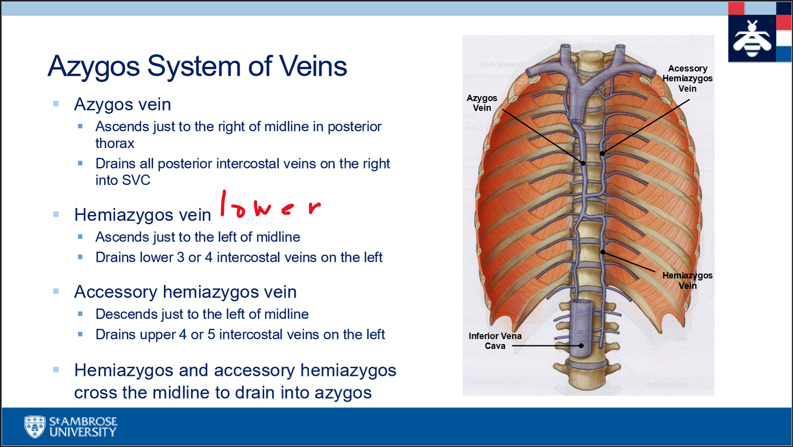
acessory =upper
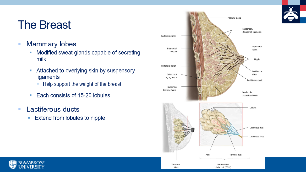
The breast
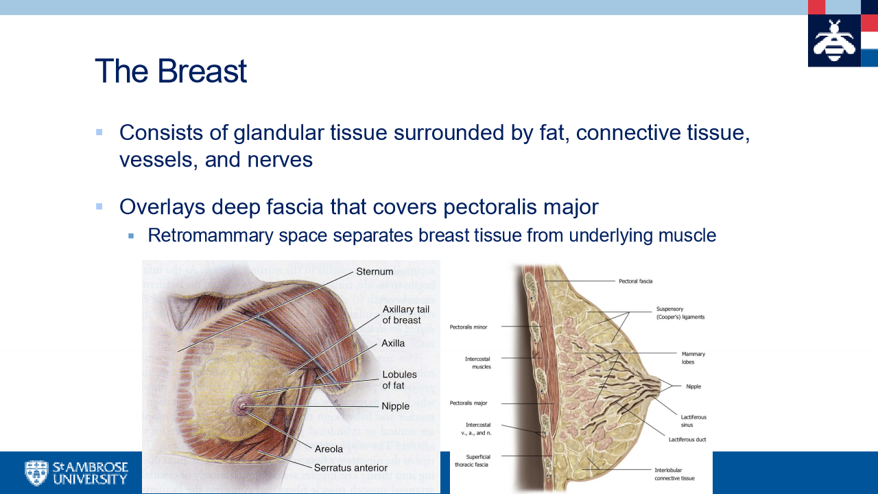
Retromamillary spcae
space separates breast tissue from underlying muscle
Mamillary lobes and lactiferous ducts
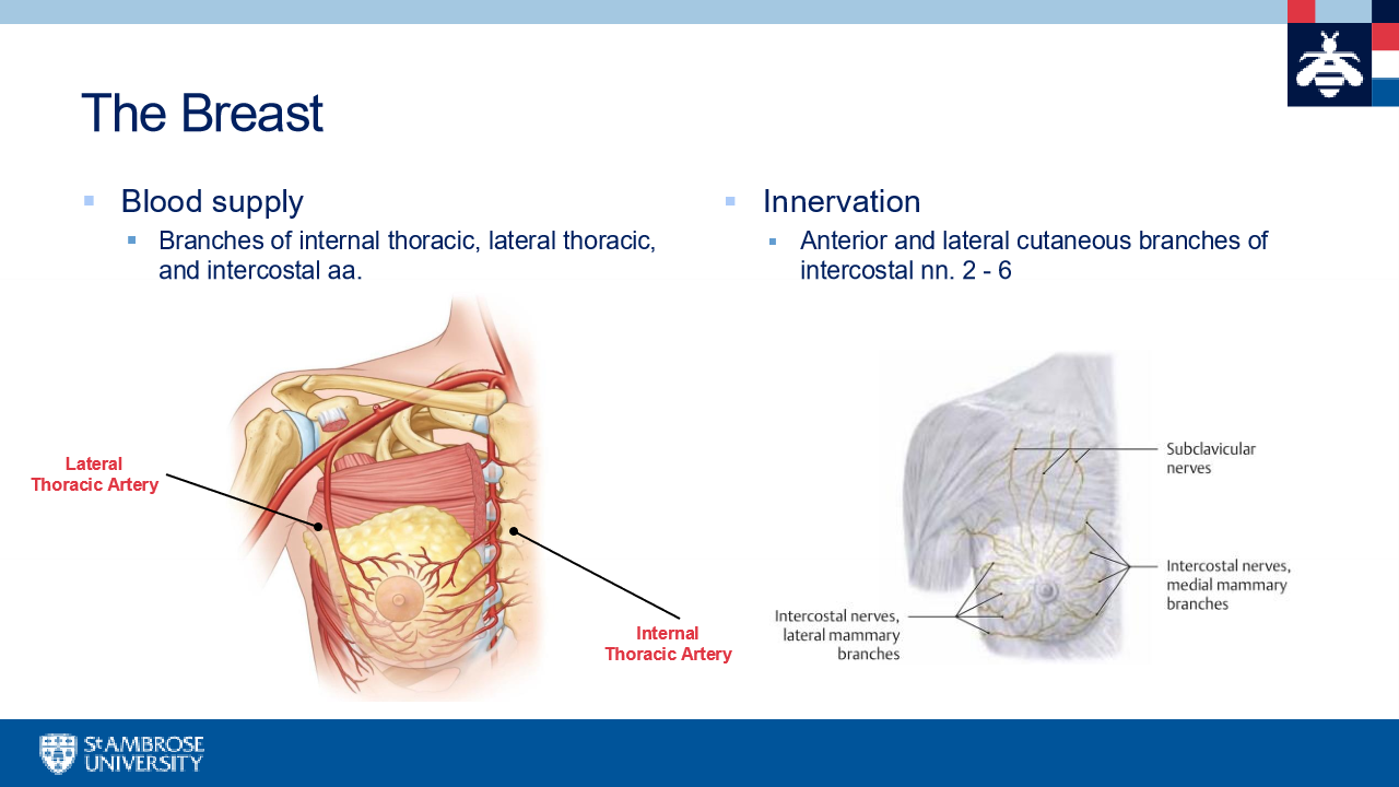
breast blood supply and innervation
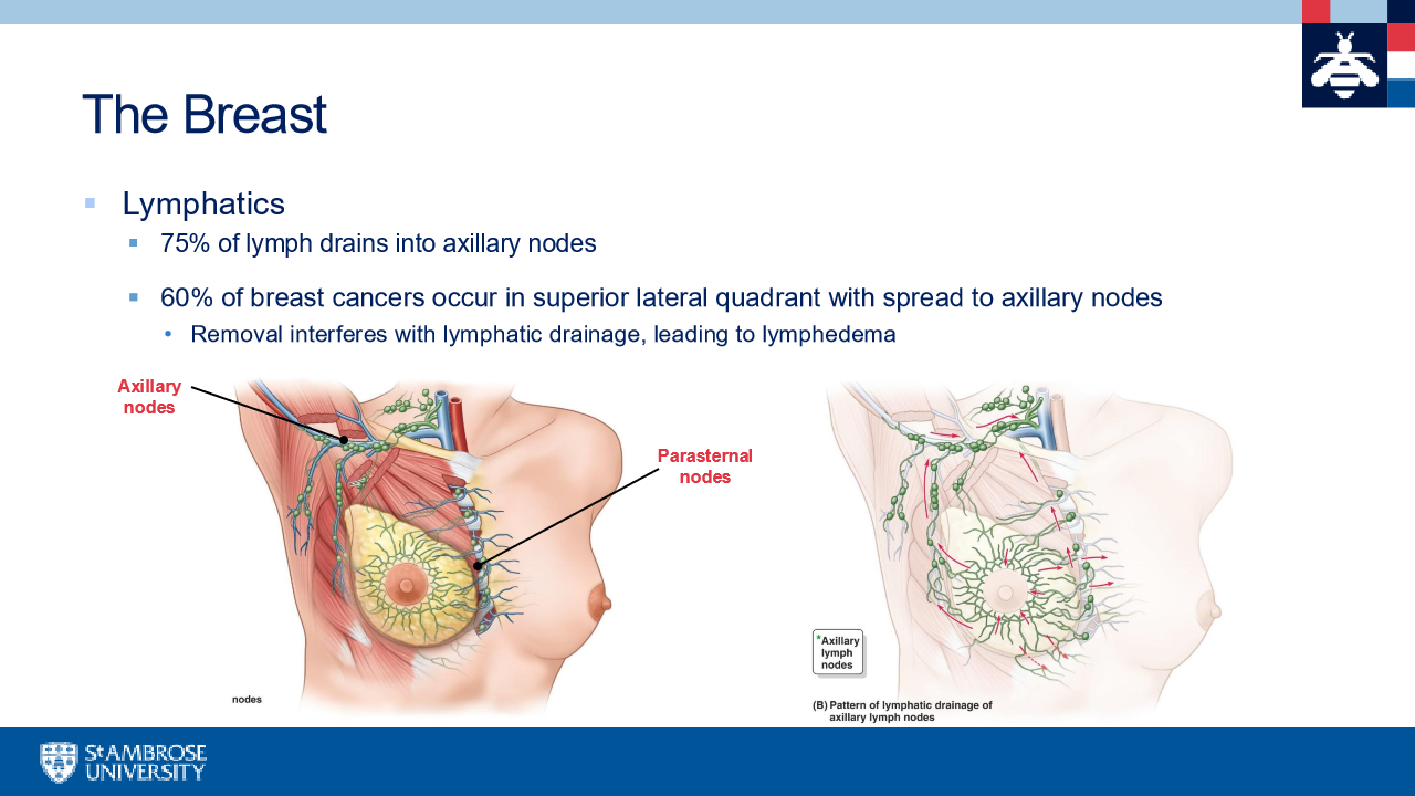
lymphatics in breast
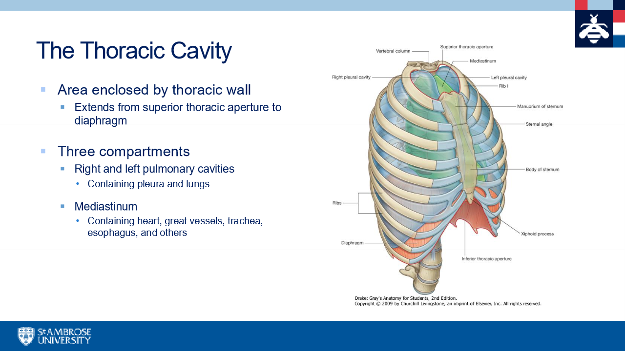
The Thoracic cavity
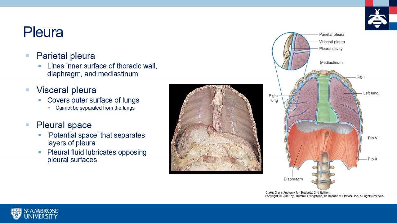
Pleura
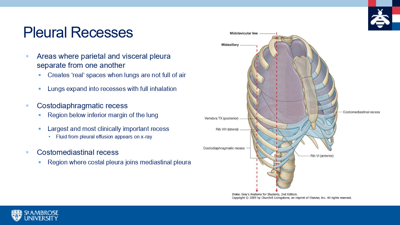
pleural recesses
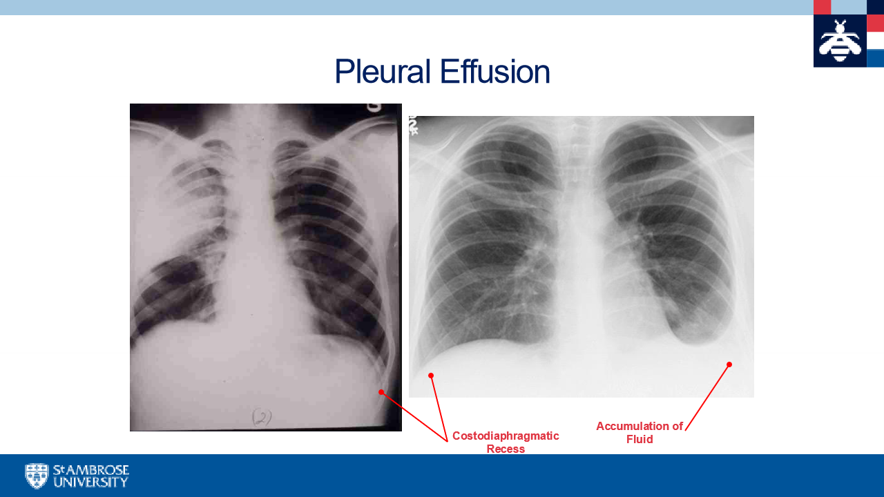
pleural effusion
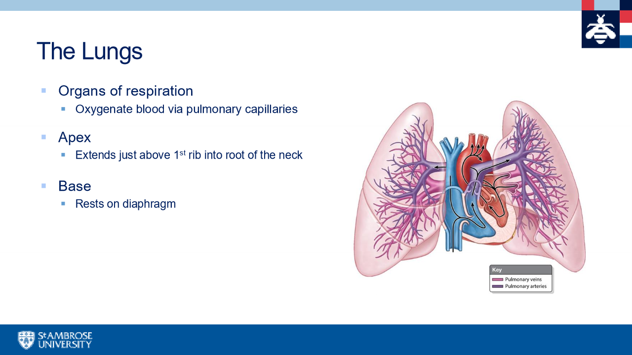
The lungs
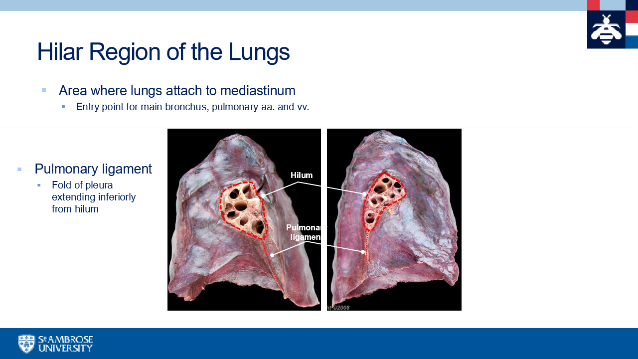
Hilar region of the lungs
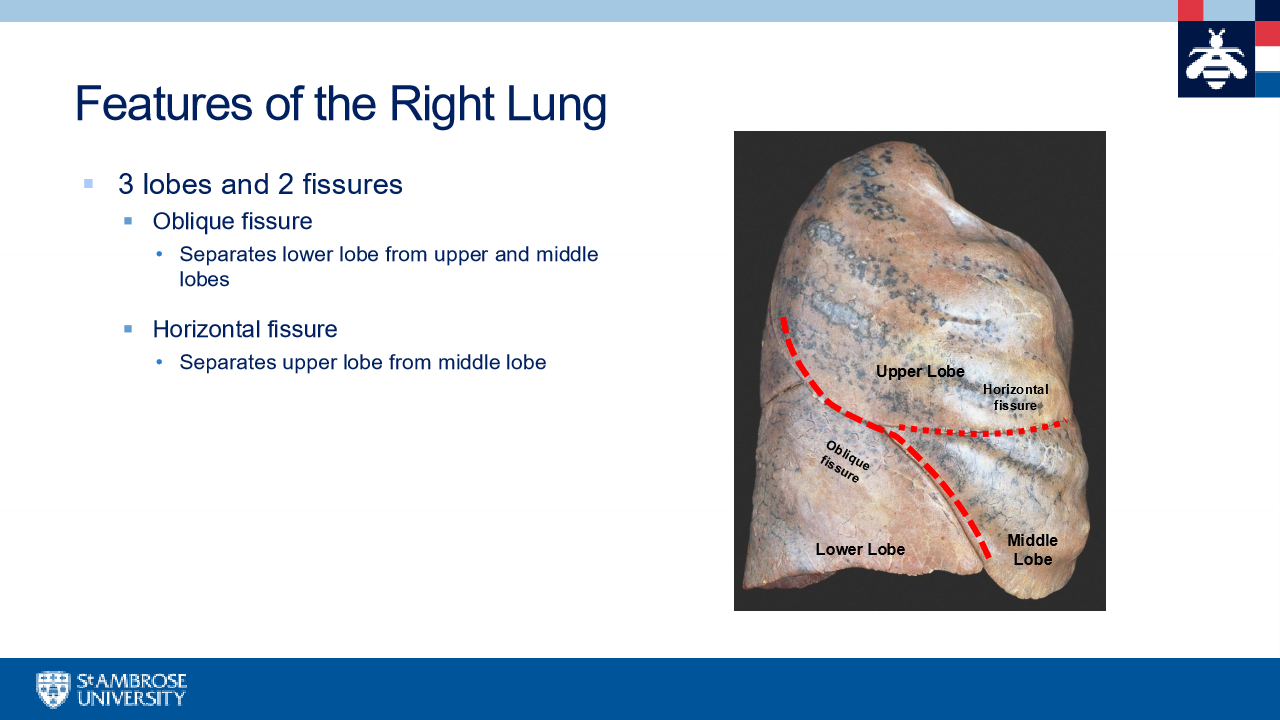
features of the right lung
costal, cardiac, esophageal, and venous impressions(IVC and arch of azygos vein)
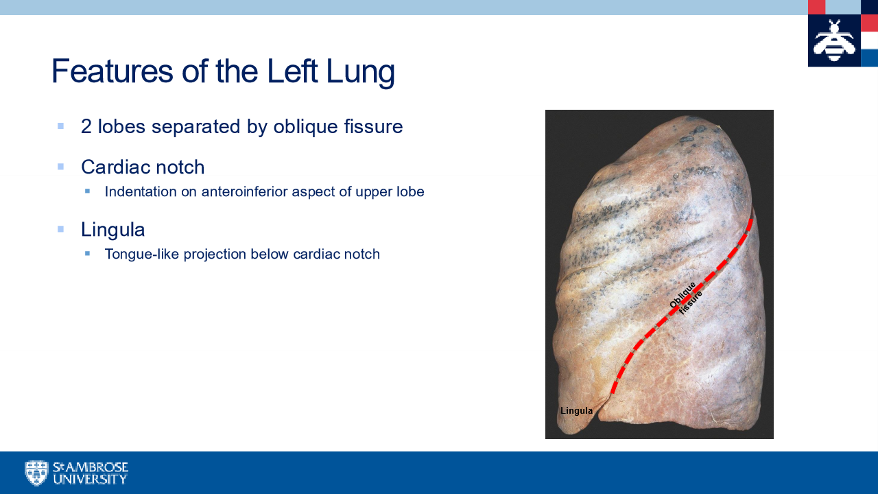
features of the left lung
costal impressions, aortic arch, thoracic aorta, cardiac impressions,
pulmonary artery is superior to bronchus in the left lung Remeber RALS
RALS:
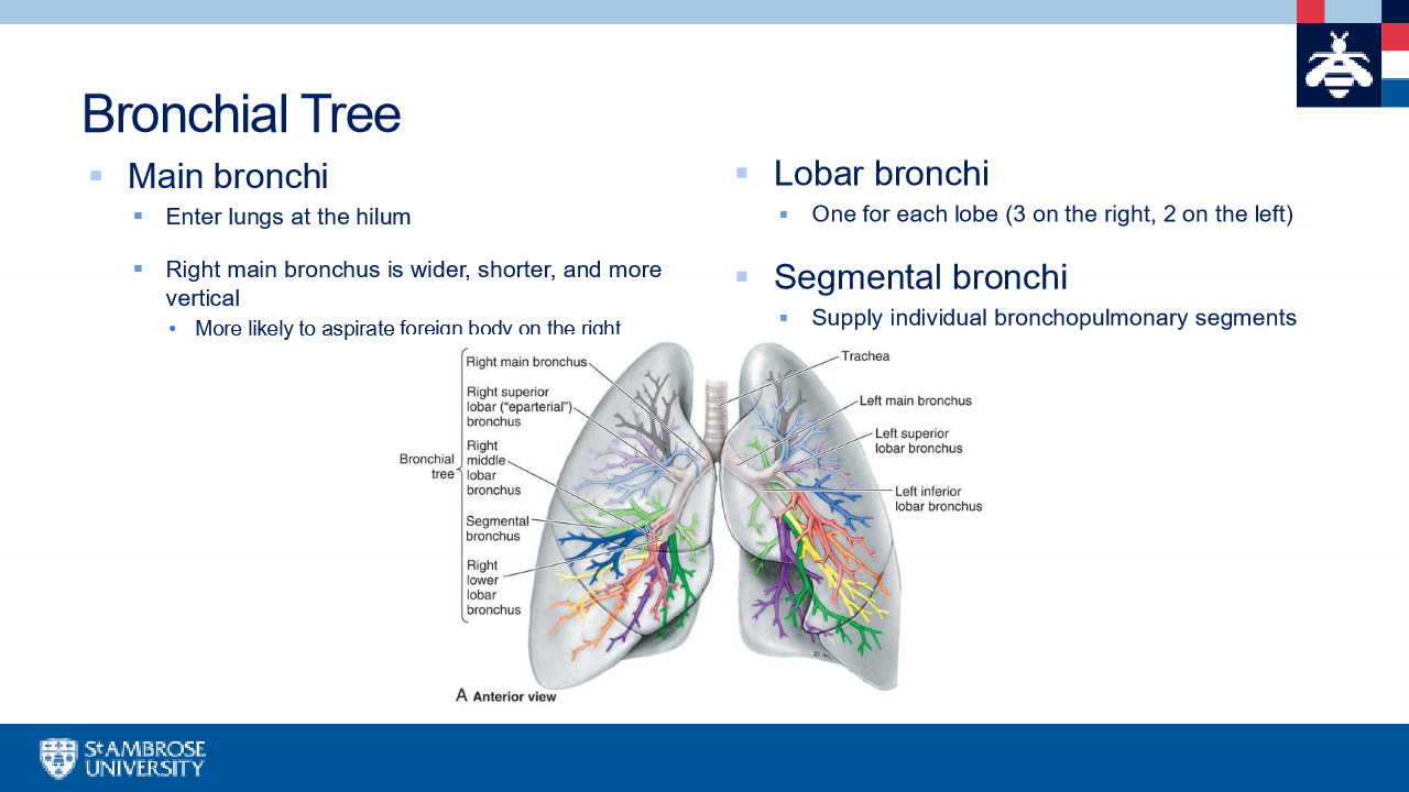
bronchial tree
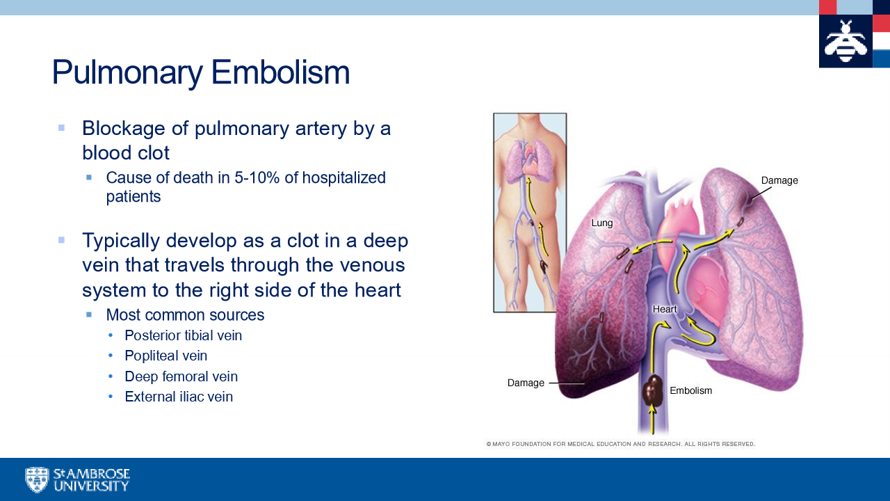
pulmonary embolism
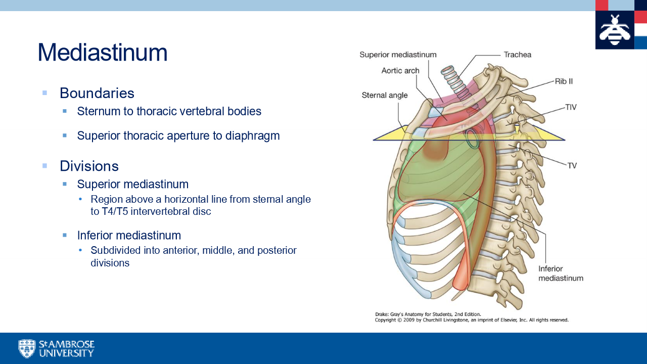
mediastinum
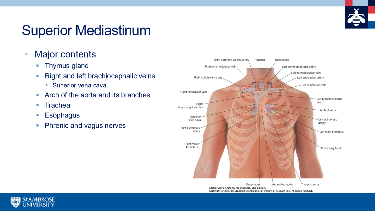
superior mediastinum
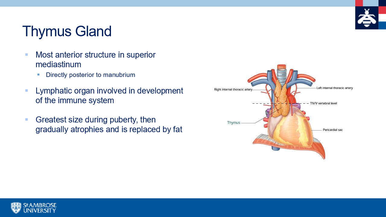
thymus gland
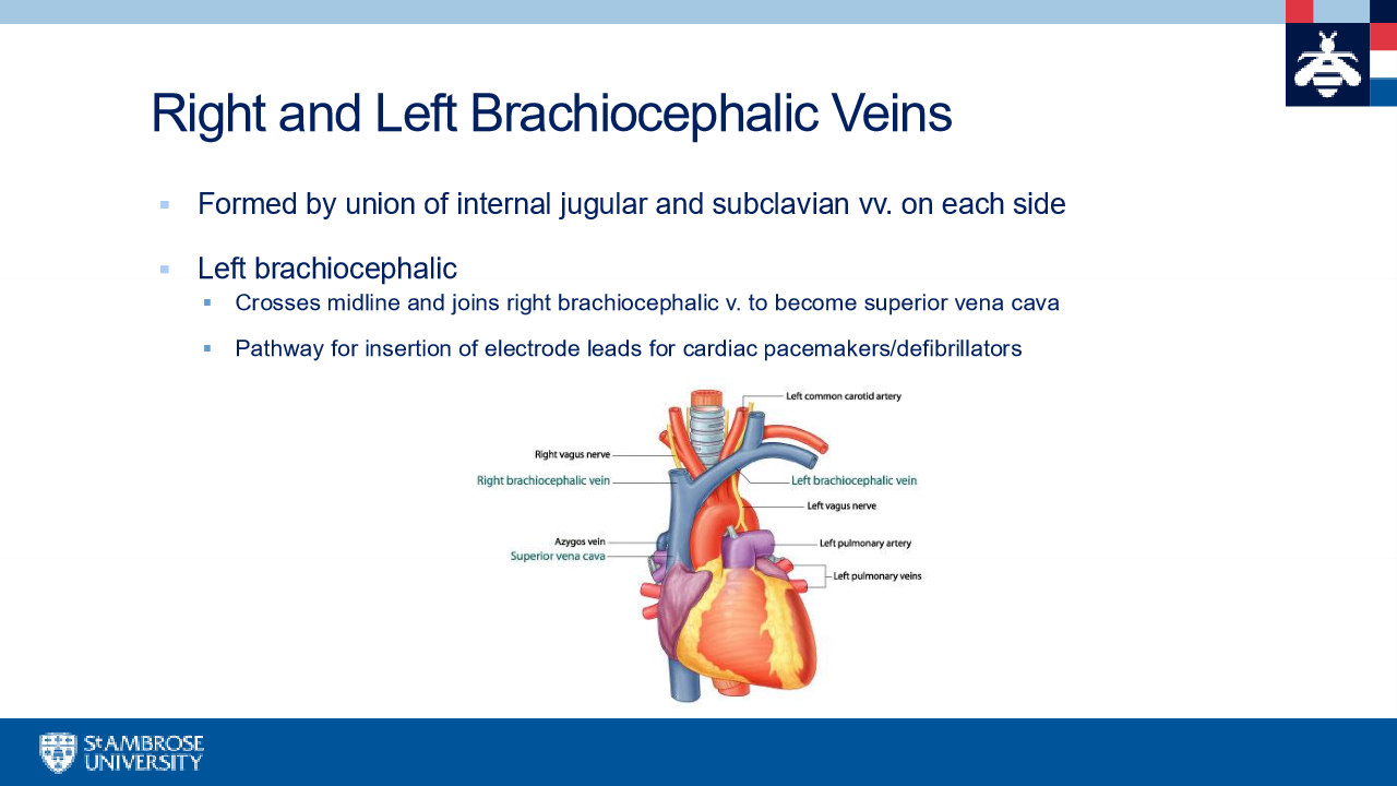
RL brachiocephalic veins
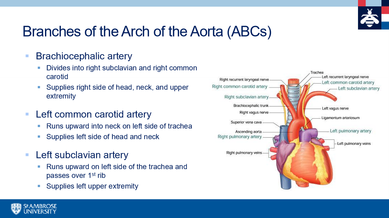
branches of arch of the aorta
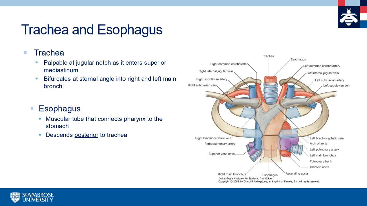
trachea and esophagus
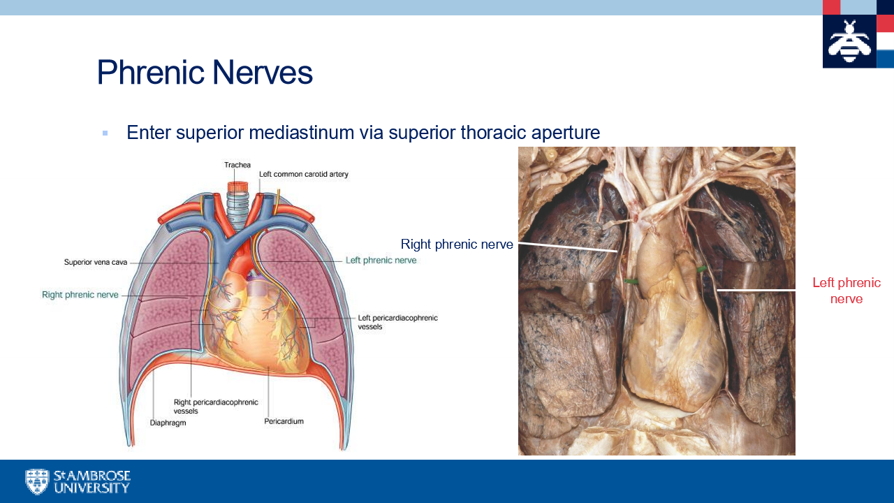
phrenic nerves
Vagus nerves (cranial nerve X)
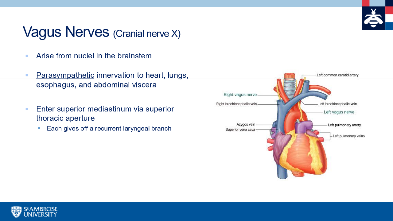
Right vagus nerve

left vagus nerve
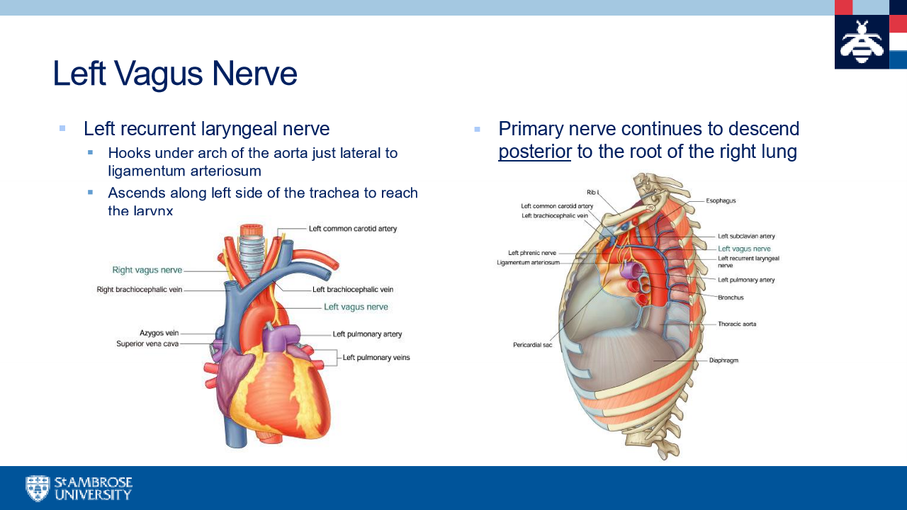
another name for the left and right vagus nerve
recurrent laryngeal nerves
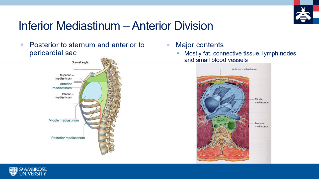
divisions of the the inferior mediastinum
anterior middle and posterior
inferior medistinum- anterior division
inferior mediastinum-middle divison
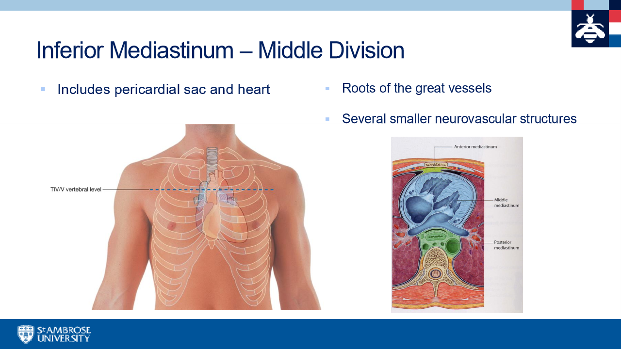
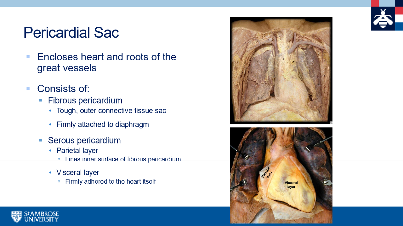
pericardial sac
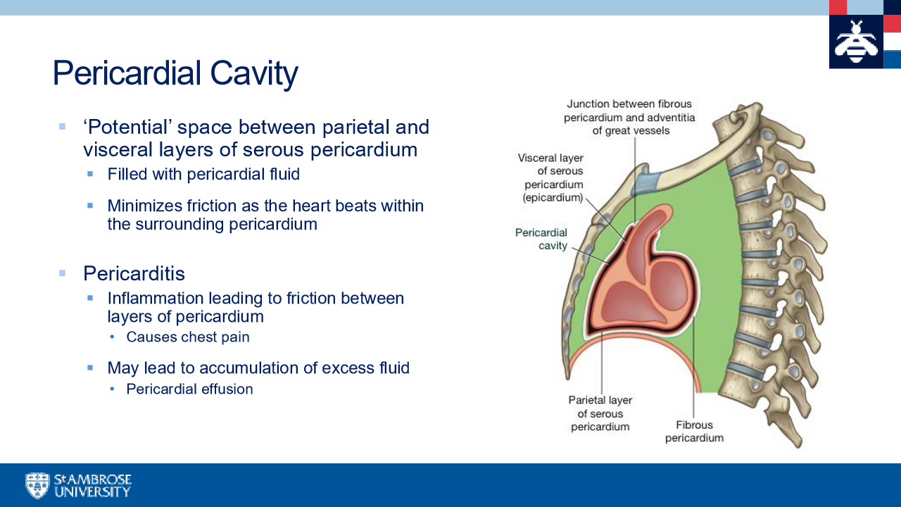
pericardial cavity
phrenic nerves
phrenic nerves
heart
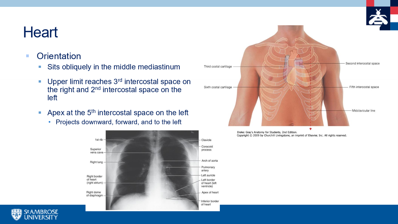
surfaces of the heart
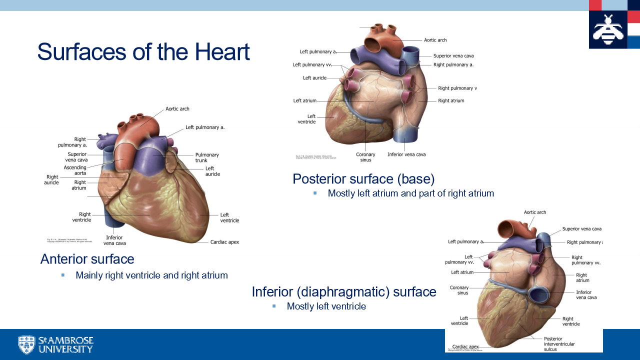
external sulcus of the heart
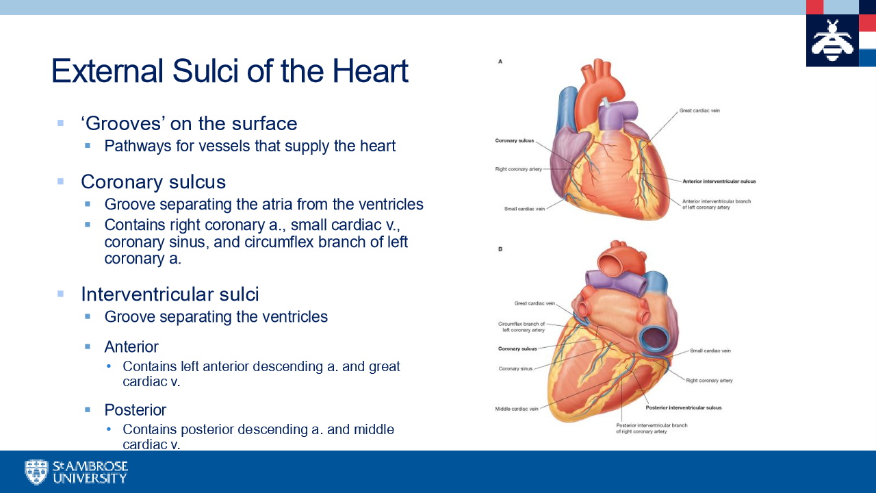
coronary circulation
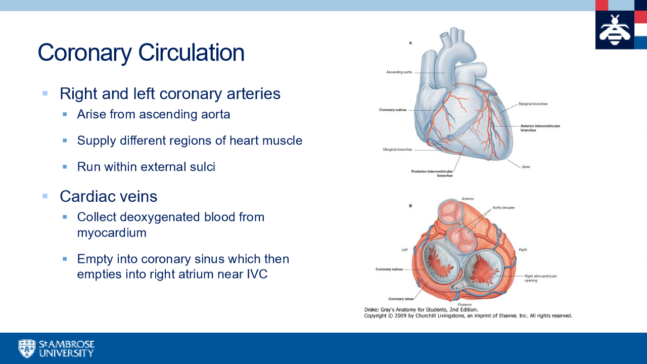
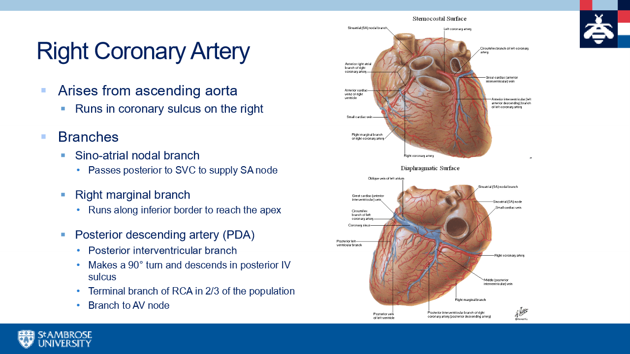
Right coronary artery
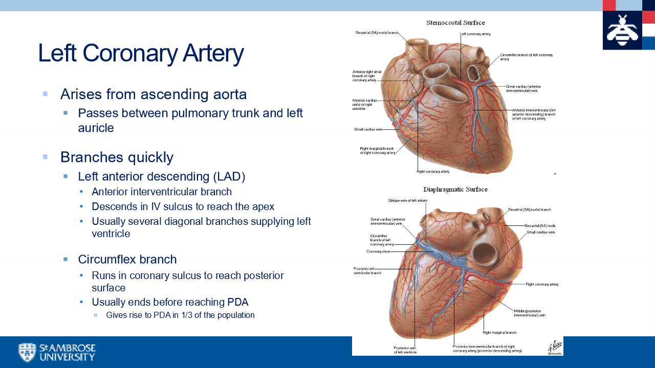
left coronary artery (realy tiny before branching)
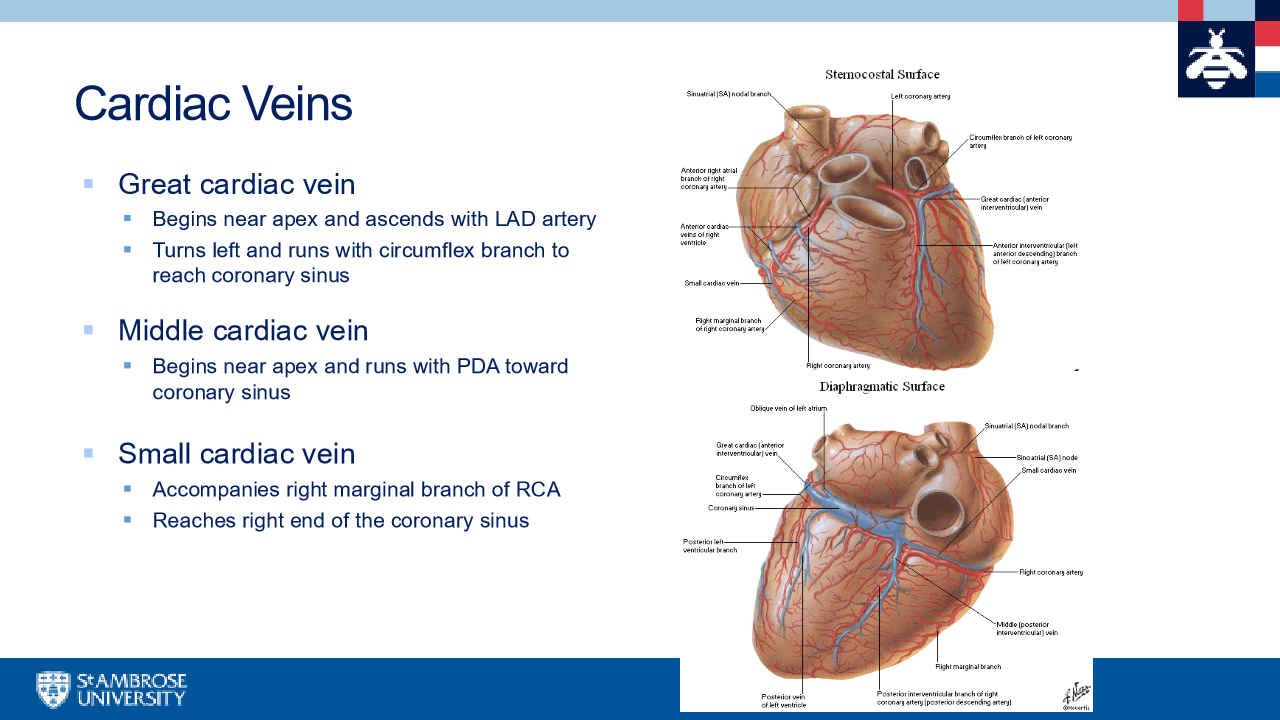
cardiac Veins
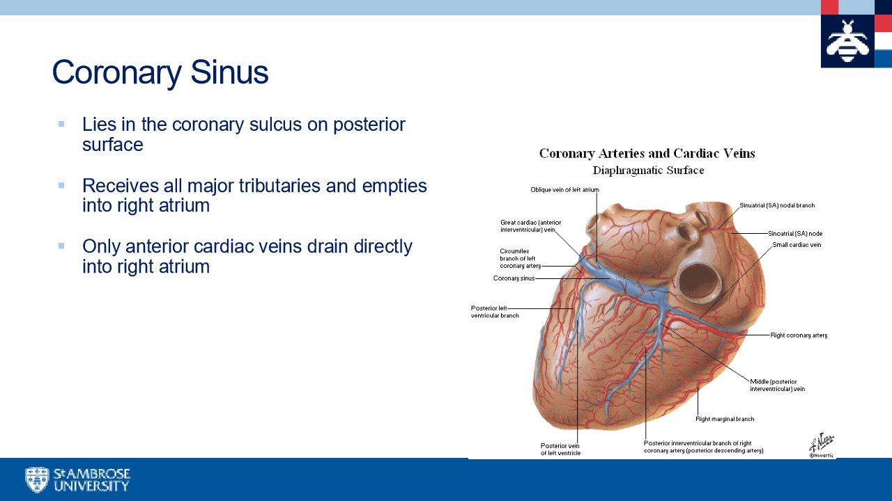
coronary sinus
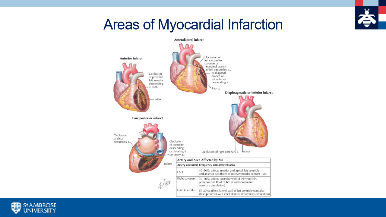
areas of myocardial infraction
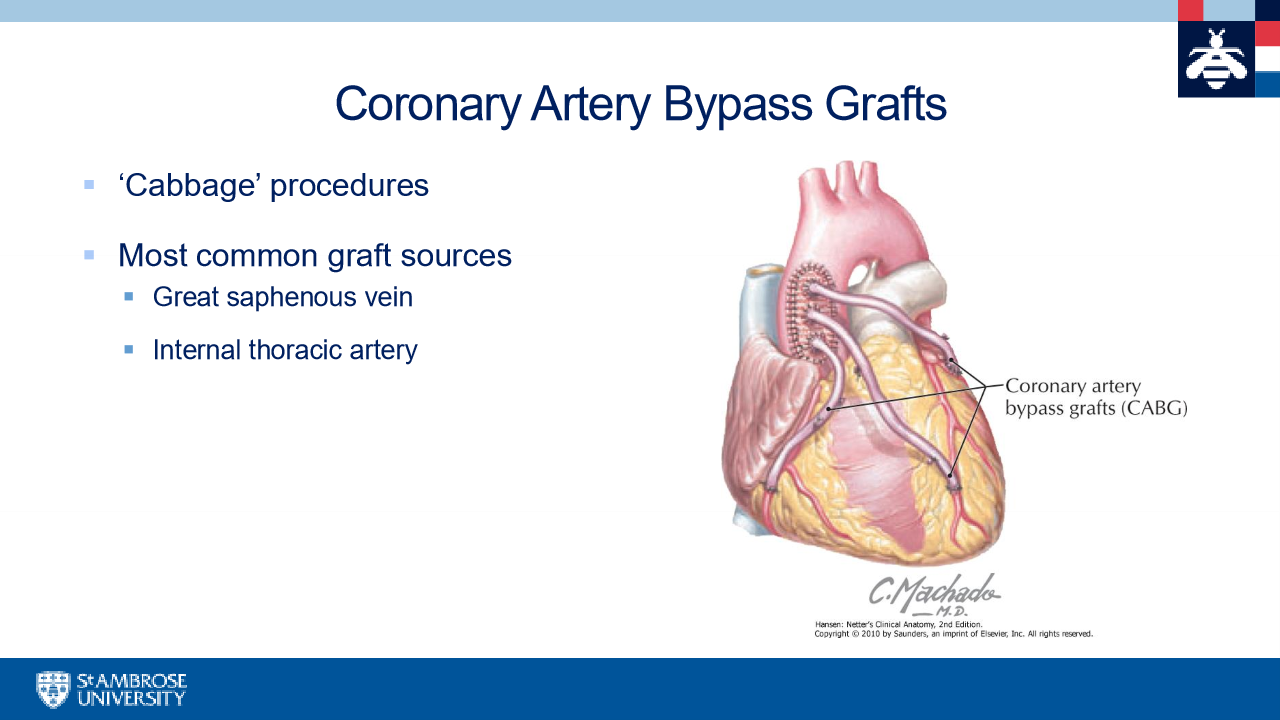
coronary artery bypass grafts
walls of the heart
epicardium, myocardium, endocardium
epicardium
thin layer formed by visceral later of pericardium
myocardium
thick middle layer
Acts as the motor for pumping action
endocardium
thin inner layer,lines chambers of the heart
chambers of the heart
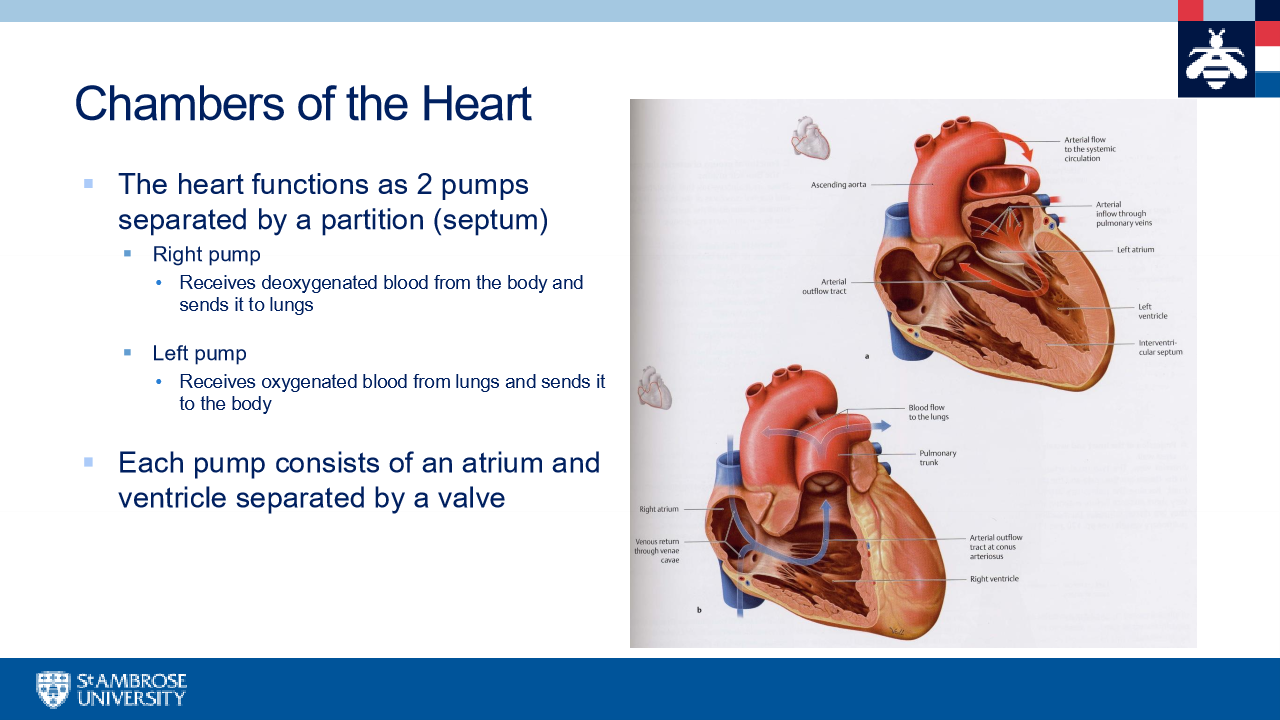
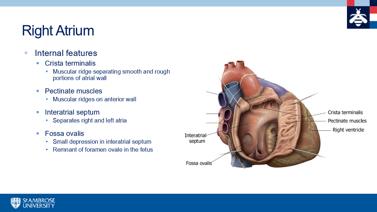
Right atrium
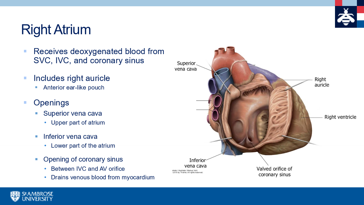
right atrium pt 2
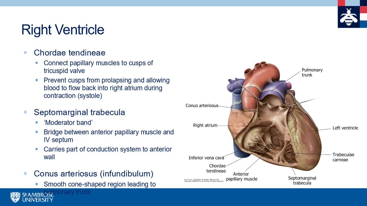
Right ventricle
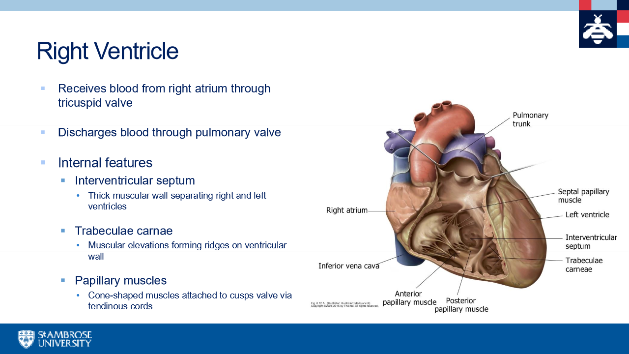
right ventricle pt 2