Alteration in Perfusion: Sinus Rhythms
 Call Kai
 Learn
Learn Practice Test
 Spaced Repetition
 Match
Match Flashcards
 Knowt Play
1/35

There's no tags or description
Looks like no tags are added yet.
Name | Mastery | Learn | Test | Matching | Spaced | Call with Kai | Chat |
|---|
No analytics yet
Send a link to your students to track their progress
36 Terms
Mechanism of Normal Sinus Rhythm
The sinus node initiates regular impulses at a normal rate. Each impulse is conducted normally to the ventricles.
Pacemaker: sinus node
Rates: 60-100 bpm
Regularity: regular
Conduction: normal; each impeller is conducted through to the ventricles
Rules for Normal Sinus Rhythm
Regularity: Regular
Rate: 60–100 bpm
P Wave: Uniform shape; one P wave in front of every QRS complex
PRI: 0.12–0.20 second and constant
QRS: Less than 0.12 second
Rules for Normal Sinus Rhythm
Regular, rate 90, P are upright (positive), sinus rhythm (60-100)
PR: 0.16
QRS: 0.08

Mechanism of Sinus Bradycardia
The sinus node is the pacemaker, firing regularly at a rate of less than 60 times per minute. Each impulse is conducted normally through the ventricles.
Pacemaker: sinus node
Rates: <60 bpm
Regularity: regular
Conduction: normal; each impeller is conducted through to the ventricles
Rules for Sinus Bradycardia
Regularity: Regular
Rate: Less than 60 bpm
P Wave: Uniform shape; one P wave in front of every QRS complex
PRI: 0.12–0.20 second and constant
QRS: Less than 0.12 second

Sinus Bradycardia
Cause: vagal stimulation- causes decreased sinus node firing.
Can reflect normal, athletic heart
Can be dangerous if suctioning patient.
Sinus Bradycardia can occur with
Can occur with bowel movements (vasovagal), inferior wall MI, ocular pressure, hypothyroidism, increased intracranial pressure, carotid massage (pressing on both carotid)
May be related to Hs and Ts
Hypoxia:
low oxygen levels in the blood
Hypovolemia
low amount of circulating blood
Hyperkalemia or hypokalemia
and related disturbances of calcium or magnesium levels.
Hypothermia/Hyperthermia
body temperature not maintained
Hydrogen ions (Acidosis):
Sodium bicarbonate or bronchodilators, vents
Hypoglycemia
Tension pneumothorax:
increased pressure in the thoracic cavity, leading to decreased venous return to the heart
Tamponade
fluid or blood in the pericardium, compressing the heart
Toxic:
chemicals, whether medication or poisoning
Thromboembolism and related mechanical obstruction
(blockage of the blood vessels to the lungs or the heart by a blood clot or other material)
Assessment/Treatment
Dizziness, syncope, confusion, hypotension, chest pain or SOB
ECG changes may occur
Treat causes: Oxygen, atropine 0.5 to 1 mg IV push (0.6 mg over 1 min) not slower
If no response and HR < 45 bpm, prepare for temporary or permanent pacemaker
Mechanism of Sinus Tachycardia
The sinus node is the pacemaker, firing regularly at a rate of greater than 100 bpm. Each impulse is conducted normally through the ventricles.
Pacemaker: sinus node
Conduction normal, each impulse is conducted through to the ventricles
Rules for Sinus Tachycardia
Regularity: Regular
Rate: Greater than 100 bpm (usually does not exceed 160 bpm)
P Wave: Uniform shape; one P wave in front of every QRS complex
PRI: 0.12–0.20 second and constant
QRS: Less than 0.12 second
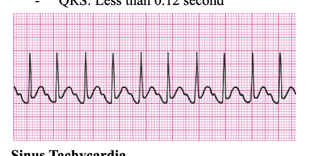
Sinus Tachycardia
Clinical associations
Physiologic stressors
Exercise
Pain
Hypovolemia
Myocardial ischemia (can extend infarct)
Heart failure (HF)
Fever
Anxiety
Sinus Tachycardia clincial manifestations
Rapid, regular pulse
Possibly asymptomatic
Palpitations, dyspnea
Dizziness and hypotension due to decreased CO
Increased myocardial oxygen consumption may lead to angina
Sinus Tachycardia treating the underlying cause
administer pain meds, reduce fever, administer oxygen
Clients on ventilators may need to be suctioned and oxygenated.
Sinus Tachy: Medications to slow heart rate
Beta blockers (olol)-propranolol, metoprolol
Ca channel blockers –nifedipine, diltiazem
Mechanism of Sinus Arrhythmia
The sinus node is the pacemaker, but impulses are initiated in an irregular pattern.
The rate increases as the patient breathes in and decreases as the patient breathes out (has to be mentioned in the questions!!!). Each impulse is conducted normally through the ventricles.
Normally in young people, can grow out of it

Rules for Sinus Arrhythmia
Regularity: Irregular
Rate: 60–100 bpm (usually)
P Wave: Uniform shape; one P wave in front of every QRS complex
PRI: 0.12–0.20 second and constant
QRS: Less than 0.12 second
Sinus Arrhythmia
Rate increases as the patient breathes in and decreases as the patient exhales.
Cyclical speeding up and slowing down of rhythm
Sinus Arrhythmia monitor strips
Monitor strip will show periods or short R-R intervals then long R-R intervals.
rarely causes symptoms, Treated when it causes symptoms
Sinus Arrhythmia is common in
children, teenagers and young adults
Collaborative Care Principles
Arrhythmias are treated when they cause (or are likely to cause) clinical symptoms.
Identify and correct hypoxia, acidosis, hypovolemia, etc.
All patients with arrhythmias (or the potential for them)
should be monitored, receive oxygen, and have a keep-open IV as a precaution.
why assess Perfusion
Assess perfusion parameters (BP, pulses, skin, etc.) to determine impact on cardiac output.
perfusion
If the heart is going too fast it isn’t pulling blood out of the heart too slow has not enough blood to the body so perfusion is altered
If there is not enough perfusion then there is not enough oxygen, nutrients, and electrolytes are missing
In addition to treating the specific arrhythmia, you may need to provide
ventilation, chest compression, and/or blood pressure support.
Matrix of Clinical Impact

A patient’s cardiac rhythm is sinus bradycardia with a heart rate of 34 beats/min. If the bradycardia is symptomatic, the nurse would expect the patient to exhibit:
A. Palpitations.
B. Hypertension.
C. Warm, flushed skin.
D. Shortness of breath.