General terms procedures 1
 Call Kai
 Learn
Learn Practice Test
 Spaced Repetition
 Match
Match Flashcards
 Knowt Play
1/123

There's no tags or description
Looks like no tags are added yet.
Name | Mastery | Learn | Test | Matching | Spaced | Call with Kai | Chat |
|---|
No analytics yet
Send a link to your students to track their progress
124 Terms
osteology
study of bone
206 bones
arthrology
study of joints
Axial skeleton
80 bones
skull
verterbral column
ribs
sternum
all bones on or near cental axis
Appendicular skeleton
126 bones
all bones of upper and lower extremities, shoulder, and pelvic girdle
image receptor (IR)
“film”
where the image pops up
Central ray (CR)
X-ray beams ar center point
Sagittal (midsagittal) plane
seperates left and right parts
coronal (midcoronal) plane
Serperates anterior and posterior parts
Horizontal (axial) plane
transverse plane
seperated top and bottom parts
flexion
decreases the angle of the joint
extension
increase the angle of the joint
radial deviation
movement toward radial side (thumb)
ulnar deviation
movement toward ulna side (pinky)
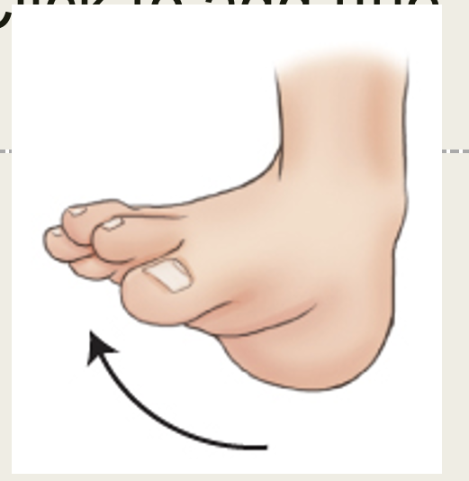
plantar flexion
point toes
dorsiflexion
toes toward knee
eversion (valgus)
forces outward from midline
away
inversion (varus)
forces inward toward midline
inside
medial rotation
turn medial
toward midline
lateral roation
turn lateral
away from midline
abduction
away from midline
adduction
toward the midline
rotation
body stays in line and part turns
tilt
body is not in line
superficial
toward surface of skin
deep
further from skin on inside of body
supine
lay on back
prone
lay on stomach
erect/upright lateral
stanings straight up
recumbent
laying down
supine/ dorsal recumbent
lay on back
prone/ ventral recumbent
lay on stomach
lateral recumbent
lay on side
trendelenburg
feet above head
fowler
feet below head
lithotomy position
legs open
lay on back
head lower then feet

Axial skeleton

Appendicular skeleton

pink plane
coronal plane

green plane
sagittal plane
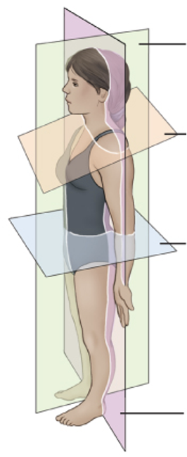
orange plane
oblique plane

blue plane
horizontal plane
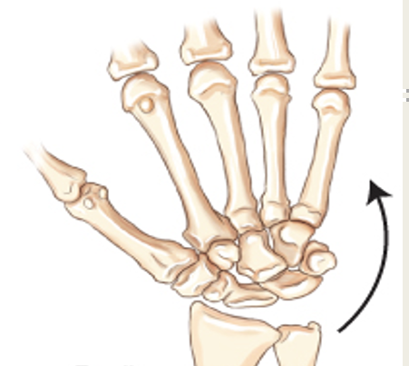
radial deviation

ulnar deviation

Plantar flexion

dorsiflexion

inversion
varus
eversion
valgus

abduction
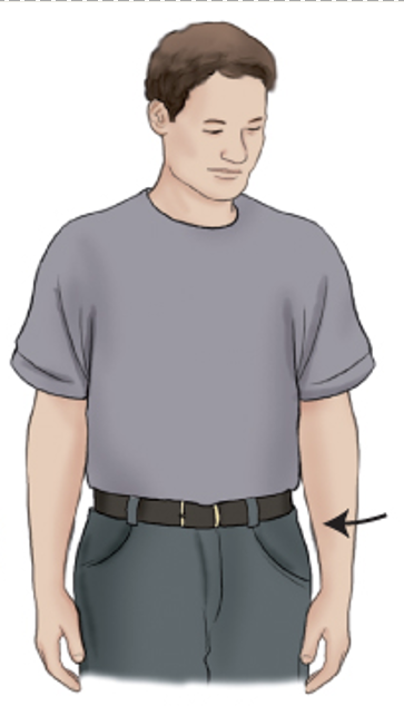
adduction

supine
dorsal recumbent

prone
ventral recumbent
Sesamoid bones
small, oval shaped bones
not part of appendicular or axial skeleton
long bone
only appendicular skeleton
extremities
short bones
wrists and ankles
flat bones
sternum, ribs, scapulae, calvaria
irregular bones
vertebrae, facial bones
synarthrosis joint
immovable
amphiarthrosis joint
limited movement
diarthrosis joint
freely movable
functional joints
synarthrosis
amohiarthrosis
diarthrosis
structral joints
fibrous
cartilaginous
synovial
palpation
process of applying loght pressure with fingertips on patient to locate landmarks
Exception to positiong rule
post reductiion for fracture alignmnet
pelvis study only AP projection unless hip injury
2 projection rule
long bones
location of foreign body
determination of alignment of fractures
3 projection rule
for joints in interest area
AP or PA
lateral
oblique
tangential projection
when CR is skimming part
Caudal CR angles
angle down
cephalic CR angle
angle up
lateromedial projection
projection from laterl to medial
mediolateral projection
projection from medial to lateral
AP projection
anterior to posterior projection
PA projection
posterior to anterior projection
LPO position
Left posterior oblique
LAO postiton
left anterior oblique position
RAO position
right anterior oblique position
RPO position
right posterior oblique postiion

Left posterior oblique AP position

Right anterior oblique PA projection
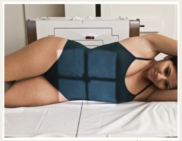
Left lateral decubitus AP projection
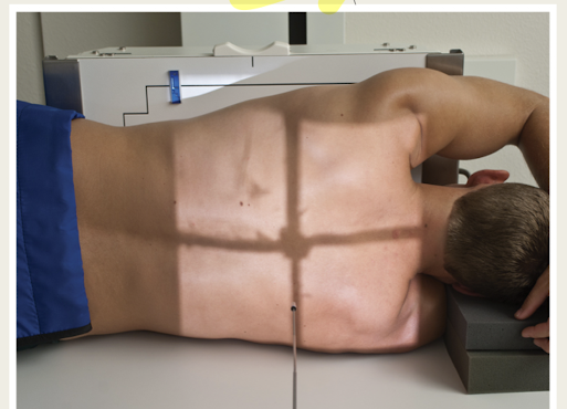
right lateral decubitis PA projection
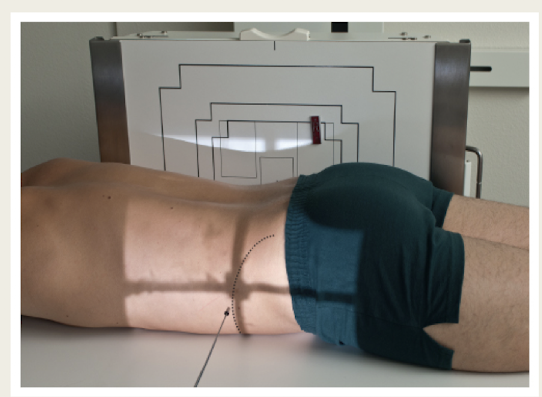
ventral decubitus lateral projection

dorsal decubitus lateral projection

PA projection

AP projection
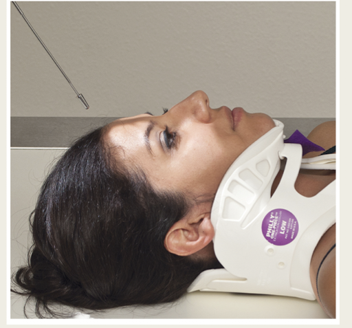
Caudal CR angle
cephalic CR angle

Sesamoid bone
specific type of small, oval shaped bone in the foot that is embedded in certain tendon
largest seasmoid bone
patella
Long bone
only appendicular
extremities
short bones
wrists and ankles
roughly cuboidal
flat bones
sternum, ribs, scapular, calvaria
irregular bones
vertebrae, facial bones, bones of the pelvis
compact bone
hard, dense bone tissue that is outer shell of most bone
body/shaft of bone
diaphysis
thicker layer of compact bone
spongy bone/trabecular bone
inside shell of compact bone
contain red bone marrow
medullary cavity
hollow portion of long bone
contain yellow marrow
periosteum
outer covering of bone
essential for bone growth
hyaline cartilage
glassy cartilage or connective tissue
on end of bone for joint
functional joints
due to relation of mobility or lack of
synarthrosis joint
immovable joint
functional joint