Obstetrics
 Call Kai
 Learn
Learn Practice Test
 Spaced Repetition
 Match
Match Flashcards
 Knowt Play
1/41

There's no tags or description
Looks like no tags are added yet.
Name | Mastery | Learn | Test | Matching | Spaced | Call with Kai | Chat |
|---|
No analytics yet
Send a link to your students to track their progress
42 Terms
Stages of Labour
Duration
Process INSIDE cow
Clinical signs
STAGE ONE:
Duration:
Cow = 3 - 8hr
Heifer = 12 - 48hr
Inside:
Foetus moves into position for delivery
Relaxation of birth canal, pelvic symphysis and soft tissue
Cervix softens, thins and dilates
Uterine contraction begins
Clinical Signs:
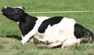
Change in behaviour = Restless, isolation, inappetence
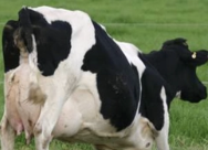
Raised tail (“sprung” tail head)
Swollen udder and engorged milk vein
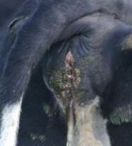
Lengthening and swelling of vulva
Clear/pale yellow mucoid discharge from vulva
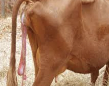
Membranes protruding from vulva
 |  |  |
 |  |
STAGE TWO:
Duration:
Cow = 30 minutes - 1hr
Heifer = 1 - 4hr
Inside:
Fluid released from around foetus as chorioallantoic membrane ruptures
Calf MAY be delivered before chorioallantoic membrane ruptures (RARE)
-ve: Calf may suffocate before cow can sufficiently lick membranes away from nostrils (image)
Clinical Signs:
Obvious abdominal straining during uterine contraction
Shifting between recumbency and standing
Slow progressive outward-movement of foetus seen at vulva
Head, shoulders and pelvis may take longer due to larger diameter
Normal for cow to take short breaks after passing head, shoulders and pelvis
Defaecation, vocalisation and milk leaking
Finished when calf completely expelled from cow

STAGE THREE:
Duration: 30 minutes - 8hr
Inside:
Placenta separates from uterus (cotyledons and maternal caruncles unbutton)
Uterus begins to shrink resulting in expulsion of placenta → Helps control haemorrhage within uterus
Cervix begins to close
Clinical Signs: Do NOT assume a cow has completed stage 2 of parturition as calf may not have been delivered OR a 2nd calf within
Placenta protruding from vulva
Small amount of haemorrhage
Fluids draining from vulva
Finished when entire placenta has been delivered
5 Positive indicators of normal birth
Active and productive contractions
Calf movement
Fluid being expelled from calf’s lungs
Pink tongue
Visualisation of 2 feet and a nose
Overview of Obstetrics
4 Methods of assisted calving
4 Major causes of dystocia
Incidence in NZ (heifers vs. cows)
Methods:
Manual traction
Foetotomy
C-section
Euthanasia
Major Causes:
Head back
Leg back
Failure to dilate/uterine inertia
Foeto-maternal disproportion #1
Incidence
Heifer = 6.5%
Cow = 3.8%
4 Consequences of dystocia
Obturator paralysis
Trauma, tearing, bruising or contusion of birth canal
Rupture of middle uterine artery → Fatal haemorrhage
Increased risk of post-calving infection and RFMs → Infertility
Describe 3 ways to prevent dystocia
Feeding → Reduced intra-pelvic fat in heifers
1st trimeter → Greatest effect on calf birth weight
Cannot easily reduce foetal GR with feed restriction
Effects visible only after 6 weeks
30 60 90 rule = Prioritise heifer growth rate monitoring
30% MWT at 6 months
50% MWT at 12 months
60% MWT at mating
90% MWT at calving
Choice of bull
Jersey bull OR AI to LIC short-gestation length bulls for 1st calving heifers
AI beef bulls strongly selected for short gestation length and calving ease
Natural service beef bulls increasingly being selected for easy-calving characteristics
Genotype cows and match appropriate bulls
Causes of Dystocia
3 Maternal causes
4 Foetal causes
MATERNAL CAUSES
Skeletal
Soft tissue
Ineffective labour
Uterine inertia
Uterine torsion
FOETAL CAUSES
Foetal oversize
Multiple foetuses
Foetal death
Foetal monsters
THREE Maternal Causes of Dystocia
Mechanism
Examples
ONE: SKELETAL CAUSES
MoA: Small pelvis → Relative foeto-maternal disproportion
Examples:
Poorly grown and under-sized heifers at calving (24 months)
Higher risk if mated over sire predisposed to producing large calves → Foeto-pelvic disproportion
Previous pelvic trauma (fractures or dislocations) from previous assisted birth or mating injuries
Genetically malformed pelvis eg. Holstein-Friesian #1 = Longer and narrower pelvis
Rare FM disproportion in beef breeds with purebred calves
Jerseys less prone to FM disproportion (despite being smaller than Holstein-Friesians)
TWO: SOFT TISSUE CAUSES
MoA: Small pelvis → Relative foeto-maternal disproportion OR incomplete dilation of cervix
Examples:
Ineffective/incomplete cervical dilation
Previous vaginal/rectal prolapse
Uterine torsion
Fibrosis of cervix from previous injury
Neoplasia/abscess → Narrowed vagina
Scar tissue in birth canal from previous dystocia or calving trauma → Reduced soft tissue stretching
Excess intra-pelvic fat → Reduced space for calf to pass
eg. Lifestyle animals and carry-over cows with high BCS
Rupture of the pre-pubic tendon (rare)
THREE: INEFFECTIVE LABOUR
Uterine Inertia = Absent contractions after stage 1 labour due to:
1˚ uterine inertia = Over-distended uterus eg. Twins or hydrallantois (hydrops)
2˚ uterine inertia due to dystocia = Myometrium becomes exhausted
Hypocalcaemia = Reduced contractility of skeletal and smooth muscle
Weak/emaciated
Overfat cow with no exercise
Uterine Torsion
What is this?
Diagnosis
3 Treatments

Ruptured Pre-Pubic Tendon (RARE)
Diagnosis: Pendulous abdomen due to herniation
Treatments:
Emergency slaughter (unless dam or calf extremely valuable)
Attempt to induce parturition (some may manage normal vaginal delivery)
± Caesarean section by ventral approach
What is this?
2 Methods of diagnosis
Treatment

Hydrallantois (Hydrops) = Excess accumulation of allantoic fluid in pregnant cow
Diagnosis:
PE = Round cow with no udder development at 6 - 7m gestation and BCS ≤ 3
Rectal exam = Massive fluid-filled uterus with no calf palpated
Treatment: Induce labour with prostaglandin and corticosteroid
Cow does NOT come into milk (salvage situation)
FOUR Foetal Causes of Dystocia
Mechanism
Examples/risks
ONE: FOETAL OVERSIZE
MoA: Absolute foetal oversize →
Foetus too big to fit through pelvic canal
Insufficient room in uterus for calf to get into correct “diving” position to parturition with anterior presentation and extended posture
Correct malpresentation → Still difficult to get out
Examples:
Paternal effects on calf size (Continental breeds or double muscling eg. Belgian Blue)
Long gestation length → Larger calf
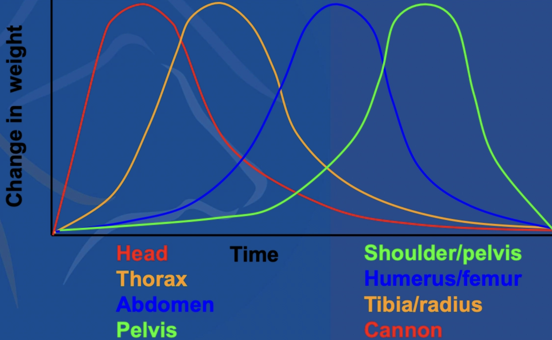
Late gestational growth favours growth of parts contributing to increased calf diameter (eg. shoulders and pelvis)
Exponential increase in foetal weight after 282d of gestation
Male calf
TWO: MULTIPLE FOETUSES
MoA:
Multiple foetuses approach the pelvic canal simultaneously
Multiple foetuses become entangled around each other
THREE: FOETAL DEATH
MoA:
Incorrect presentation to pelvic canal (NOT active during birth process) → Delays normal cervix dilation (even smaller aborted foetuses cannot assume normal position)
Early rupture of membranes → Loss of natural lubrication required for delivery
Decomposition (emphysematous “fizzers”) → Accumulation of SC gas → Calf increases in size significantly → Foetotomy required for delivery
Maternal fatigue and uterine atony
FOUR: FOETAL MONSTERS
MoA: Genetic defects → Misshapen and deformed calves which require more intervention than the standard malpresentation (eg. foetotomy or C-section)
Examples:
Hydrocephalus
Schistosomus reflexus
Foetal ascites
Conjoined twins
Foeto-Maternal Disproportion
2 Types
When to intervene?
Types:
Relative = Calf and pelvis mismatch (eg. small heifer)
Absolute = Calf too big irrespective of pelvis size
Intervention:
Cow = Amniotic sac broken 3hr with no progress
Heifer = Amniotic sac broken 2hr with no progress
5 Ways to determine if the calf is dead
Smell
Anterior presentation → Gently push finger into eye (elicit blink) or squeeze leg (kick)
Posterior presentation → Squeeze leg or push finger into anus to elicit response
Head back → Usually dead
Umbilicus still attached
Hydrocephalus
Diagnosis
2 Treatments
Diagnosis: Difficult to palpate as head not present normally in birth canal
Treatments: Depends on size of skull
Foetotomy (small dome)
C-section (large dome)
Schistosomus Reflexus
Definition
Aetiology
2 Treatments
Definition: Acute angulation of spinal column dorsally and incomplete closure of abdominal wall (“inside-out” calf) → Tail next to head and viscera free-floating in amnion ± ankylosis
Aetiology: Genetic component during embryonic development
Treatment: May present longitudinally OR transversely (visceral OR limb presentation)
Visceral presentation may loos like intestinal hernia of cow
Foetotomy
C-section
Foetal Ascites
Definition
2 Causes
Treatment
Definition: Late-term abortion with ascites and oedematous intestinal walls (1% of calvings)
Dead calf delivered normally until shoulder engage → No further progression causing the abdomen and intestinal walls to fill with fluid
Causes:
Foetal infection
Developmental defects
Treatment:
Rupture calf abdomen to release fluid (40 - 50L) via thoracic inlet and diaphragm with alkathene pipe
Foetotomy to remove front legs and smash through ribs
Describe 3 P’s of Calving (+ normal)
Calves must be positioned properly to be delivered unassisted (unlike lambs and piglets which can be delivered unassisted in several different birth presentations)
ONE: PRESENTATION
Describes the spinal axis of the foetus relative to the long axis of the dam AND which part of the foetus is entering the birth canal first
Anterior (longitudinal) = Head and FL first
Posterior (longitudinal) (5%)
Rump and HL first
Breech = Sub-category of posterior presentation where both hips are flexed and held under calf’s torso
Transverse (RARE)
TWO: POSITION
Describes position of calf’s dorsum relative to the maternal pelvis
Dorsosacral
Dorsoiliac (L/R)
Dorsopubic
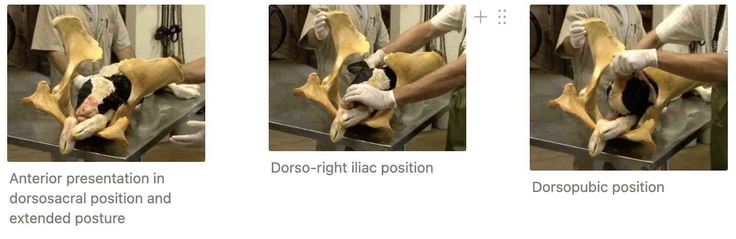
THREE: POSTURE
Describes the extremities of the foetus relative to the trunk
Extended
Flexed (retained at shoulder OR head OR hip)
Normal: Anterior longitudinal and dorsosacral with head and front legs extended into the birth canal

Describe FIVE steps of dystocia intervention
ONE: PREPARATION
Vet Checklist
PPE
Calving gown and bib
Gumboots
Warm hat
Gloves
Rectal gloves
Short gloves over top
± Thick rubber gloves for rotten calves
Silicon barrier cream to protect skin
Obstetrics Kit
Chain/ropes
Pulley/Vink calving jack
Krey hook and eye hook = Useful tool when no anchor points available to place chains or ropes on. Placed on bone, hide and limb stumps to assist in providing traction
Foetotomy equipment
Halter and casting rope
Lube (+ pump)
Medications
Epidural kit (clippers, meths, swabs, syringes/needles, lignocaine)
Tocolytic (clenbuterol = Planipart)
Antibiotics
NSAIDs
Oxytocin
Metabolic treatments (ketol, electrolyte sachet, bags, starter drench) AND Agger’s pump
Surgical Kit
Cleaning Equipment
Antiseptic
Clean scrub brush
Clean bucket
Farm Checklist
Warm water in bucket
Cow in yards
Good facilities
TWO: HISTORY
Age of dam and parity
Previous problems (eg. calving difficulties last season?)
Expected due date
Mated to AB or natural service? Issues with sire?
Full/slack udder? (eg. late abortion)
Calf still alive?
Duration of labour and evidence of straining?
Any farmer intervention? Attempted delivery and medications/metabolic treatments?
THREE: ASSESS FACILITIES
Clean with good lighting
Safe for cow, farmer and vet?
Understand how to operate head bale and open side gate in event of emergency
Make escape plan
Place chain/bar in case cows slips out of head bale (or use halter)
Tie halter low in case she goes down

FOUR: ASSESS PATIENT
Ataxia → Milk fever or nerve damage
Dehydration > 5%
HR > 100bpm → Toxaemic shock
Brick red/cyanotic MM
Demeanour (pain = groaning)
Significant blood loss?
Hindson’s traction ratio (TR)
→ Consider euthanasia if signs above are presented which indicate the cow is suffering
FIVE: SELECT METHOD
If making no progress with a method (10 - 20 minutes) → Select different method OR tool

Planipart
Active ingredient
MoA
Onset of action
2 Indications
Dose rate
WHP
Active: Clenbuterol
MoA: Tocolytic = Sympathomimetic amine (β-2-agonist) with high degree of selectively for lungs and uterus → Induces relaxation of uterine musculature → Dilation of birth canal
Onset: 20 minutes of IM administration
Indications:
C-section to exteriorise uterus
Dystocia management to create more space where calf can be more easily manipulated
Dose Rate: 2mL/100kg BWT IM or IV
WHP: 12d meat and 60hr milk (also applied to calf → important if bobby)
Method Selection for Dystocia Intervention
Calf oversized AND alive (or fresh dead) → C-section
Calf oversized AND dead → Foetotomy
Calf appropriate size → Traction
Sick cow → Euthanasia


3 Ways to determine if feet presenting at the birth canal are FL or HL
Soles of feet
Facing UP = HL (or FL in dorsopubic position)
Facing DOWN = FL (or HL in dorsopubic position)
Palpate joint proximal to fetlock
HL = Hocks more angular and bend OPPOSITE planes as fetlock
FL = Bend in SAME plane as fetlock
Reach further to find tail OR head
Hindson’s Traction Ratio (TR)
Use interischial distance and the diameter of the calf’s digit to assess best method of dystocia treatment
TR > 2.5 → Traction sufficient
TR < 2.5 → C-section or foetotomy required

6 Steps of traction
Lubricate and thoroughly clean with warm water
Administer epidural ± tocolytic
Manipulate calf to assume correct presentation/position
Place chains on feet ± head (if anterior)
Use appropriate force and calving aids
Angle downwards to pull calf through natural curve of birth canal
Rotate calf as chest emerges to minimise presenting diameter (exploit “box-like” shape of pelvis)
Put hand back in to check for “spares and tears”
2nd calf or full-thickness uterine tear
Cow and calf aftercare

Placement of calving chains and head rope
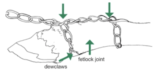
CALVING CHAINS
ALWAYS apply a chain to the leg with a loop above the fetlock (first joint) with a half hitch below the fetlock (below dew claws, above bulbs) → Prevent fracture and torn claws by dispersing pressure along limb
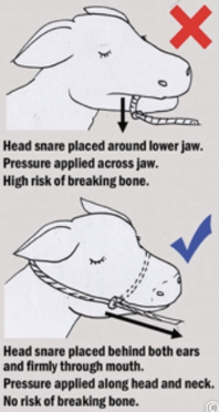
HEAD ROPE
Place snare behind BOTH ears and around mouth
Do NOT place head rope around lower jaw → Risk of breaking bone with forceful traction
Do NOT place rope around neck → Head becomes out of alignment with spinal column making job more difficult (even if dead)
5 Functions of a GYNstick
Correction of uterine torsion
Correction of upside down presentation of calf
Limb retrieval
Repulsion of calf back into uterus
Ramrod to assist breakdown of diaphragm and internal structures while debulking the calf (esp. for foetal ascites)

Calving Pulleys vs. Calving Jack
Advantages
Disadvantages
CALVING PULLEYS
+ve:
Flexible and dynamic → Can be used by single operator
With ample anchoring points, it provides more control over angle and tension of pull
If tension is rapidly released, there is little risk from pulleys injuring a person
Rapid release mechanism which can be reached even when standing directly behind the cow
Take up little room in the back of a vehicle
-ve:
Become tangled and jammed if set up incorrectly
Must place truck/tractor in correct position to be used as an anchoring point for paddock calving
Too much force can be applied if multiple people pulling
Can smell if not cleaned and dried thoroughly

VINK CALVING JACK
+ve:
Does not require additional anchoring points (U-shaped piece braces against back end of cow)
Can be used by ONE person to deliver a calf
Some models have a double ratchet movement which brings one leg forwards at time to “walk” the calf’s shoulder through the pelvic canal
-ve:
Fixed angle of pole can make getting correct angle of pull for a down cow challenging
If cow unsettled and swinging hind end → Pole can cause injury to people
Tension handle can become stuck if too much tension is applied and handle cannot be replaced all the way to the horizontal position

Hiplock
Definition
2 Treatments
Definition: Complication of anterior presentation where head and thorax is delivered BUT foetal pelvis/greater trochanters jam into the dam’s pelvis
Treatments: Do NOT continue to pull → Damage to cow
Rotate calf by twisting FL (50% success)
Partial foetotomy
Trunk → Evisceration → Split hips

Describe how to manipulate the calf for
FL back
Head back
Posterior presentation
True breech
Anterior dorsopubic
FL BACK
Inside: Only ONE of the limbs has successfully extended into the birth canal as the other has become caught behind the brim of the pelvis (retained at the shoulder, carpus or fetlock)
Occasionally, the limb can be crossed over or entangled with the extended limb or head
Diagnosis:
ONE set of toes and head protruding from vulva
Only one limb and head within pelvis on vaginal exam → Deeper examination to locate and position the 2nd forelimb
Treatment: Cannot deliver without correcting limb position
Slide arm into uterus and ID which forelimb is retained + which joint (eg. shoulder)
Slide arm down limb and grasp forelimb at the humerus
Pull limb forwards until carpus is retained now
Slide arms down and UNDER limb to grasp metacarpal
Pull metacarpal up (clear pelvic inlet with foot) until fetlock is retained now
Take other hand and slide under your arm so that it is cupping the claws
Length of limb to carpus is longer than the birth canal diameter → Attempts to straight limb forcefully results in uterine tearing
Repel the carpus back into the uterus to clear space to bring the foot into the birth canal

HEAD BACK
Inside: 2 front limbs extended normally into pelvic canal BUT head is trapped behind the pelvic inlet
Pressure from uterine contractions can exacerbate malpresentation by causing the head to move further away from the birth canal as shoulder become jammed into the pelvic inlet
Diagnosis:
2 front limbs extended normally outside vulva
No palpable nose or head → Deeper palpation reveals shoulder present at the pelvic inlet
Palpate in direction which head lies and double check it is not a posterior presentation
Treatment: One of the most difficult types of dystocia to correct (esp. when head is forcefully pushed away from birth canal)
Slide arm into uterus and ID head back presentation (may need to pull calf into birth canal to reach head)
Hook the bony orbit of the eye OR commissures of the mouth with your fingers → Gives control of head
Bring head into normal position by repelling calf into uterus and AWAY from head to replace shoulders of calf with head
Can extend hand over face of calf to rotate up and into the birth canal

POSTERIOR PRESENTATION
Inside: Calf presented to birth canal backwards with 2 hindlimbs extended into birth canal
Cervix may NOT dilate normally due to incorrect pressure applied (no gradual expansion of the cervix with forelimbs and head to shoulders)
Diagnosis:
2 feet extended from vulva with soles facing the sky
Anterior presentation with dorsopubic position OR uterine torsion
Posterior presentation
Palpation reveals opposite flexion of distal joints and no head or neck palpated ± tail identified
Treatment: Can be delivered in posterior presentation BUT difficult
Ensure cow is fully dilated at the pelvic inlet to avoid uterine tearing
Place calving chains and slowly add traction
Pull at a level angle to the cow’s pelvis until the hips have passed through the birth canal → Keeps hindlimbs and pelvis in straight line
Downward traction on hindlimbs → Forces pelvis up into the spine of the cow
After hips through the vulva, angle the traction lower than the pelvis to assist delivery of shoulders and head
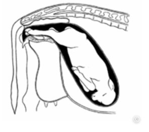
TRUE BREECH
Inside: Calf presenting into pelvic canal backwards with all four hooves pointing towards the head of the cow (i.e. tail coming first)
Diagnosis:
Cow not progressing in delivery often only sign
± Calf tail visible outside cow vulva
Treatment:
Slide hand in to grasp the tibia of the calf
Pull the leg towards the birth canal while flexing the hock
Reposition hand and grasp the metatarsal
Pull leg towards birth canal while repelling calf back into the uterus with other hand
Take other hand and slide under your arm so that it is cupping the claws
Significant length between the hock and the distal limb → Longer than diameter of birth canal and risk of uterine tearing
Repel metatarsus into the uterus while pulling the foot into the birth canal at a diagonal (widest width of the birth canal)
Repeat on the other hindlimb
Deliver via posterior presentation after cervix has fully dilated
Breech = High incidence of twin deliveries → Check for 2nd calf which makes job difficult
Twin adds pressure behind calf and limits room to repel calf

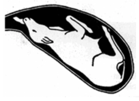
ANTERIOR DORSOPUBIC PRESENTATION
Inside: Both forelimbs extended and head in pelvic canal BUT dorsal part of skull is facing the pubic bone of the cow’s pelvis
May indicate uterine torsion
Diagnosis:
Two limbs extended out of vulva with soles facing up
Vaginal palpation to determine if limbs are forelimbs vs. hindlimbs (all 3 distal joints bend in the same direction) → Head and neck palpated (may be difficult if head is dropped ventrally)
Rectal exam → Rule out uterine torsion
Treatment: Presentation and posture correct, position incorrect (dorsopubic)
Repel most the calf back into the uterus
Turn calf over to correct position (consider criss-crossing legs until calf’s body is rotated
Overview of Caesarean Section
Endogenous contamination
Prognosis
7 Causes of mortality
Long-term effects
Endogenous Contamination: 83% of uterine environments are heavily contaminated → Wound contamination is inevitable
Aim to avoid contamination of abdominal cavity
Prognosis: Influenced by calf condition
Live calf prognosis for cow → 86%
Dead calf prognosis for cow → 79% (33% when emphysematous)
Mortality:
Peritonitis
Shock
Metritis
Concurrent disease (eg. milk fever)
Acute haemorrhage
Increased surgical time
Post-op adhesions
Long-Term Effects: No significant effects on lactation
71% pregnancy rate in dairy and 91% pregnancy rate in beef
+18.5d from calving to conception

12 Steps of Caesarean Section
Location and Restraint
Clean, light and dust-free
Ideally perform while standing (exteriorise uterus)
Pre-Op Drugs
Epidural
Clenbuterol IM
Local Anaesthesia
LEFT laparotomy
Options: Line block, inverted L-block, paravertebral block #1
Clip and Surgically Prep Site
Use clean water and scrub 3x with surgical scrub solution working outwards from intended incision site (≥5 minute contact time) → Rinse between scrubs
Wipe off last scrub with tincture and sterile swabs
Surgeon Preparation
Place sterile surgical pack in clean and safe location
Ensure hands and arms (up to armpits) are surgically scrubbed and wear surgical gloves
Have clean assistant with scrubbed hands
Incision
Location: Halfway between tuber coxae and 13th rib
Hand width below lumbar vertebrae, leaving ~1/3 of trunk un-incised
~50cm incision
Structures: Skin → SC → 3 muscle layers → Peritoneum

Exteriorise Uterus
Locate part of uterus containing a hind leg
Grasp calf’s metatarsal and steadily pull uterus up into abdominal incision
Place one hand under hock and other on dorsal pastern to lock foot into abdominal incision
→ Incise into greater curvature of uterine horn beginning at hock (avoid cotyledons and blood vessels where possible)
Grasp leg and ID 2nd leg to exteriorise
With help of assistant, lift upwards and then pull backwards along the flank to remove the calf
Calf Care and Rescrub
Uterus Closure = Continuous inverting (Utrecht) in 2 layers
Peritonitis comes from inside out → Ensure uterine serosal contact for adhesion formation and minimise exposed suture (wick bacteria)
Material: 6M chromic catgut on curved round bodied needle
Abdominal Closure = Muscle and peritoneum → SC → Skin (Ford interlocking)
Additional Treatments and Aftercare
Penicillin IM 5d
Meloxicam SC
Recheck 3 - 4d for emphysema, peritonitis and metritis
Sutures Out 2 - 3w
Overview of Foetotomy
Definition
6 Ways to protect the cow
2 Methods
Definition: Dissection of foetus into ≥1 parts while it is still inside the uterus and/or vagina
Achieved by cutting through skin, muscles, ligaments and bone with braided wire
If done correctly → No risk of uterus damage
Protect Cow:
ALWAYS epidural
Consider tocolytic agents to abolish uterine contractions
Copious amounts of obstetrical lubricant
Use proper equipment (tighter the weave = higher the quality)
Careful technique and anatomical considerations of cut location to minimise sharp edges of foetus on uterus
Cup hand over sharp points with towel in between when removing parts → Avoid laceration of uterus and vagina
Methods:
Malposition → 1 - 2 cuts
+ve: Quicker than C-section
Complete foetotomy → 3 - 6 cuts which require forethought in cut placement
~2hr if experienced
-ve: Prolonged foetotomy → Increased cow and vet exhaustion
TWO Types of Foetotomy Cuts
Definition
Indications
Method
REVERSE CUT
Definition: Head of foetotome further in → Cutting backwards
Indications: Remove head and FL OR HL
Method:
Load BOTH channels of foetotome and pass wire around head and FL (OR around HL)
Advance foetotome until its head is dorsolateral behind point of scapula and wire is seated in axilla (OR anterior to tuber coxae and wire between tail and opposite tuber ischii)
Apply traction to leg and anchor chain into notched base plate

END-ON CUT
Definition: Straight-forwards cut with head of foetotome further out
Indications: Head back and splitting pelvis of hiplock
Method:
Thread ONE channel of foetotome and pass wire around part you want to remove with an introducer
Thread other channel
Advance foetotome until hard up against calf

7 Pieces of equipment required for foetotomy ONLY
Foetotome (notched base plate for anchoring chains)
Threader (thread wire onto foetotome)
Wire handles
Krey’s hook
Introducer
Sharp cutting pliers
Embryotomy wire (tight weave = higher quality)
Cut 3.5x length of foetotome

10 Steps of general foetotomy
Restrain cow and administer epidural and tocolytic
Administer generous lube
Place calving chains
Foetotome set-up
ID end that goes into cow (mushroom-shaped and opposite to handle end)
Embryotomy wire is passed through the fetotome using a threader by pulling from the cow-end to vet-end
Attach handles to vet-end by looping wire around each alligator clip (x2) OR attaching via surgeon’s knot
Wire placed around the anatomy to be removed (may require an introducer)
Attach introducer to cow-end of wire and pass round calf
Thread 2nd channel of the fetotome with the threader from cow-end to vet-end
Get foetotome into position for end-on or reverse cut
Apply traction to calving chains and lock over base plate notch
Wire placement checked to ensure correct anatomy is being removed and no maternal anatomy included in cut
One person with hand over foetotome head to keep wires in place by keeping head of foetotome still
Good communication with person cutting
Person pulling wires is prepared to stop immediately if required
Initial short strokes to imbed wire → Longer smooth strokes to cut through skin and bone
Fell “pop” when through
ALWAYS debulk calf
Remove calf’s heart and lungs (may need finger knife)
Burrow through diaphragm to remove all intestines in reach
Lawrence Butt Cut
Indication
Method
Indication: Calf in true breech position with both hips flexed
Method:
Use extra wire (4x fetotome) and thread both channels
Pass loop around rear end of calf just below tuber ischii
Advance head of fetotome until dorsal to thoracolumbar junction
→ Calf sliced in FOUR
2 hind legs cut through proximal femur
Pelvis and spine up to ribs
Anterior portion of ribs, head and front leg
Pull pelvis → legs out with Krey hook

7 Steps of cow aftercare and 6 steps of calf aftercare (alive)
COW
± Antibiotics = 3 - 5d penicillin IM (emphysematous calf or vaginal tear)
Oxytocin
NSAID
Ketoprofen
Meloxicam (when AB given due to WHP)
PO starter drenches (eg. calcium, molases, propylene glycol)
PO fluids
Coat and nursing
Arrange metricheck 2 - 3w post-calving
CALF
Lift HL over rail to drain fluid from chest (esp. posterior-presenting calves as they breathe in fluid)
Stimulate breathing with straw or mouth-to-nose resuscitation
NSAIDs
± Penicillin 3d
Dress navel
Tube with 2L fresh colostrum
Overview of Uterine Prolapse
Aetiology
Sequelae
Prevalence
Predisposing risks
Young cow (4)
Older cow (5c)
Aetiology: Continual abdominal contractions expel uterus through vagina
Sequelae: EMERGENCY
Shock
Severe haemorrhage
Prevalence: 0.02% in NZ
Risks:
Young Cow = Foetal oversize, nerve damage, assisted calving, farm contour
Older Cow = Milk fever, nerve damage, poor BCS, assisted calving, farm contour
Describe SEVEN steps of uterine prolapse treatment
ONE: PHONE INSTRUCTIONS
Need warm water, 2 buckets and shed aprons or clean sheet
Where is the cow? Will drive to cow (do NOT bring to shed)
Administer SC calcium
Need 1 - 2 helpers to retrain cow and replace uterus
Do NOT walk excessive distance if cow up and mobile
TWO: HISTORY
Age of cow and parity (milk fever?)
When did the cow calf?
Was the calving assisted? (excess traction → traumatic uterine prolapse)
AI or natural mating (oversize?)
Any metabolic treatment (no response → other pathology)
Cow up since calved? (existing milk fever or nerve damage a potential complication?)
THREE: PATIENT ASSESSMENT
Milk fever → Give Ca (not too much to prevent mobilisation of cow)
Haemorrhage → ± Transfusion after replacing uterus OR euthanasia ASAP
Uterine damage → Amputation OR euthanasia
Mastitis → Euthanasia if severe
FOUR: PRE-REPLACEMENT TREATMENT
Roll into sternal
Administer sedative if fractious (0.1mg/kg xylazine IV)
Epidural ± tocolytic
Position cow in frog-leg position OR restrain in narrow race if up
Frog legged = Ideal position for down cow with prolapse to tilt the pelvis down towards the cow’s head (replace with gravity)
Thoroughly clean with warm water and iodine
Remove placenta ± trim
Use plenty of lube

FIVE: TREATMENT
Support weight of uterus with sling or on thighs
Do NOT use sugar to shrink uterus by osmosis
Use flat hands or firsts and begin at top → Sweep hands around internal rim of vulva (breaststroke action)
Use clean lubed bottle to push out tips of uterus
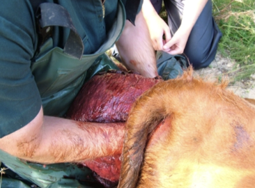
SIX: REFRACTORY
Check bladder if retroflexed within uterus → Requires drainage with needle
Feels like a firm ball or large swelling inside the uterus
Check no haemorrhage/haematoma preventing replacement
Uterus not sufficiently elevated → Sedate cow and shackle back legs to fore end loader → Hoist cow rear ~1m above growth and try to replace
Uterus not relaxed → Try tocolytic clenbuterol IV
Check pushing uterus back into correct hole
SEVEN: AFTERCARE
Do NOT stich vulva
Aftercare as for dystocia = OT, Ca2+, AB, NSAIDs, propylene glycol, metricheck
Overview of Uterine Torsion
Timing
Most common direction of twist
4 Methods of diagnosis
4 Methods of correction
Timing: 90% at parturition = Late 1st stage of labour
Does NOT occur in Bos indicus → Different broad ligament anatomy
Twist: 90 - 360˚ and 75% anticlockwise (observe from behind cow)
Diagnosis:
History = Failure for delivery to progress beyond 1st stage of labour (farmer may report no cervical dilation)
± Deviated angle of vulva
Vaginal palpation = Narrowed and twisted vaginal and cervical lumen
DDx: Incomplete cervical dilation
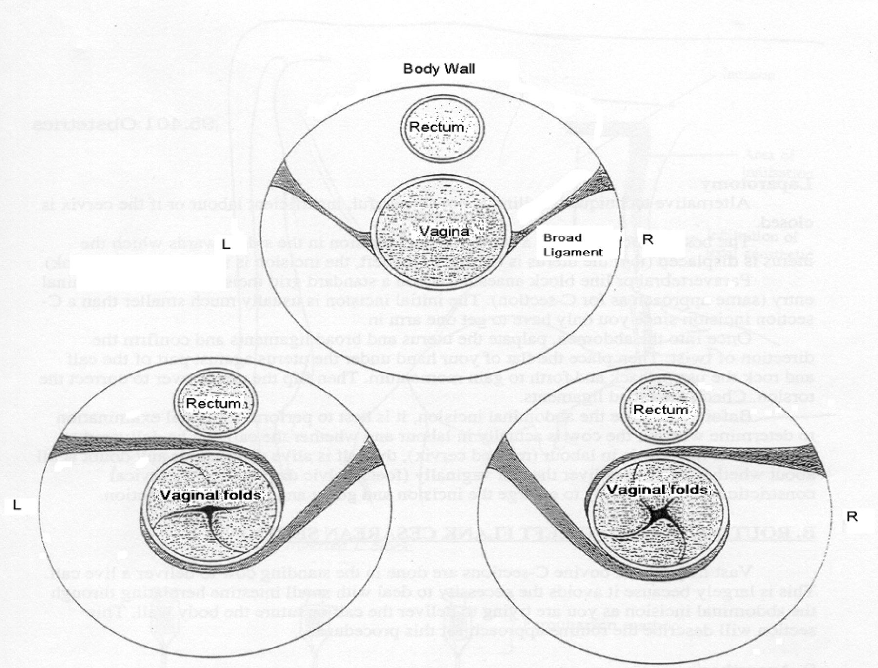
Rectal palpation = Broad ligaments spiralling over top of uterine body → Confirms diagnosis ± Oversized calf
→ Determine which direct the twist is occurring via vaginal and/or rectal palpation
Twist occurs in the SAME direction as the curve across the top of the vagina
Hand swept to right → Clockwise rotation
Hand swept to left → Anti-clockwise rotation
Treatments:
Create momentum
GYNstick/Torsion bar
Rolling cow
C-section
FOUR Treatments of Uterine Torsion
Indication
Disadvantage
Method
ONE: CREATE MOMENTUM
Indication: Limbs MUST be palpated in birth canal
-ve:
Physically impossible in some cases
Risk of injury to vet
Method: Use calf to rock uterus back and forth in abdomen → Create sufficient momentum to flip calf AND uterus back to correct the twist
TWO: GYNSTICK/TORSION BAR
Indication: Limbs MUST be palpated in birth canal
Method: Ropes applied to both limbs of calf → Ropes cross through loop in torsion bar → Bar rotated in opposite direction of the twist to use the body of the calf to pull the uterus back into the correct position

THREE: ROLL COW
Indication: Cannot get hand through cervix to reach limbs
Roll cow so she catches up with her uterus (twist the cow about her uterus which remains stationary)
Equipment:
Long rope
Long and wide plank
≥3 adult helpers
Method: Roll cow in SAME direct as twist while anchoring uterus stationary (eg. anticlockwise twist to left → left lateral recumbency)
Cast cow using long rope and Reuff’s method and tie legs together
Place plank over gravid uterus = Between wing of ilium and ribs
Someone stands on plank → Surf the cow
Plank fixes position of uterus while cow is unrolled around it
Have 1 person in charge of hindlimbs, 1 person in charge of forelimbs and 1 person in charge of head
Ideally keep hand in vagina to ensure there is no tightening
Roll cow through 180˚ and keep her on the ground to check for progress
Often need to repeat rolling to fully correct
Keep cow rolling in same direction
Do NOT allow her to get to her feet
→ Replace plank and roll again

FOUR: C-SECTION
Method: Depending on type, direction and degree of rotation, the uterus can be corrected first OR calf removed first
+ve:
Less space and assistance required
Vet can visualise uterus to ensure complete correction
Saves time when cervix is not dilated after uterine torsion correction
Uterine Torsion After Treatment
Indicators of correction
5 Additional treatments after torsion is corrected
Correction: Passage of uterine fluids and urine
Vaginal palpation to assess progress
Additional Treatments: Often not fully dilated cervix
Prostaglandin/steroid to continue cervical dilation
Ideally place calf legs through cervix to initiate contractions
Recheck for delivery in 3 - 4hr
± Veterinary assistance for delivery (oversized calf)
± Caesarean or cut into cervix (may not dilate)
Therapeutic Induction of Calving
Indication
Disadvantage
Method
Indication: Late gestation as a solution for poor bull selection
Easy when mating date known
-ve: May negatively affect lactation (BUT cow welfare #1 priority)
Method: Inducing heifers/cows of unknown expected calving date
Examine udders and select cows that have entered the pre-calving mammary growth spurt
Give long-acting corticosteroid
Re-examine at 7 - 10 days
Give 2nd short-acting corticosteroid if final udder growth has taken place OR more long-acting and wait
Check through mob of heifers weekly