The Fetal Gastrointestinal System
 Call Kai
 Learn
Learn Practice Test
 Spaced Repetition
 Match
Match Flashcards
 Knowt Play
1/51

There's no tags or description
Looks like no tags are added yet.
Name | Mastery | Learn | Test | Matching | Spaced | Call with Kai | Chat |
|---|
No analytics yet
Send a link to your students to track their progress
52 Terms
The fetal gut develops at the end of the fifth menstrual week and can be divided into the
foregut, midgut, and hindgut
The esophagus may be visualized when needed as
several parallel echogenic lines within the thorax
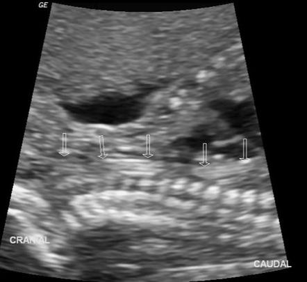
the fetal stomach can be visualized as early as 8 weeks’ gestation, but most certainly should be seen by
14 weeks in the left upper quadrant as an anechoic, circular organ.
The diameter of small bowel is
smaller than the colon and does not typically exceed 5 mm
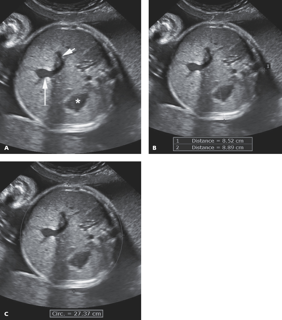
abdominal circumference
axial view of the fetus and should include the fetal stomach, transverse thoracic spine, and intrahepatic portion of the umbilical vein and its junction with the left portal vein
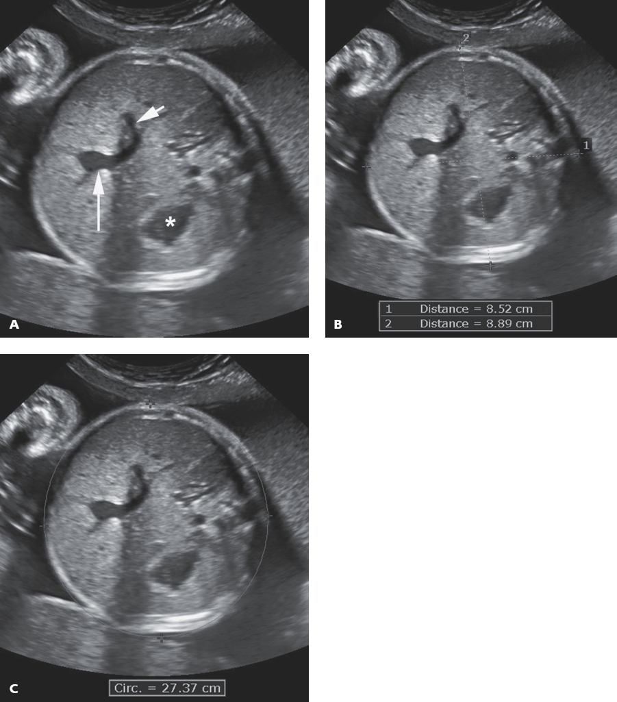
abdominal diameter
AC = 1.57 × (AD1 + AD2).
What is Polyhydramnios
excessive amniotic fluid,
AFI > 24 cm
DVP >8CM
in later gestation, somewhere around 9 weeks, the fetal kidneys begin to produce urine, a liquid that eventually comprises
most of the amniotic fluid.
The fluid passes through the esophagus, into the stomach, and travels through
the small bowel and into the colon, where absorption takes place
Polyhydramnios results when there is an
obstruction or disturbance to the normal flow and absorption of amniotic fluid. Consequently, there is a buildup of amniotic fluid resulting from the continual production of urine by the fetal kidneys
The congenital absence of part of the esophagus is termed
esophageal atresia
Consequently, the esophagus and the trachea often form an abnormal connection known as a
tracheoesophageal fistula (90% of the time)
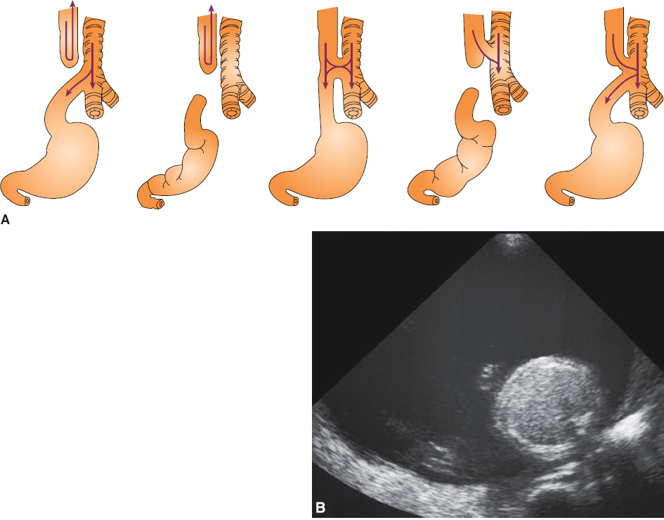
Esophageal Atresia is associated with
duodenal atresia, VACTERL association, trisomy 21, intrauterine growth restriction, and trisomy 18/edward (E FOR EDWARDS).
Sonographic Findings of Esophageal Atresia
Absent or small stomach
Polyhydramnios
Intrauterine growth restriction
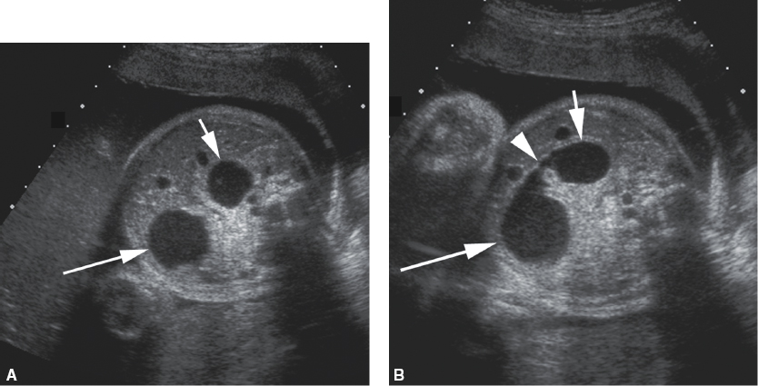
The congenital maldevelopment or absence of the proximal portion of the small bowel, the duodenum, is termed
duodenal atresia
Duodenal atresia classically presents sonographically as a
dilated, fluid-filled anechoic stomach and an anechoic fluid-filled proximal duodenum, offering the “double bubble” sign
Duodenal atresia has a proven association with
trisomy 21; thus, additional sonographic markers of trisomy 21 should be aggressively investigated during the examination.
Also esophageal atresia, VACTERL association, intrauterine growth restriction, and cardiac anomalies.
Sonographic Findings of Duodenal Atresia
“Double bubble” sign
Polyhydramnios
Intrauterine growth restriction
It is essential to remember that in the fetus, the left lobe of the liver is typically
larger than the right lobe
What is the most common abnormality of the fetal liver.
hepatomegaly
Hepatomegaly may occur as a result of
intrauterine infections, fetal anemia (Rh incompatibility), or be seen with Beckwith–Wiedemann syndrome.
Enlargement of the fetal spleen—splenomegaly—can accompany hepatomegaly and may be suggestive of
intrauterine infections or Rh incompatibility with hydrops
Fetal gallstones (cholelithiasis) and sludge within the gallbladder may be noted in utero, most often in the third trimester. Gallstones appear sonographically as
echogenic foci in the right upper quadrant of the fetus that may or may not produce posterior shadowing
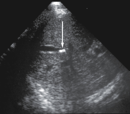
An additional rare abnormality of the biliary tree is the
choledochal cyst
There are four different types of choledochal cysts, with the most common being described as the
cystic dilatation of the common bile duct.
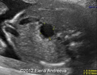
Choledochal cysts can lead to
cholangitis (swelling of bile duct) , portal hypertension, pancreatitis, and liver failure.
In general, the echogenicity of the small intestine should not be
isoechoic to or greater than that of fetal bone
if a higher frequency transducer suggests echogenic bowel, that the sonographer should
decrease the frequency to 5 MHz or less and decrease the overall gain
Echogenic bowel has been linked with
trisomy 21, cystic fibrosis, growth restriction, fetal demise, congenital infections such as cytomegalovirus, and gastrointestinal obstructions.
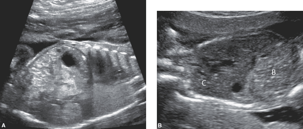
what is Hirschsprung disease and what is it caused by
a functional fetal bowel obstruction, is caused by the absence of nerves within the bowel wall.
Hirschsprung disease is more common in
males
Hirschsprung disease has a strong association with
trisomy 21
The sonographic finding of dilated loops of bowel within the fetal abdomen is indicative of
a fetal bowel obstruction.
These dilated loops of bowel should not exceed
7 mm in diameter or measure greater than 15 mm in length
Obstruction of the fetal bowel most often occurs when there is a meconium plug causing the barrier, a condition referred to as
meconium plug syndrome.
The most common type of colonic atresia that will lead to a bowel obstruction is
anorectal atresia
Anorectal atresia may be linked with
VACTERL association and chromosomal abnormalities
Anorectal atresia will most often lead to the visualization of a
dilated fetal rectum.
what are the two of the most common ventral abdominal wall defects
Gastroschisis and omphalocele
it is important to note that MSAFP levels have been shown to be much higher in gastroschisis than in
omphalocele.
As a part of normal fetal development during the first trimester, the midgut herniates into the base of the umbilical cord; this is termed
physiologic bowel herniation
The intestines return to the abdomen by the
12th gestational week.
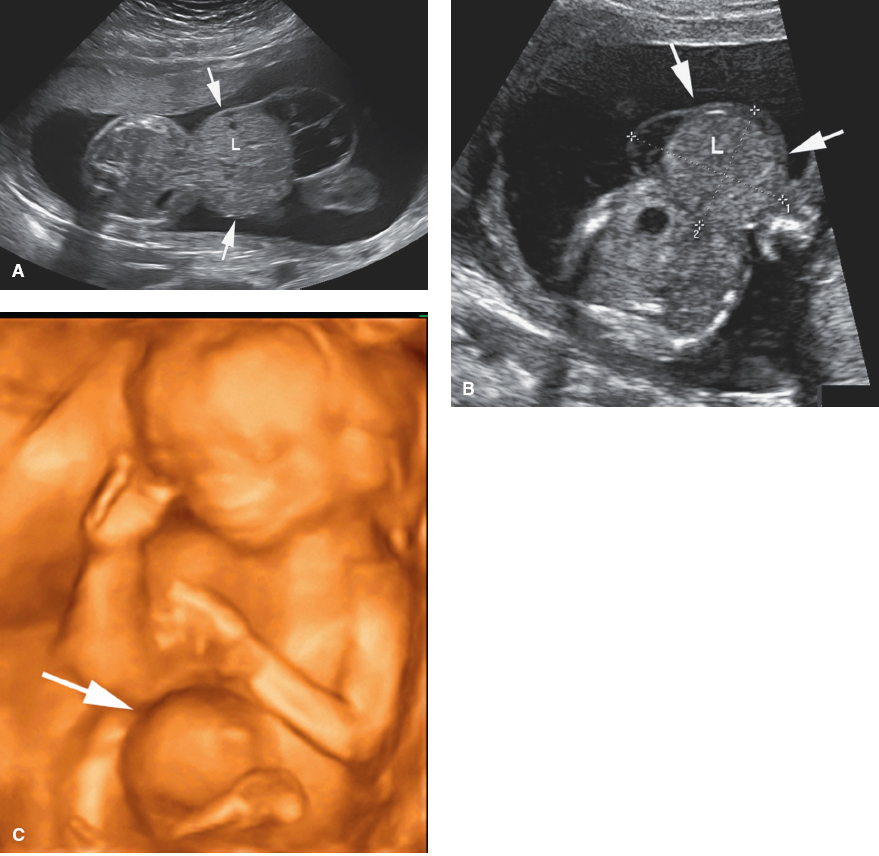
Gastroschisis is the
herniation of abdominal contents through a right-sided, periumbilical abdominal wall defect
Gastroschisis is thought to be caused by a vascular incident occurring to either the
right umbilical vein or the omphalomesenteric artery
Gastroschisis, unlike omphalocele,
does not have a strong association with chromosomal abnormalities
Clinical Findings of Gastroschisis
Elevated MSAFP
Sonographic Findings of Gastroschisis
Normal cord insertion
Periumbilical, right-sided mass
Recognizable loops of bowel outside the abdomen
Intrauterine growth restriction
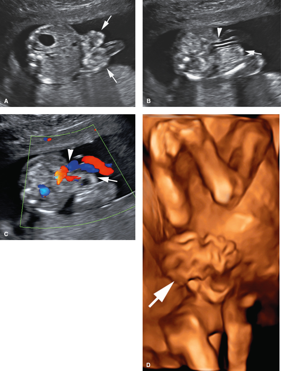
The evidence of persistent herniation of the bowel, and potentially other abdominal organs, into the base of the umbilical cord leads to the diagnosis of an
omphalocele
what is associated with omphaloceles?
Trisomy 18, trisomy 13, Turner syndrome, and Beckwith–Wiedemann syndrome
. Pentalogy of Cantrell is another group of anomalies that include an
omphalocele, along with ectopic cordis, cleft sternum, anterior diaphragmatic defect, and pericardial defects
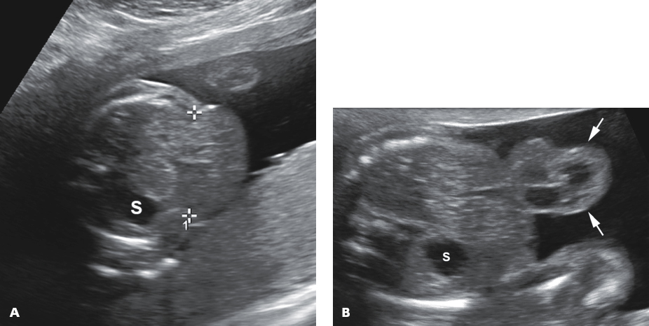
Clinical Findings of Omphalocele
Elevated MSAFP
Sonographic Findings of Omphalocele
Midline abdominal mass at the base of the umbilical cord that contains bowel, the liver, and/or other abdominal organs
Abnormal cord insertion into the midline abdominal mass
Multiple associated anomalies