Neurobiology Test 1 (PPT 1/6) (BIO 462)
1/126
There's no tags or description
Looks like no tags are added yet.
Name | Mastery | Learn | Test | Matching | Spaced |
|---|
No study sessions yet.
127 Terms
what is the definition of neurobiology?
the study of neurons and how they are organized into functional circuits that process information to mediate alterations in behavior and/or physiology (in response to internal and external cues)
what makes neuroscience different from neurobiology?
neuroscience includes application..... it is the study of biological and chemical processes in the brain; study of the mechanisms of brain function and disease
what are the goals of neuroscience?
understand structure and function relationships and how those mediate behavior
what are the major aspects of neuroscience?
dev bio, genetics, all strong bios
what is the importance of neuroscience?
existence, necessary for addressing disease
what are the two origins of neurobiology?
first origin is 600-400 B.C. Greek philosophers and much earlier indigenous peoples (evidence in Peru); they thought that the effects of brain damage was the fault of spiritual beings................. second origin is Hippocrates (Hippocratic corpus)- 5th century BC to 1st and 2nd century AD; thought that supernatural influence don't play a dominant role and the brain is important in consciousness, feelings, perception, moral judgment; there was anatomical exploration of the brain and understanding of neurological diseases and brain/spine injury..................... at this time, there was also convergence of information from early experimental sciences and clinical application (anatomy, embryology, physiology, pharmacology, psychology)
what was early work on the brain called?
trepanation: scrape the skull to get down to the dura to release pressure..... dating back to neolithic times or earlier
What was the ventricular theory and who was responsible for it?
Claudius Galen; it states that sensation and movement are initiated as fluid is moved to or from the ventricles via nerves= brain fluid hollow tubes
who was responsible for phrenology? when was it coined? what does it mean?
Franz Joseph Gall; 1700; it is the study of mapping out personality traits on a particular origins/regions of the brain (did move the field closer to modern times though bc they knew structure was important for function)
what are the three important dates in origins of cellular connection and what happened in each?
1800s- Camillo Golgi vs. Ramon y Cajal and the great debate of reticular theory and the neuronal doctrine......... 1897- Sherrington identifies a synapse and reticular theory falls out of favor..... 1950s- use of the electron microscope in neurobiology
what did Golgi believe?
reticular theory.....he thought neurons were a network.... he made the golgi stain to differentiate cells in the brain at interconnected places..... does have some relevance with glial cells
what did Ramon y Cajal believe?
neuronal doctrine.....he said that neurons weren't a network, they are distinct units.... he used golgi's stain to look at non-interconnected places
what is the modern view of the nervous system?
cellular connectionism/cell theory of neuroanatomy..... individual neurons are the signaling units of the nervous system, they are arranged in functional groups that are connected to one another in a precise fashion........different behaviors are produced by specific brain regions that are connected to each other by distinct neural pathways (K. Wernicke)
which field of neuroscience studies how molecules/proteins allow cellular function?
molecular neuroscience
which field of neuroscience studies how coordination of molecular signaling results in cellular specialization?
cellular neuroscience
which field of neuroscience studies how neural circuits of cells result in specific function?
systems neuroscience
which field of neuroscience studies how neural systems create integrated behaviors?
behavioral neuroscience
which field of neuroscience studies how brain activity creates the mind?
cognitive neuroscience
in terms of evolution, how did we get to our current brain?
nerve net (cnidaria), cephalization (round worms), then our current brain
what is the cindria and the nerve net it has?
its neurons are all interconnected (which works with reticular theory).... this changed with animals became motile
what is the round worm and the cephalization?
it means that more complex behaviors are available... there is localization of neurons in anterior region of the worm with distinct, localized functions
is it clear or unclear if there is a common ancestor for vertebrae and invertebrate?
unclear
what defines a vertebrae in terms of neuroscience?
specialization of brain regions for a specific function and Neopallial (cortical) expansion
what is the carboniferous period?
when the neocortex first emerges in replies
what is neopallial (cortical expansion)?
an increase in the size of the cortex and of structures within that region..... by adding cells, you increase the thickness of the cortex, and you add differentiation
when did the 6 layers emerge in early mammals?
the triassic/jurassic transition
what is the encephalization quotient?
the ratio of brain size to body weight tells you about the intelligence of an animal
talk through the evolution of the neocortex and the fish to mammals/primates to humans
fish: hindbrain (major areas concerned with motor reflexes), midbrain (optic lobes), and forebrain (olfactory and cerebellum)..... there is then continued specialization and expansion of the cortex within vertebrates....... there is folding of the neocortex (sculpting of the brain)....... we increase in # of cells in brain overall AND size of prefrontal cortex (humans also have greater surface area bc of more folding)
what are highly conserved across catarrhini primates?
cortical genes regulatory networks and cell types
what do lineage specific adaptations alter?
transcription, development, and neurotransmission
compare to a NHP (non human primate), do humans have more or less layers of the prefrontal cortex? overall cells in the brain? folding of neocortex? cortical interneurons?
more, more, more, more
in what layer of the prefrontal cortex does the proportion of excitatory neurons increase? what about the layer in which inhibitory neurons increase in diversity? the layer in which inhibitory neurons increase in general?
L4, L5, L2/L3
in terms of transcriptomic modifications, in humans (vs NHP), what happens in microglia?
upregulation of FOXP2
in terms of post-translational modifications, in humans (vs NHP), what are the classes of interneurons?
human interneurons or either all SST or TH...... NHP can either be SST + TH mix or just SST
are there more or less proteomic modifications (alterations after synthesis) in humans than NHP?
more
when looking at lineage-specific genetic changes, what do humans have in their genome? what about NHP?
humans have a deletion and a supplication..... NHP have a single-nucleotide change, insertion, and an inversion from
do humans have more or complex regulatory mechanisms than NHP?
more
differentiate between spinogenesis and genomic changes between humans and NHP
in humans, there is NO SOX5 binding to the enhancer and this allows for CBLN 2 (cerebellin 2) activity to happen which increases spinogenesis in the prefrontal cortex (this means that the PFC will be larger, more involved, and will have a deeper layered cortex)- also, with the increase in spinogenesis, we increase the amount of surface area on dendrites which means that we increase synaptic connections and signaling
in primates, SOX5 binds to the enhancer which blocks CBLN 2 activity which means that there will be an increase spinogenesis in the PFC (this means that the PFC will not have deep layers, will have decreased surface area, and decreased synaptic connections and signaling
fill in the blank: approximately ___________ of the __________ human genes are expressed in the developing and/or adult brain
14,000.... 20,000
what animal has more genes than us?
mice
what amphibian has been recently used to help map vertebrate brain evolution and diversification?
the axolot (because their CNS can regenerate)
what happens in severe microsephely?
there is a mutation in the aspm (abnormal spindle-like microcephaly associated gene)...... this causes difficulties in motor and cognitive function due to an underdeveloped cerebral cortex
what are the two types of cells of the nervous system?
neurons and glial cells
about how many neurons are there in the brain? what about glia?
100 billion neurons in the brain....... 50 times more glia than neurons
what are the two types of glia?
macroglia and microglia
what is shown in A?
cortical neurons stained using the Golgi method (impregnation with silver salts)

what is shown in B?
Golgi-stained Purkinje cells in the cerebellum....... Purkinje cells have a single, highly branched apical dendrite

what is shown in C?
Intracellular injection of fluorescent dye labels two retinal neurons that vary dramatically in the size and extent of their dendritic arborizations
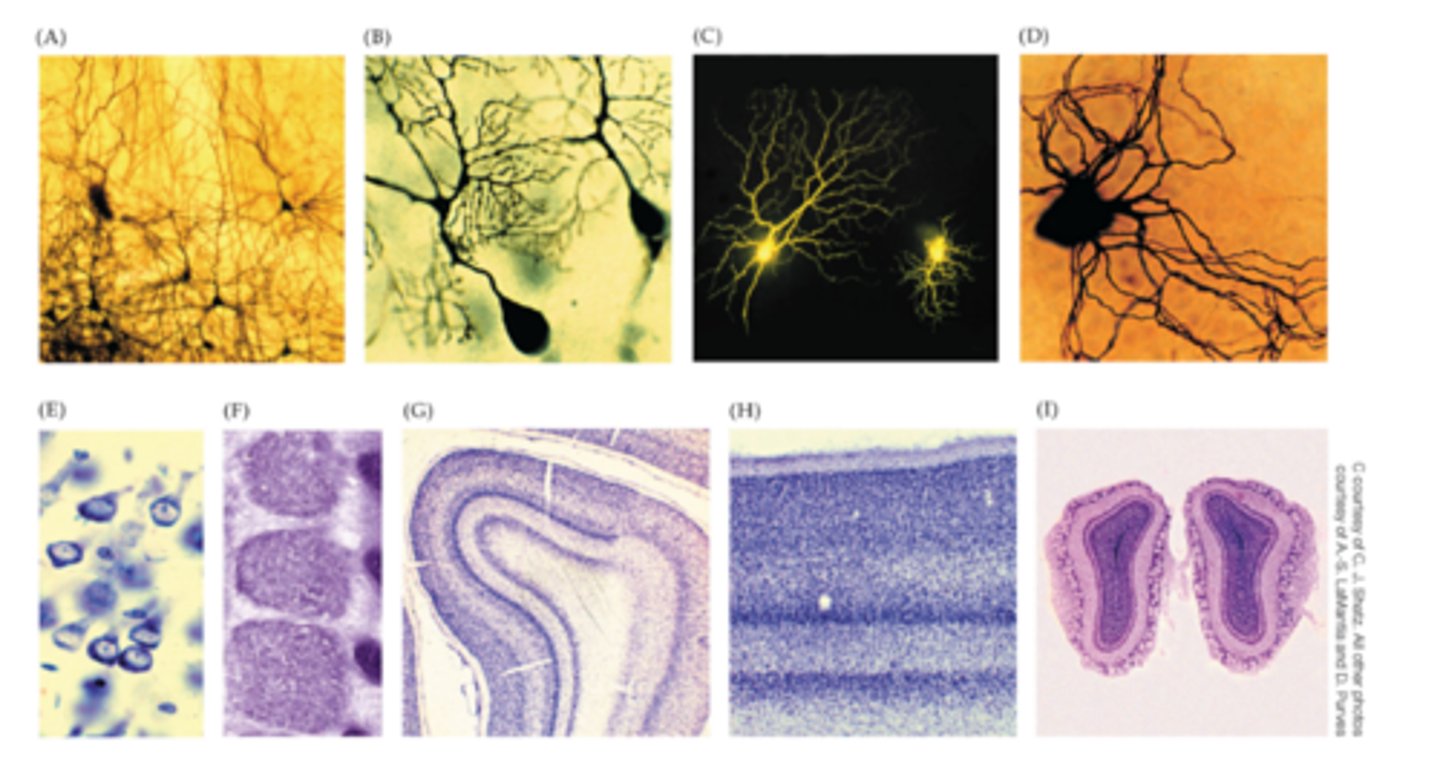
what is shown in D?
Intracellular injection of an enzyme labels a neuron in a ganglion of the autonomic (involuntary control of internal organs) nervous system

what is shown in E?
the dye cresyl violet stains RNA in all cells in a tissue, labeling the nucleolus (but not the nucleus) as well as the ribosome-rich endoplasmic reticulum.... dendrites and axons are not labeled, which explains the "blank" spaces between these neurons

what is shown in F?
nissl-stained section of the cerebral cortex reveals lamination: cell bodies arranged in layers of differing densities........ the different laminar densities define boundaries between cortical areas with distinct functions

what is shown in G?
higher magnification of the primary visual cortex....differences in cell density define the laminae of the primary visual cortex and differentiate this region from other cerebral cortical areas

what is shown in H?
nissl stain of the olfactory bulbs reveals a distinctive distribution of cell bodies, particularly those arranged in rings on each bulb's outer surface.... these structures, including the cell-sparse tissued contained with each ring, are called glomeruli

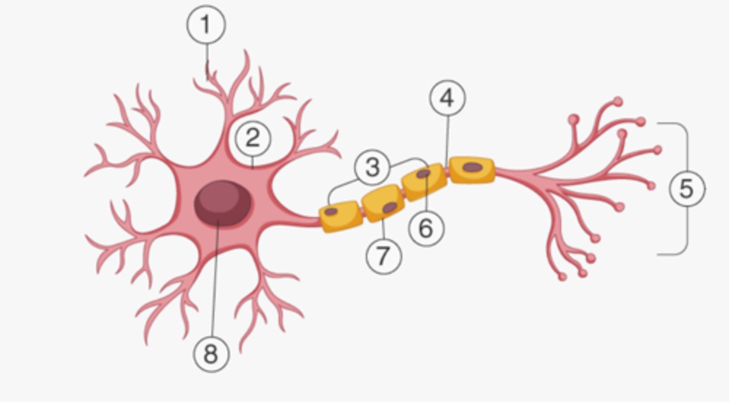
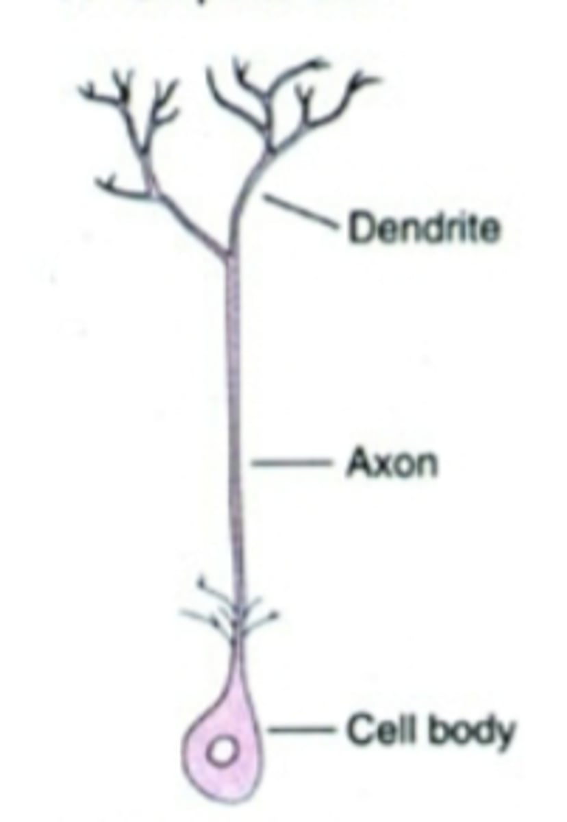
What is 1?
dendrite

what is 2?
soma

what is 3?
axon

what is 4?
node of ranvier

what is 5?
axon terminal

what is 6?
schwann cell

what is 7?
myelin sheath

what is 8?
nucleus
what did Ramon y Cajal and the Principle of Dynamic Polarization set the stage for?
understanding electric potential and knowing that information flows across a neuron unidirectionally
what is the basic info about a neuron?
neurons are the primary functional unit of the nervous system......they are specialized cells that make complex connections with one another to send and receive information from the spinal cord....... a single neuron can make connections with 100s of neurons
how many different subtypes of neurons? do they have the same morphology?
10,000 different subtypes....no! they're all different which means that they all have a slightly different function
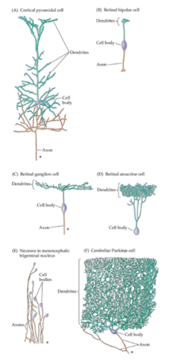
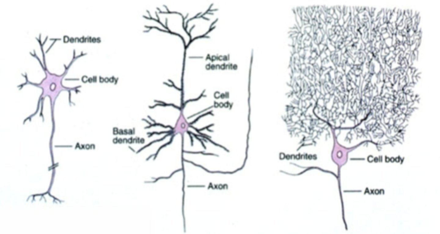
note the features of a cortical pyramidal cell
they have BOTH pia (toward outside) and basal (closer to cell body and inner structures) dendrites..... not super common to have basal dendrites, long axon

note the features of a retinal bipolar cell
small, dendrites are small as well and very contained, axon is NOT long

note the features of a retinal ganglion cell
about the same size as the retinal bipolar cell but longer, but the it DOES have a long axon....... the dendrites are not as contained

note the features of a retinal amacrine cell
no axon..... dendrites are long, but they are very contained in one big cluster
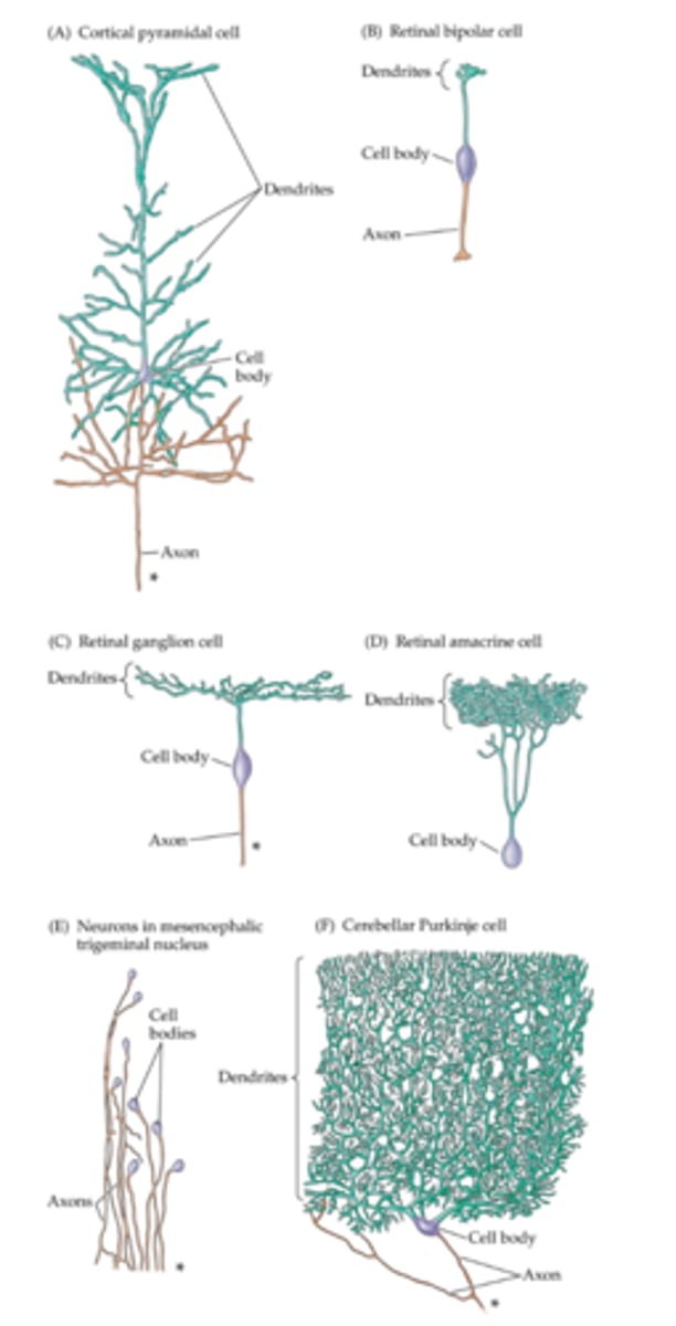
note the features of the neurons in the mesencephalic nucleus of cranial nerve V
pseudounipolar structure (The "axon" in this context refers to the entire neuronal process that carries sensory information)... dendrites cannot be seen.... this means that dendrites aren't needed)

note the features of the cerebellar purkinje cells
these neurons have a large dendritic tree and this is needed to integrate vast amounts of sensory information and facilitate motor coordination and learning
describe what is being shown in A
this is a diagram of the nerve cell and its component parts

describe what is being shown in B
axon initial segment (blue), the region of the axon that emerges from the neuronal cell body, entering a myelin sheath (gold)

describe what is being shown in C
terminal buttons (blue) loaded with synaptic vesicles (arrowheads) forming synapses (arrows) with dendrite (purple)

describe what is being shown in D
transverse section of axons (blue) ensheathed by the processes of oligodendrocytes (gold); the surrounding myelin is black

describe what is being shown in F
nerve cell bodies (purple) occupied by large round nuclei, dense networks of endoplasmic reticulum in the cytoplasm, and numerous mitochondria (dark tubular structures)
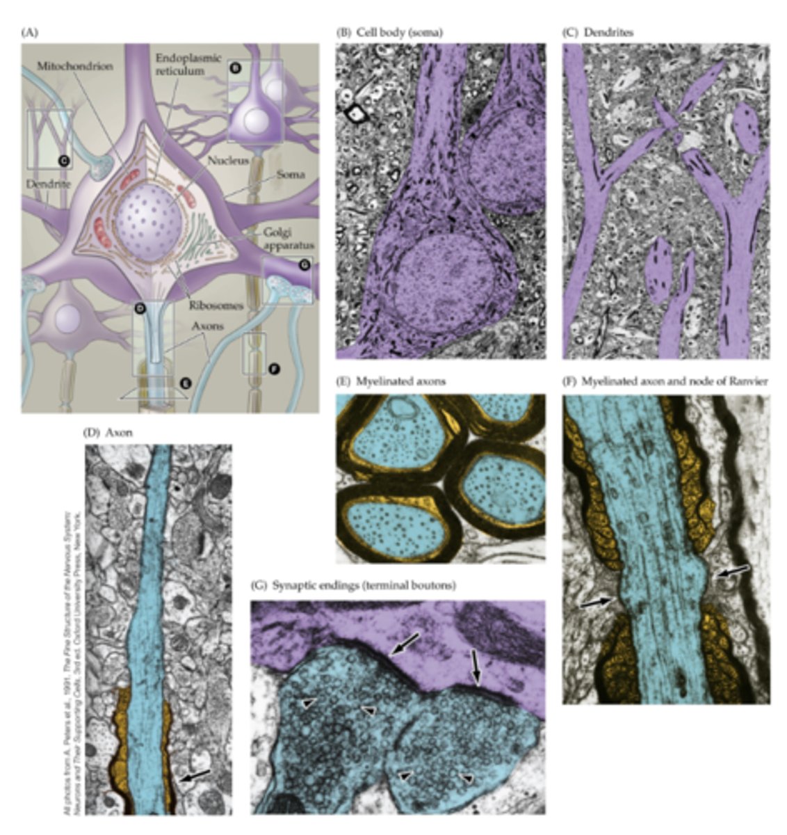
describe what is being shown in G
portion of a myelinated axon (blue) illustrating the intervals that occur between adjacent segments of myelin (gold and black) referred to as nodes of Ranvier (arrows)

describe what is being shown in E
apical dendrites (purple) of cortical pyramidal cells

what are dendrites (and the soma) rich in? what do they cause lots of in these areas?
ribosomes, protein production
in the nodes of Ranvier, what are the node and paranode regions?
node regions are those with Na+ channels and paranode regions are those with K+ channels
what is morphological similar between most/all neurons?
cell body/soma, dendrites, axons, presynaptic terminal, synapse, growth cone, and presynaptic bouton
how do dendrites help with an AP?
they turn ligand binding into the flow of ions which creates electrical flow
what is the purpose of a growth cone?
they are found at the tip of axonal projections, are the sensory and motile organelles of developing neurons that enable axon pathfinding and target recognition for precise wiring of neural circuitry
what do neurons with growth cones have lots of?
mitochondria for growth
how does a growth cone determine how a neuron will grow?
the fillipodia (on the outside of the dendrites) will send signals to the lamellipodia to determine how the neuron will grow........... the filopodia are thin, finger-like protrusions that act as sensors, detecting environmental cues to guide the growth cone's path.......lamellipodia are broader, sheet-like structures that extend from the growth cone's leading edge and are thought to generate the force for its movement
what is the presynaptic button?
also called the axon terminal or synaptic bouton, is the specialized, knob-like structure at the end of a neuron's axon that releases neurotransmitters to communicate with other neurons or target cells. It contains synaptic vesicles filled with neurotransmitters, as well as numerous mitochondria to provide the energy needed for the release process
how were some of the early morphological differences discovered?
through the work of Ramon y Cajal/the Neuronal Doctrine and the link between morphology and function
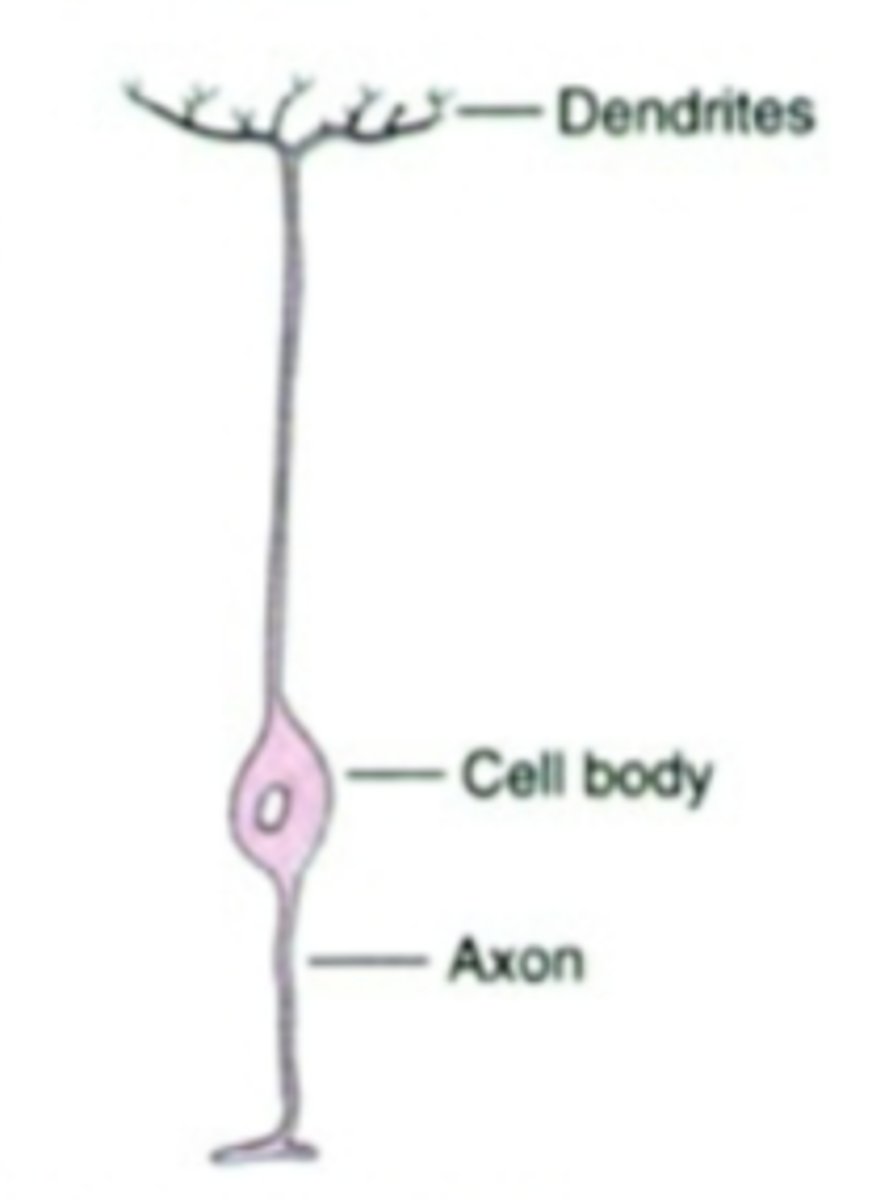
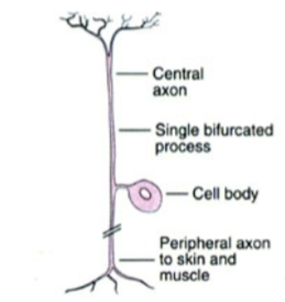
neurons are usually grouped by what? and what are the different classes of that?
morphology..... unipolar, pseudo-unipolar, bipolar, and multipolar
what type of cell is shown in the photo and what is an example?
unipolar cell... invertebrate neuron.... secretory cells
what type of cell is shown in the photo and what is an example?
bipolar cell.... bipolar cell of retina..... sensory cells in mammalian/vertebrate nervous system
what type of cell is shown in the photo and what is an example?
pseudo-unipolar cell; ganglion cell of dorsal root (sensory cell, outside of spinal cord)
what types of cell is shown are the photo and what are examples?
multipolar cells (which means multiple dendritic processes)...... motor neuron of spinal cord, pyramidal cell of the hippocampus, purkinje cell of cerebellum
true or false: there is distinctive arrangement of cytoskeletal elements in neurons
true
what would try to stain to identify an astrocyte?
GFAP
what would you try to stain to identify microglia?
P2Y12 and CD11B
what would you try to stain to identify axons?
TAU
what would you try to stain to identify dendrites
MAP2
what are the glial cell types?
astrocytes, oligodendrocytes, and microglia cells
describe astrocytes
they are structurally firm, express GFAP
describe oligodendrocytes
they produces myelin proteins so in order to see them, we can try to stain for phospholipids that would be present in the myelin........ oligodendrocytes have more myelin basic protein than schwaan cells and they have a protein called no go A.....both of these proteins are important for wrapping of the myelin and when the myelin becomes disrupted and the growth cone is exposed, then the growth cone is inhibited by no go A
why does the PNS regenerate and the CNS doesn't
schwaan cells have different proteins than PNS.... Schwaan cells lay down laminin (an adhesive substrate for growth cones) and this gets regeneration