The Fetal Spine and Musculoskeletal System
 Call Kai
 Learn
Learn Practice Test
 Spaced Repetition
 Match
Match Flashcards
 Knowt Play
1/71

There's no tags or description
Looks like no tags are added yet.
Name | Mastery | Learn | Test | Matching | Spaced | Call with Kai | Chat |
|---|
No analytics yet
Send a link to your students to track their progress
72 Terms
Axial skeleton begins to form between & weeks. It consists the bones of the ____ & _____
6 & 8 weeks, cranium & spine
As bones ossify it allows sonographers to readily visualize these structures as echogenic reflections that produce
acoustic shadowing
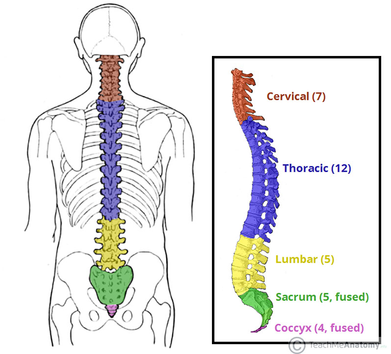
The spine consists of five sections:
cervical, thoracic, lumbar, sacrum, and coccyx.
Each fetal vertebra consists of three echogenic ossification centers:
one centrum and two neural processes
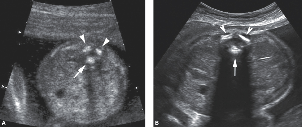
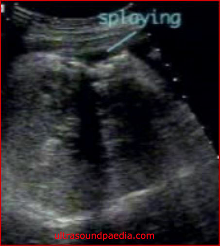
The echogenic laminae are normally angled inward, whereas with spina bifida, the defective laminae will be angled
outward, or splayed
Among the list of neural tube defects are
cephaloceles, anencephaly, and spina bifida. Anencephaly and spina bifida are the most common neural defects,
because of the skin covering, which conidition is not associated with elevated MSAFP?
closed spina bifida
folic acid significantly reduces the likelihood of her fetus developing spina bifida and other neural tube defects, how many mg a day?
0.4 mg. For high-risk, as high as 4 mg may be prescribed
what is spina bifida
AKA spinal dysraphism, meningocele, and meningomyelocele (myelomeningocele). spina bifida is a neural tube defect that occurs when the embryonic neural tube fails to close.
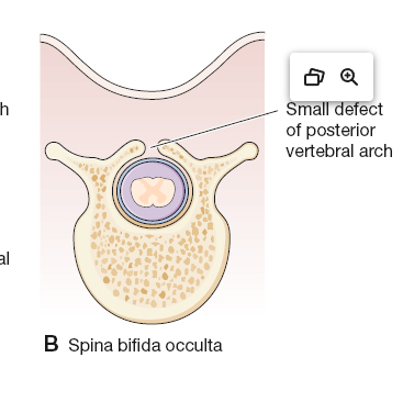
Spina bifida occulta characteristics:
Closed defect
Skin surface abnormality noted on postnatal physical examination can be a sacral dimple, tuft of hair, hemangioma, or lipoma
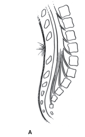
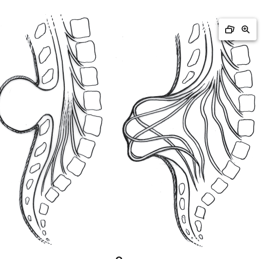
Spina bifida aperta (most common version of spina bifida) characteristics
Typically, an open defect
May also be referred to as spina bifida cystica
Mass is referred to as a meningocele or meningomyelocele (myelomeningocele) depending upon contents
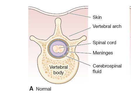
cross sectional of normal vertebra and spinal cord
cross sectional of spina bifida occulta
cross sectional of spina bifida aperta
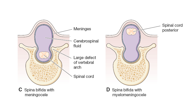
Keep in mind that spina bifida is associated with two yellow fruits
banana sign and lemon sign
With open spina bifida, the exposure of the delicate spinal nerves to the amniotic fluid during fetal life is thought to be one of the causes of
neurologic impairment
Open fetal surgery can be performed on the fetus with spina bifida when a mass is identified on the spine, even as early as
16 weeks
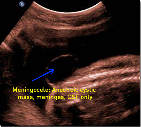
meningocele
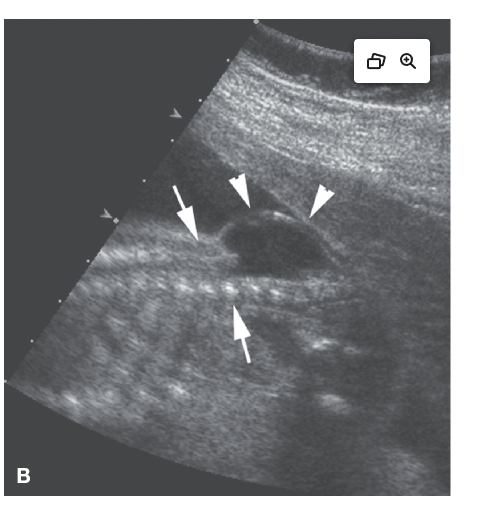
myelomeningocele
Clinical Findings of Open Spina Bifida Aperta (Open)
Elevated MSAFP
Sonographic Findings of Spina Bifida Aperta (Open)
Splaying of the laminae in the area of the defect
Cystic mass (meningocele) or complex mass (myelomeningocele) protruding from the spine
Lemon sign—lemon-shaped cranium with flattened frontal bones
Banana sign—banana-shaped cerebellum
Obliterated cisterna magna
Colpocephaly
Hydrocephalus
Clinical Findings of Spina Bifida Occulta
In utero—normal laboratory values
Postnatal—sacral dimple, hemangioma, lipoma, or excessive hair is identified directly over the distal spine
Sonographic Appearance of Scoliosis
Lateral curvature of the spine
S-shaped spine
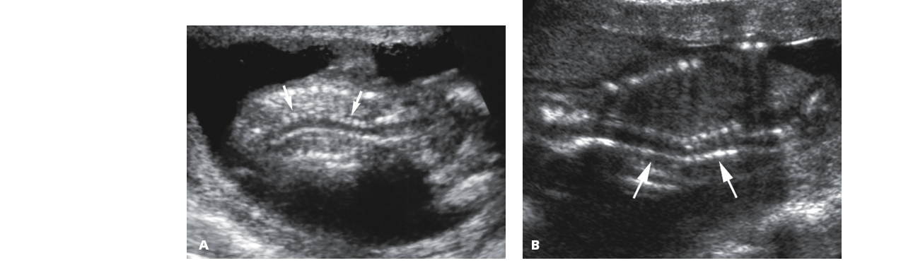
Sonographic Appearance of Kyphosis
Abnormal posterior curvature of the spine
Although these abnormalities may be the only anomaly noted during a fetal sonogram, distortion of the spine can be seen with
hemivertebrae, myelomeningoceles, amniotic band syndrome, and limb–body wall complex (LBWC)
Scoliosis and kyphosis are also often associated with additional anomalies in other systems, as seen in
VACTERL association/ VATER association
LIMB–BODY WALL COMPLEX (LBWC)
AKA body stalk anomaly or short umbilical cord syndrome, is a rare group of fetal defects
There are three postulated causes for this fatal condition LBWC :
vascular occlusion, amnion rupture, or embryonic dysgenesis
Clinical Findings of Limb–Body Wall Complex
Elevated MSAFP
Sonographic Findings of Limb–Body Wall Complex
Short or absent umbilical cord
Marked scoliosis
Various other anomalies including craniofacial and limb defects
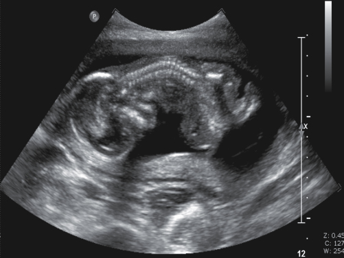
Dysplasia denotes the
abnormal development of a structure.
Skeletal dysplasias exist as a large group of
abnormalities of the skeletal system
The four most common skeletal dysplasias are as
achondroplasia, achondrogenesis, osteogenesis imperfecta, and thanatophoric dysplasia
What is the most common nonlethal skeletal dysplasia
Heterozygous achondroplasia
Heterozygous achondroplasia is
a type of dwarfism in which the proximal portions of the limbs, the humeri and femurs, are much shorter than the distal portion of the limbs, a condition known as rhizomelia.
Heterozygous achondroplasia is an autosomal dominant disorder, although, many times, it is the result of a
spontaneous genetic mutation
Rhizomelia is typically detected when a notable difference in the gestational age measurements between the
biparietal diameter and the femur length is discovered, typically in the mid to late second trimester.
Homozygous achondroplasia, which can occur when both parents are dwarfs, is usually
fatal within the first 2 years of life.
Sonographic Findings of Achondroplasia
Macrocrania
Frontal bossing
Flattened nasal bridge
Micromelia (resulting from rhizomelia)
Trident hand
what is Achondrogenesis
“not producing cartilage”; it is a rare, lethal condition, resulting in absent mineralization of the skeletal bones
Sonographic Findings of Achondrogenesis
Severely shortened limbs (micromelia)
Absent mineralization of the skull, spine, pelvis, and limbs
Large skull
Narrow chest and distended abdomen
Polyhydramnios
Osteogenesis Imperfecta
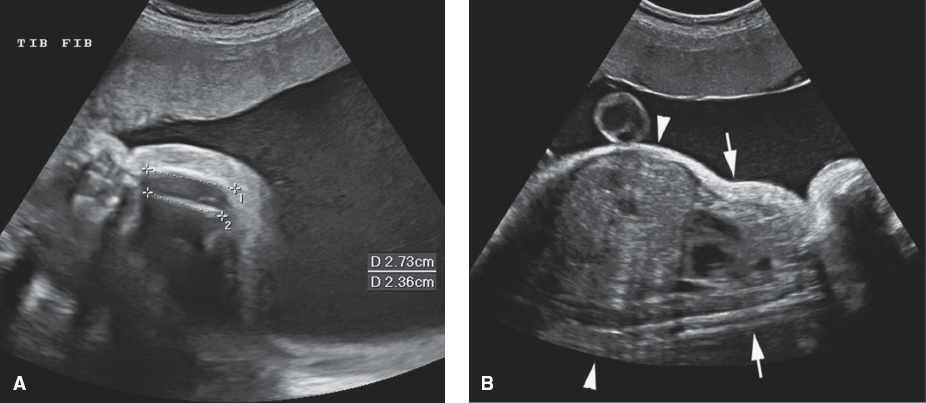
3D findings of achondrogenesis
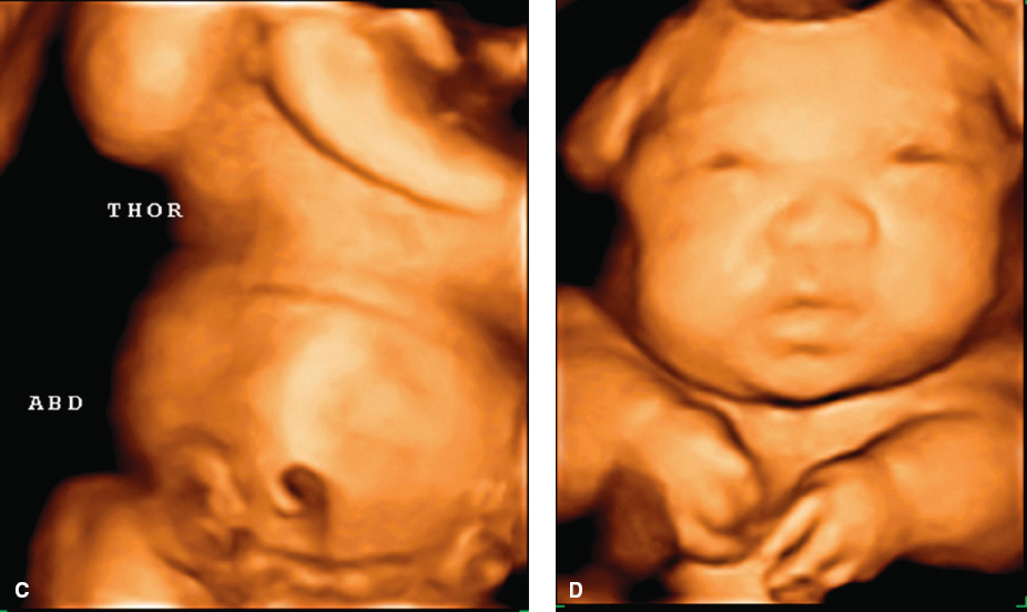
what is Osteogenesis imperfecta,
brittle bone disease, is a group of disorders that results in multiple fractures that can occur in utero
There is 4 types of osteogenesis imperfecta, type 1, 3, and 4 are typically diagnosed after birth. Type 2 is the most severe type and results in
multiple fractures in utero, skull demineralization (recognized by a lack of posterior shadowing), bell-shaped chest, and decreased fetal movement.
One distinctive finding of osteogenesis imperfecta type 2 is
when transducer pressure is applied to the skull, the shape of the “soft” skull can be distorted
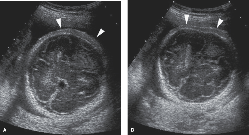
sonographic findings of osteogenesis imperfecta
Demineralization of the skull (transducer pressure can alter the shape of the skull)
Multiple fractures
Bell-shaped chest
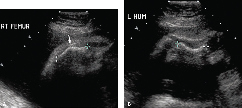
what is Thanatophoric Dysplasia
“death-bearing”; most common lethal skeletal dysplasia. Fetuses typically die shortly after birth, succumbing most often to respiratory distress as a result of pulmonary hypoplasia.
Sonographic Findings of Thanatophoric Dysplasia
Cloverleaf skull
Hydrocephalus
Depressed nasal bridge
Bell-shaped chest (narrow thorax)
Polyhydramnios
Redundant soft tissue
Telephone receiver–shaped long bones
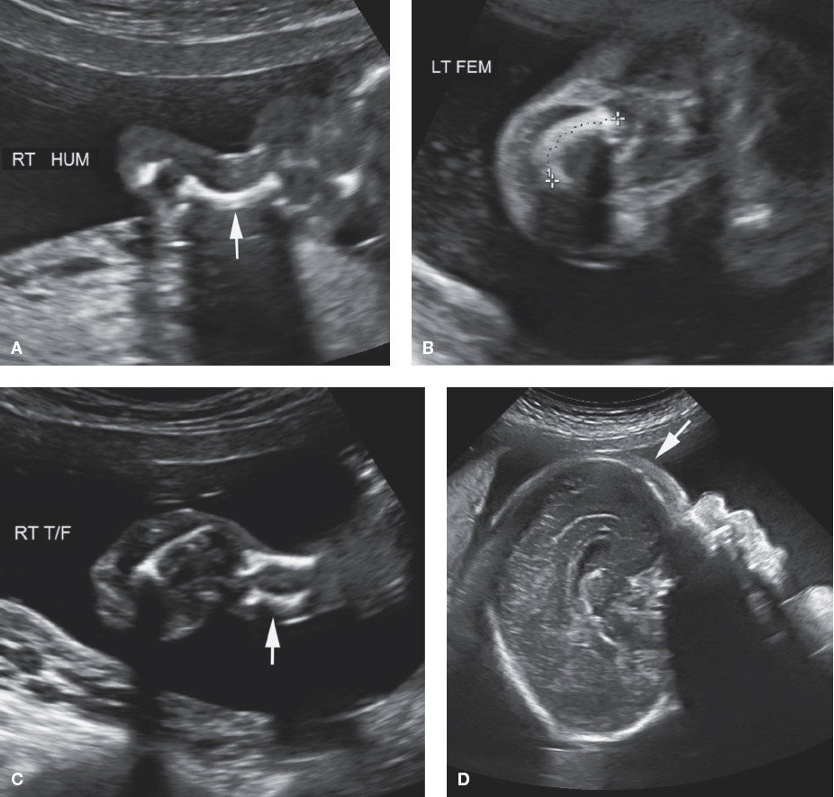
what is Caudal regression syndrome
aka sacral agenesis; Uncontrolled maternal pregestational diabetes has a strong association with caudal regression syndrome.
Clinical Findings of Caudal Regression Syndrome
Uncontrolled maternal pregestational diabetes
Sonographic Findings of Caudal Regression Syndrome
Absent sacrum (sacral agenesis) and possibly part of the lumbar vertebra
Possible abnormalities in the lower extremities like clubfeet
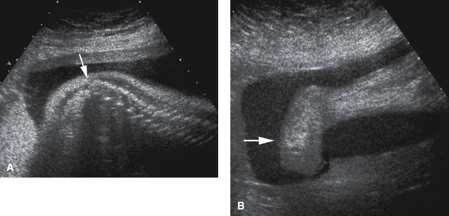
what is Sirenomelia
aka mermaid syndrome because of the fusion of the lower extremities
Clinical Findings of Sirenomelia
Uncontrolled pregestational maternal diabetes
Sonographic Findings of Sirenomelia
Fusion of the lower extremities
Bilateral renal agenesis therefore leading to
Oligohydramnios (possibly anhydramnios)
what is SACROCOCCYGEAL TERATOMA
a germ cell tumor which contains the three different germ cell layers: endoderm, mesoderm, and ectoderm. SCT has been cited as the most common congenital neoplasm and is more frequently found in females
Large SCTs have
malignant potential
Sonographic Findings of Sacrococcygeal Teratoma
Complex mass extending from the distal fetal spine
Mass can be highly vascular
Hydronephrosis may be present (when mass invades the pelvis)
Fetal hydrops may be present
Cardiomegaly
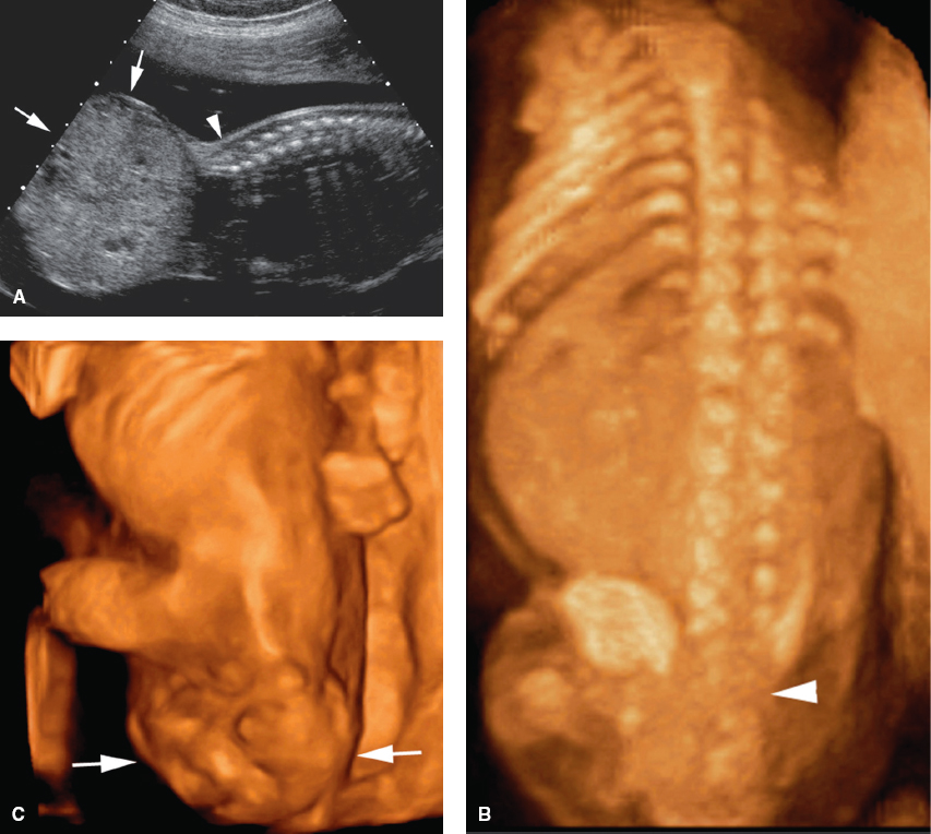
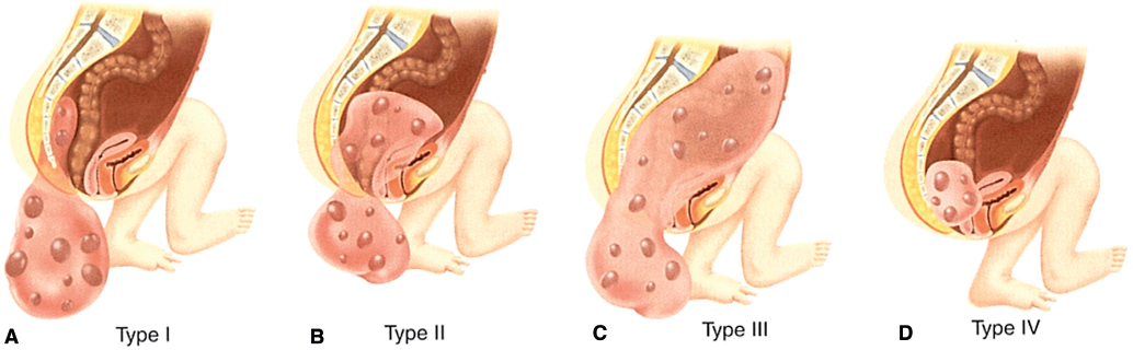
types of sct
Similar to the axial skeleton, the appendicular skeleton begins to form between the
sixth and eighth menstrual weeks
The appendicular skeleton includes the bones of the
upper extremities, lower extremities, and pelvic girdle.
The sonographic determination of the shortening of a limb is made when the
long bones measure more than four standard deviations below the norm for gestational age
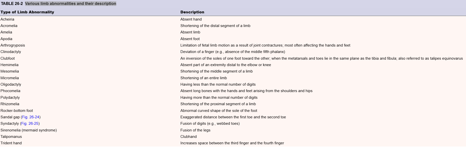
Various limb abnormalities and their description
sandal gap
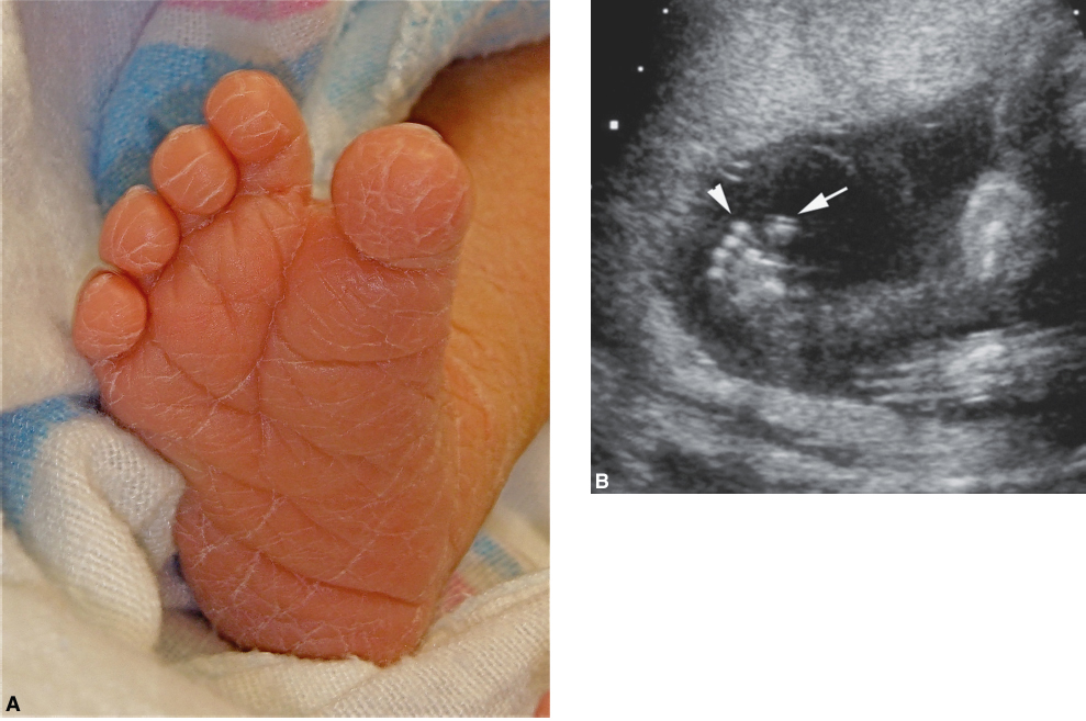
syndactyly
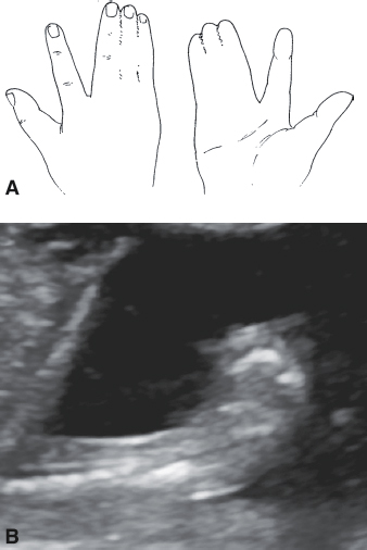
what is Radial ray defect
uncommon and described as the absence (aplasia) or underdevelopment (hypoplasia) of the radius. This abnormality can be seen in the presence of trisomy 13, trisomy 18, and several other syndromes.
Sonographic Findings of Radial Ray Defect
Absent or hypoplastic radius
Various defects in other body systems: cardiac and VACTERL association
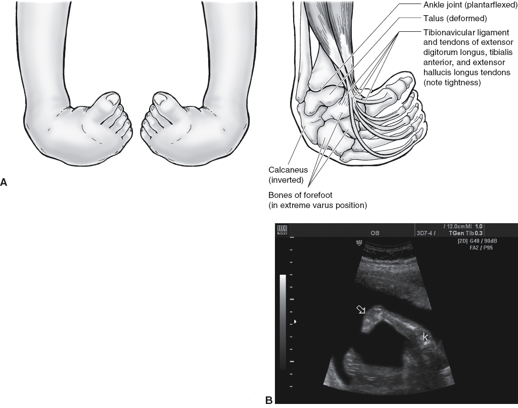
what is Clubfoot,
aka talipes or talipes equinovarus;malformation of the bones of the foot. The foot is most often inverted and rotated medially
. If an abnormal foot shape is suspected, a foot length measurement can be obtained by measuring from the skin edge of the heel (calcaneus) to the distal end of the longest toe. Foot length has been used for gestational dating as well, and it has been shown to be nearly equivalent to the
resulting in a femur-to-foot ratio of approximately 1.0 after 14 weeks’ gestation
Sonographic Findings of Clubfoot
Metatarsals and toes lie in the same plane as the tibia and fibula
Limb reduction can be caused by
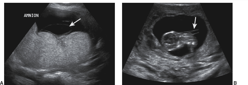
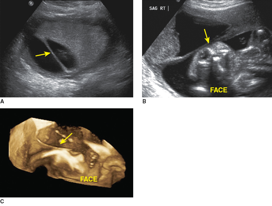
amniotic band syndrome, also referred to as amniotic band sequence. Sticky bands result from the rupture of the amnion.
amniotic bands should not be confused with
uterine synechiae; which may be recognized as linear, thin membranes with a broad base crossing the amniotic sac
Sonographic Findings of Amniotic Band Syndrome
Amputation of fetal parts or severe edema in the affected area
Thin, linear bands may be seen
Facial clefting