The Nervous System: Structure, function, and cellular components
1/104
There's no tags or description
Looks like no tags are added yet.
Name | Mastery | Learn | Test | Matching | Spaced | Call with Kai |
|---|
No analytics yet
Send a link to your students to track their progress
105 Terms
What is the Central nervous system (CNS) composed of?
Composed of the brain and spinal cord
What protects the CNS?
Protected by skull and vertebral column
What is the CNS responsible for?
Processes and integrates all neural information
What is the CNS’s function?
Central command center for bodily functions
What is the Peripheral nervous system (PNS) composed of?
All nerves and neural components outside the CNS
What does the PNS connect?
Connects the CNS to the rest of the body
What does the PNS include?
Sensory and motor pathways
What is the PNS divided into?
Divided into somatic and autonomic divisisions
Define afferent neurons (it’s function, where they are located, and what they are also known as)
Information flows from the periphery to the CNS
Carry sensory information (touch, pain, temperature)
Cell bodies located in dorsal root ganglia
Also known as sensory neurons
Define efferent neurons (it’s function, where they are located, and what they are also known as)
Information flows from the CNS to the periphery
Carry motor commands to muscles and glands
Cell bodies located in ventral horn of spinal cord
Also called motor neurons pathways
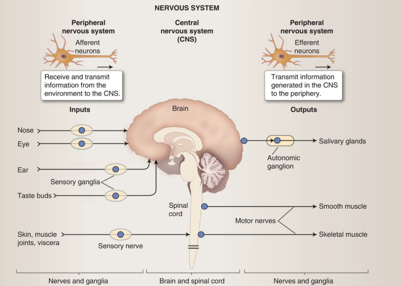
Image to review
How many neurons are in the human brain?
100 billion
Each neuron is connected to how many others?
Each neuron connects with approximately 1,000 other neurons
How are neurons organized?
Neurons are organized into circuits and networks that process specific information
What are the cellular components that form the foundation of all neural activity, enabling everything from basic reflexes to complex cognitive functions like memory, learning, and consciousness?
Scale, connectivity and organization
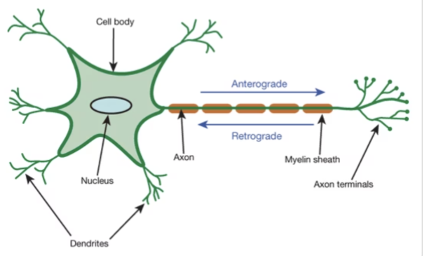
What does the soma (cell body) contain?
Contains the cell nucleus
What is the site of protein synthesis?
Soma (cell body)
What does the soma (Cell body) produce?
Produces neurotransmitters and hormones
What is the soma (cell body) also known as?
Metabolic center of the neuron
What are dendrites?
Branching extensions from the cell body
What does the dendrites receive?
Receives synaptic input (Synaptic input refers to the signals a neuron receives from other neurons at synapses)
What does the dendrites do?
Conduct signals toward soma and increase surface area for connections
Describe the location of the axon and where does it begin?
It is a long projection from the soma
It begins at the axon hillock
What does the axon do?
Conducts action potentials and make synaptic contacts
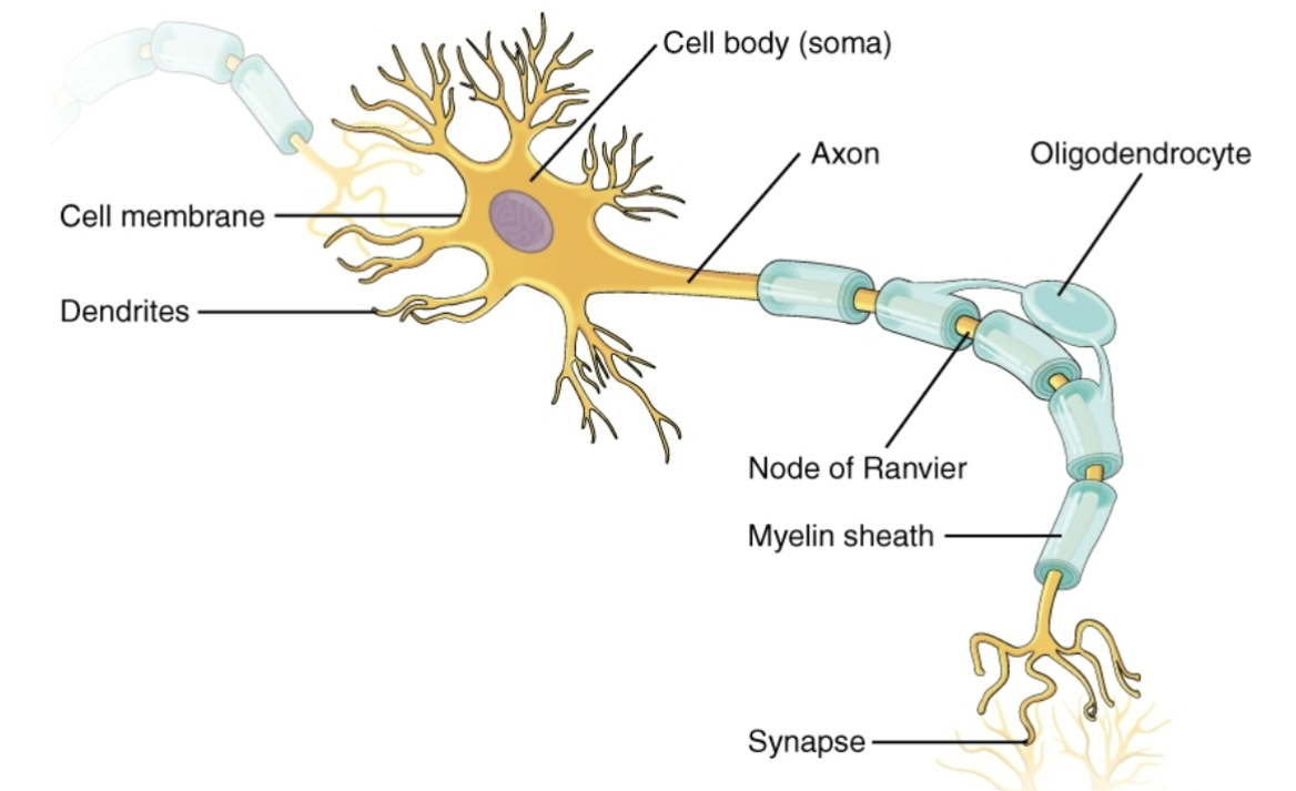
Image to review
How can neurons be classified?
By function, morphology, and neurotransmitters
What are some examples of neurons being classified by it’s function?
Sensory (afferent) neurons
Motor (efferent) neurons
Interneurons (local circuit)
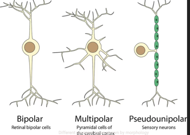
What are some examples of neurons being classified by it’s morphology?
Morphology are it’s shape BTW
Multipolar neurons
Bipolar neurons
Pseudounipolar neurons
What are some examples of neurons being classified by it’s neurotransmitters?
Cholinergic neurons
GABAergic neurons
Dopaminergic neurons
Glutamatergic neurons
What is the most abundant type of neuron in the CNS?
Multipolar neuron
What does a multipolar neuron look like?
Multiple dendrites and single axon
Where is the multipolar neuron found and what are some examples?
They are found on brain and spinal cord
Examples: motor neurons, pyramidal cells
Where are the pseudounipolar neurons found?
Found primarily in spinal ganglia
What does a pseudounipolar neurons look like?
It is one process that divides into two, one going to the periphery and the other to the CNS
No dendrites. The axon serves both functions
What does the pseudounipolar neurons do?
Relay sensory information to CNS
Where are the bipolar neurons found?
Found in retina and olfactory epithelium
What does the bipolar neuron look like?
Single dendrite and single axon
What does the bipolar neuron do?
Specialized for sensory transduction (they are responsible for directly converting a stimulus into a neural signal)
Linear transmission of information (the signal flows in a straight way)
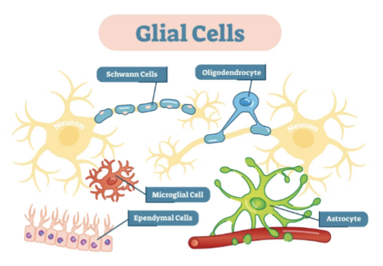
What are glial cells also called?
The supporting cast
How are neurons compared to glial cells?
While neurons are the primary signaling cells of the nervous system, glial cells outnumber neurons by approximately 10:1 and perform essential supportive functions
What do glial cells do?
Support and protect neurons
Provide the stem cell pool within the nervous system
Maintain homeostasis of the neural environment
Mediate immune responses to inflammation and injury
Participate in synaptic signaling and modulation
Guide neuronal development and migration
What are astroglia cells also called?
The neural caretakers
What are the types of astroglia cells?
Fibrous astrocytes - found in white matter
Protoplasmic astrocytes - found in gray matter
Müller cells - specialized astroglia in retina
What do astroglia cells do?
Provide physical and metabolic support to neurons
Regulate blood flow to active neural regions
Supply nutrients and remove waste products
What are the homeostatic functions for astroglia cells?
Maintain ion balance at synapses
Recycle neurotransmitters after synaptic transmission
Form essential component of blood-brain barrier
What do Myelinating Glial Cells form?
The Oligodendrocytes (CNS) and Schwann Cells (PNS) both form the myelin sheath
Define oligodendrocytes (where it’s found, function, and fun fact)
Found exclusively in central nervous system
Create myelin sheaths around CNS axons
Less capable of regeneration than Schwann cells
Define schwann cells (where it’s found, function, and fun fact)
Found exclusively in peripheral nervous system
Act as phagocytes to clear debris after injury
Regulate neurotransmitter levels at neuromuscular junction
What is the functional significance of myelin?
Myelin increases the speed of action potential conduction through saltatory conduction (when it jumps from one gap to the next) allowing for rapid neural communication across long distance
Demylenating conditions like multiple sclerosis highlight how important these glial cells are
What are microglia also called as?
The neural immune system
Microglia are the resident immune cells of the central nervous system
What do the microglia cells do?
Constant surveillance of neural environment
Phagocytosis (eliminating) of cellular debris and pathogens
Release inflammatory molecules (cytokines, chemokines)
Synaptic pruning during development (synaptic pruning= eliminating weak or unused synaptic connections between neurons)
Modulation of neuronal circuits (aka, changing the strength, timing, or overall activity of a neural circuit to alter its function or the information it processes)
What happens when microglial activity are not regulated?
It can cause microglia cells to become dysfunctional and contributing to the onset and worsening of neurodegenerative diseases like Alzheimer's, Parkinson's, and ALS
What are the two type of specialized glial cells?
Ependymal cells and polydendrocytes
What does the ependymal cells do?
Line the ventricles of the brain
Create barrier between CSF and neural tissue
Contribute to choroid plexus structure
Ciliated cells help circulate CSF
May retain neural stem cell properties
What does the polydendrocytes do?
Function as neural stem/progenitor cells
Generate both neurons and glial cells
Respond to neural injury
Why are ependymal cells and polydendrocytes important?
They perform unique functions in specific regions of the nervous system, highlighting the complexity and specialization of neural support systems
What are synapses also known as?
Communication junctions
Synapses are specialized junctions where neurons communicate with other cells. Information processing in the nervous system depends on these precise connections
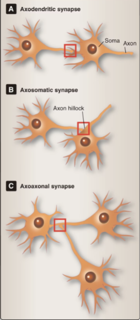
Define axodendritic
The most common type of synapses
Axon terminals will contact with dendrites of another neuron
The dendritic tree will receive thousands of axodendritic synaptic inputs.
Thousands of these inputs allow for temporal and spatial summation. If enough signal adds up, the neuron will dire an action potential (temporal summation= signals coming close together in time pile up, spatial summation = signals coming from different spots on the dendrites combine)
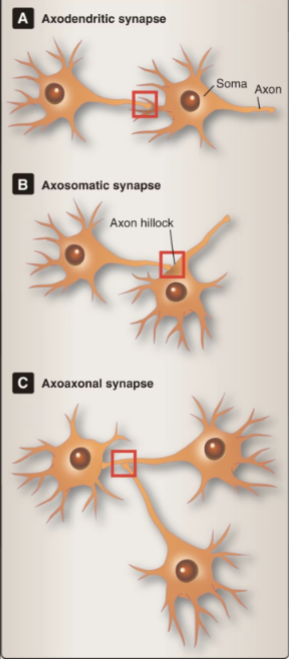
Define axosomatic
Axon terminals contact directly on cell soma.
Less common but powerful, especially near axon hillock, where action potentials originate
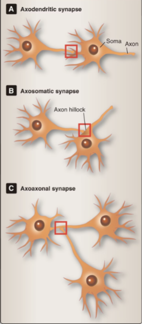
Define axoaxonic
Axon terminals contact another axon.
These can powerfully modulate neurotransmitter release at the terminal, providing presynaptic control
Define blood-brain barrier (BBB)
The blood-brain barrier (BBB) is a highly selective semipermeable border that separates the circulating blood from the brain and extracellular fluid in the central nervous system
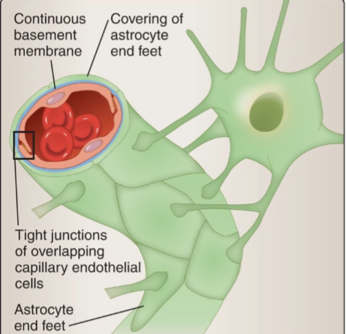
What are the key components to the blood-brain barrier?
Endothelial cells with tight junctions
Basement membrane
Astrocytic end-fee
What is the function of the blood-brain barrier?
Protects brain from pathogens and toxins
Regulates ion balance for optimal neural function
Controls selective transport of nutrients
Prevents most medications from reaching brain tissue
Define ions
Charged particles that create electrical potentials across cell
membranes
Define homeostasis
Homeostasis maintains stable internal conditions despite external changes.
Define membrane potential
Difference in electrical charge across a membrane. Resting potential is the stable voltage when a neuron is inactive
Define polarization
Normal state
Define depolarization
Membrane potential becomes LESS negative (moving closer to zero)
Action Potential more likely to occur
Define hyperpolarization
Membrane potential becomes MORE negative
Action potential less likely to occur
Define action potential
Electrical signal that travels along a neuron. Propagation is the movement of this signal along the axon
Define leak channels
Passive channels that allow specific ions to diffuse across the membrane continuously
Define mechanically-gated
Sensitive to physical forces like pressure, stretch, or vibration
Define ligand-gated
Open or close in response to specific neurotransmitters or chemical signals.
Define voltage-gated
Respond to changes in membrane potential; critical for action potential generation.
Define thermally-gated
Activated by temperature changes; important for sensory perception
What does membrane potential represent?
The membrane potential represents the electrical charge difference across a neuron's membrane. This potential changes during neural activity
Define resting potential
Homeostatic state
Maintained at approximately -70mV with the inside negative and outside positive.
ATP-powered ion pumps maintain this differential
Define local potential
Initial changes from sensory input or synaptic activity.
May be excitatory or inhibitory.
Define action potential and what takes place
The propagating wave of depolarization that transmits information along the axon.
Occurs when threshold is reached.
Depolarizing wave in consistent direction
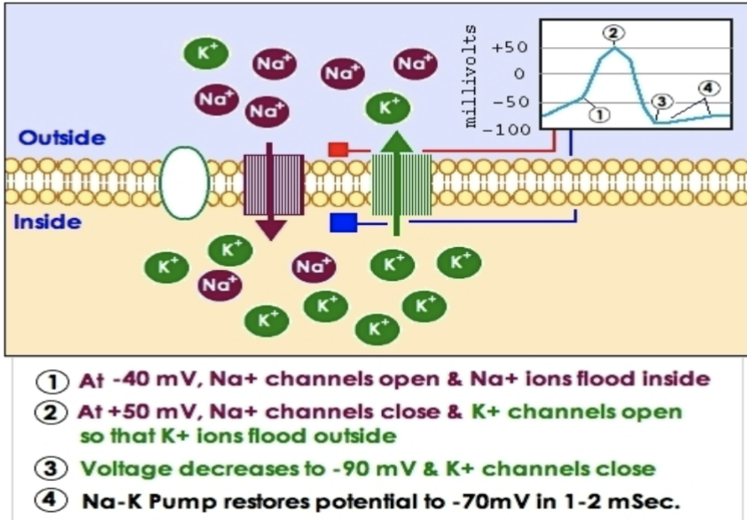
Describe how action potential step-by-step
At it’s resting state, the inside of neuron is negative (-70mV)
-Sodium (+) is mostly on the outside and potassium (+) mostly inside
-The gates for sodium and potassium gates are closed
Stimulus occur, which means depolarization begins
-Stimulus opens up some Na+ channels
-Na+ will rush in, making inside less negative
-If it reaches threshold, action potential will start (-55mV)
Rising phase (depolarization)
-More Na+ channels open
-Huge influx of Na+ makes inside positive
Falling phase (repolarization)
-Na+ channels closes
-K+ channels opens, K+ rushes out, making the inside negative again
Undershoot (hyperpolarization)
-K+ channels stay open a bit too long
-Inside becomes extra negative
Return to resting potential
-K+ channels close
-Sodium-potasium pump restores normal balance (3 Na+ pumped out and 2 K+ pumped in)
What are the negatively charged ions?
Chlorine and anions
What are the positively charged ions?
Sodium, potassium, and calcium
Describe how action potential is an all or none response
Once threshold is reached, full action potential always occurs
Action potentials function as binary signals - they either occur completely or not at all. This "all-or-none" principle ensures reliable signal transmission throughout the nervous system.
Define action potential
Action potentials are electrical impulses that travel along the neuron's membrane. They represent the fundamental mechanism of information transmission in the nervous system
When are some key components to action potential?
Changes in membrane permeability for different ions drive the
action potential
Na+ influx initiates depolarization
K+ efflux drives repolarization
Enables communication between neurons
What happens if there’s a sodium influx in action potential?
Influx means moving into the cell
Na+ channels open in response to reaching threshold potential.
Sodium ions rush into the cell, causing depolarization as the
membrane potential becomes more positive.
What happens if there’s a potassium efflux in action potential?
Efflux means moving out of the cells
Na+ channels inactivate quickly. K+ channels open more slowly,
allowing potassium to flow out of the cell, causing repolarization as
the membrane potential returns to negative.
What is restoration in action potential?
Na+/K+ ATPase pumps work to restore original ion concentrations,
moving Na+ out and K+ in, reestablishing the resting membrane
potential.
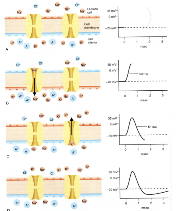
What are the phases of action potential?
A. Resting Membrane Potential
B. Rapid depolarization due to influx of Na+ channels
C. Rapid depolarization due to efflux of K+ channels
D. RMP restored-diffusion of ions
Define the all of none principle in action potential
If threshold is reached – Cell will fire
If depolarization reaches threshold (typically -55mV), a full
action potential is generated
If threshold is not reached, no action potential occurs
The magnitude of an action potential is constant regardless of
stimulus strength
Stronger stimuli do not produce larger action potentials - they
may produce more frequent ones
This binary nature ensures reliable signal transmission throughout the nervous system and prevents signal degradation over long distances.
Define absolute refractory period
During this phase, the neuron cannot generate another action potential regardless of stimulus strength.
No response
Typically lasts 1-2 milliseconds
Ensures unidirectional propagation (ensures that an action potential travels only one direction down the axon)
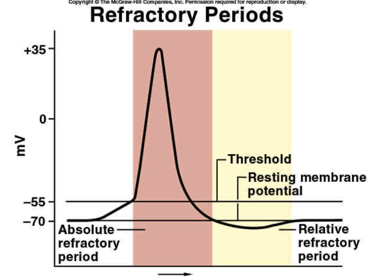
Define relative refractory period
During this phase, a stronger-than- normal stimulus may trigger another
action potential
Decreased response
Requires higher threshold for firing
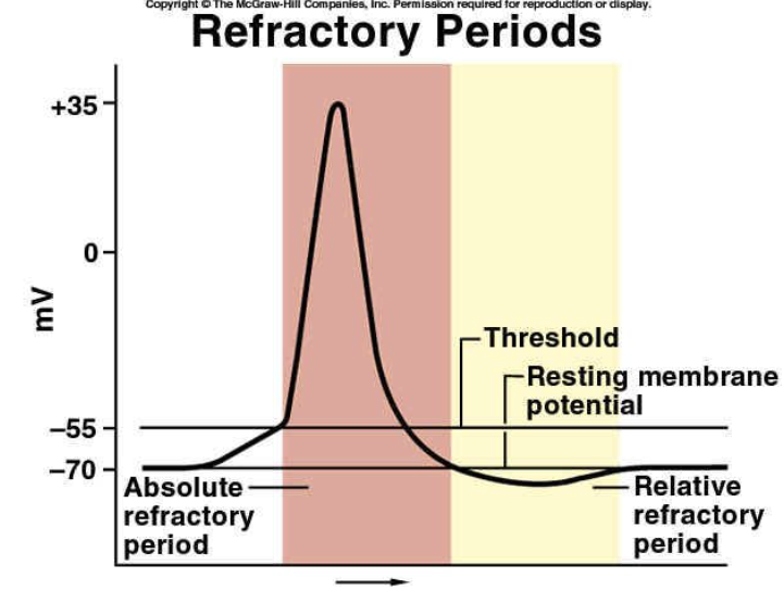
Why is the refractory period important?
The refractory period is critical for proper neural function. It limits the maximum frequency of action potentials, controls information transmission rates, and prevents backflow of the signal
What are the types of neural conduction?
Unmyelinated and myelinated axons
Define unmyelinated axons in regards to action potential
In unmyelinated axons, action potentials propagate continuously along the entire membrane:
Na+ channels distributed throughout axon length
Signal travels as continuous wave
Slower conduction velocity
Common in autonomic nervous system
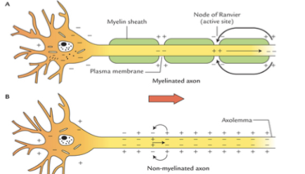
Define myelinated axons in regards to action potential
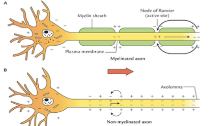
In myelinated axons, action potentials "jump" between nodes of Ranvier:
Na+ channels concentrated at nodes
Signal "jumps" between nodes (saltatory conduction)
Much faster conduction velocity
More energy efficient
The more myelin and larger the axon diameter, the faster the signal transmission
What are factors affecting signal speed and integration
Axon diameter, myelination, temporal summation, and spatial summation
How does axon diameter affect signal speed and integration
Larger diameter axons conduct signals faster due to decreased internal
resistance. This is why giant squid axons (up to 1mm diameter) were
crucial for early neurophysiology research
How does myelination affect signal speed and integration
Myelin sheaths created by Oligodendrocytes (CNS) or Schwann cells
(PNS) insulate axons and enable saltatory conduction, increasing
transmission speed up to 100 times
How does temporal summation affect signal speed and integration?
Integration of multiple signals arriving in rapid succession at a single
location. If multiple small signals arrive within milliseconds, their effects
add up