7 Medical Treatment Algorithms
 Call Kai
 Learn
Learn Practice Test
 Spaced Repetition
 Match
Match Flashcards
 Knowt Play
1/96

There's no tags or description
Looks like no tags are added yet.
Name | Mastery | Learn | Test | Matching | Spaced | Call with Kai | Chat |
|---|
No analytics yet
Send a link to your students to track their progress
97 Terms
Does lowering intraocular pressure (IOP) actually prevent or slow glaucoma?
Yes. Lowering IOP is the only proven treatment strategy shown to:
Reduce conversion of ocular hypertension (OHT) to primary open-angle glaucoma (POAG) (OHTS)
Decrease the risk of glaucoma progression and optic nerve damage in patients who already have glaucoma (EMGT)
What is the relationship between IOP and glaucoma progression risk?
Higher IOP is associated with a higher risk of progression, while lowering IOP reduces risk.
Each mmHg reduction in IOP decreases the likelihood of glaucoma progression.
IOP is the primary modifiable risk factor in glaucoma management.
What factors should be considered when deciding whether to start treatment for ocular hypertension (OHT)?
Treatment decisions are individualized and should consider:
Current IOP level
Risk of developing POAG
Expected benefit from therapy
Burden/costs of lifelong treatment
Overall health status and life expectancy
Why is the decision to begin hypotensive therapy in glaucoma considered complex?
Because glaucoma diagnosis and management depend on multiple variables, not IOP alone:
Intraocular pressure
Structural damage (optic nerve/RNFL changes)
Functional damage (visual field defects)
Individual patient risk factors and prognosis
What structural and functional findings are evaluated when considering glaucoma treatment?
Structural findings:
Optic nerve head changes (cupping)
Retinal nerve fiber layer (RNFL) loss
OCT abnormalities
Functional findings:
Visual field defects/perimetry changes
What evidence of progression should prompt reconsideration or escalation of glaucoma treatment?
Evidence of structural or functional worsening over time:
Progressive RNFL thinning on OCT
Worsening optic nerve cupping/neuroretinal rim loss
New or enlarging visual field defects
Increasing rate of change on trend analyses
Why are serial OCT and visual field tests important in glaucoma management?
Glaucoma is a progressive disease, so management relies on comparing exams over time to detect:
Structural progression (optic nerve/RNFL loss)
Functional progression (visual field deterioration)
How do disease severity, age, and life expectancy influence treatment decisions in glaucoma?
Treatment aggressiveness should be based on the likelihood that glaucoma will cause meaningful visual disability during the patient's lifetime.
Younger patients: Often treated more aggressively because they have more years to accumulate damage.
Older patients with limited life expectancy: May require less aggressive treatment if progression is slow.
What are the general goals of glaucoma treatment?
To reduce the risk of progressive optic neuropathy in:
High-risk patients who have not yet developed glaucoma (e.g., OHT, strong family history, systemic risk factors)
Patients with established glaucoma and existing optic nerve damage
What is the ultimate goal of glaucoma therapy?
Prevent (further) vision loss.
Glaucoma treatment slows or prevents progression.
Existing optic nerve damage is generally irreversible.
Lost visual function is typically not recoverable.
What is the most important counseling point for patients starting glaucoma treatment?
Glaucoma therapy is designed to preserve current vision and prevent future loss, not to improve vision that has already been lost.
What are the two specific IOP-related goals of glaucoma treatment?
Achieve an adequate percentage reduction in IOP relative to the highest untreated IOP (Tmax).
Maintain a consistent, acceptable IOP with minimal diurnal (daily) fluctuation.
What is Tmax, and why is it important in glaucoma management?
Tmax is the patient's highest measured untreated IOP.
Used as the baseline for determining treatment effectiveness.
Target IOP is typically set as a percentage reduction from Tmax.
An inaccurately low Tmax may result in undertreatment.
Why should multiple pretreatment IOP measurements be obtained before establishing a target IOP?
Because IOP fluctuates throughout the day.
Multiple measurements:
Better estimate the patient's true untreated Tmax
Identify the degree of diurnal fluctuation
Help establish a more accurate treatment target
How is a patient's true Tmax best identified?
Measure IOP at different times of day before treatment.
What three factors are required to maintain a stable, acceptable IOP long term?
Appropriate medication selection and dosing regimen
Appropriate follow-up testing to monitor stability or progression
Patient adherence/compliance with therapy and follow-up
Why is patient adherence one of the most important determinants of glaucoma treatment success?
Even highly effective medications cannot prevent progression if they are not used consistently.
Poor adherence can lead to:
Increased IOP fluctuations
Inadequate pressure reduction
Increased risk of optic nerve damage and progression
What is the Medication Possession Ratio (MPR), and what does an MPR < 1.0 suggest?
MPR measures how consistently patients obtain their medications.
MPR < 1.0 suggests the patient does not have medication available for every day prescribed.
Indicates likely nonadherence/noncompliance with glaucoma therapy.
What are the most common barriers to glaucoma medication adherence?
Difficulty instilling eye drops
Forgetfulness
Low confidence in administering drops correctly
Difficulty following the dosing schedule
What factors determine whether a patient can maintain a stable, acceptable IOP over time?
Proper medication selection and dosing
Appropriate follow-up testing to assess stability/progression
Patient adherence to medications and appointments
What is a target pressure in glaucoma management?
A target pressure is the IOP that clinicians aim to achieve with treatment because it is believed to be low enough to minimize future glaucomatous damage and progression.
Based on the patient's disease severity and risk profile
Can be expressed as a number or range (e.g., <18 mmHg, 18-21 mmHg, mid-teens)
How is a target IOP selected?
Target IOP is determined by considering:
Tmax (highest untreated IOP)
Severity of glaucoma
Evidence of progression
Age and life expectancy
Risk factors for future vision loss
What did the Advanced Glaucoma Intervention Study (AGIS) demonstrate regarding target pressures?
AGIS found that patients with advanced glaucoma experienced less visual field progression when IOP was maintained below 18 mmHg at every visit.
Why is it incorrect to interpret AGIS as proving that every glaucoma patient only needs an IOP <18 mmHg?
Because the patients who maintained IOP <18 mmHg at every visit actually had an average IOP of approximately 12 mmHg.
How does glaucoma severity affect target IOP selection?
As disease severity increases, target IOP generally decreases.
Typical approach:
Mild glaucoma → upper teens acceptable
Moderate glaucoma → mid-teens
Severe/advanced glaucoma → low teens or lower
What did the Early Manifest Glaucoma Trial (EMGT) teach about target IOP reduction?
EMGT demonstrated that a 25% reduction in IOP decreases glaucoma progression risk, but does not eliminate it.
Approximately 45% of patients still progressed despite a 25% IOP reduction.
Therefore, some patients require more aggressive pressure lowering.
Why can't the same target IOP be used for every glaucoma patient?
Patients differ in:
Baseline (Tmax) IOP
Severity of optic nerve damage
Rate of progression
Risk factors
Remaining visual lifespan
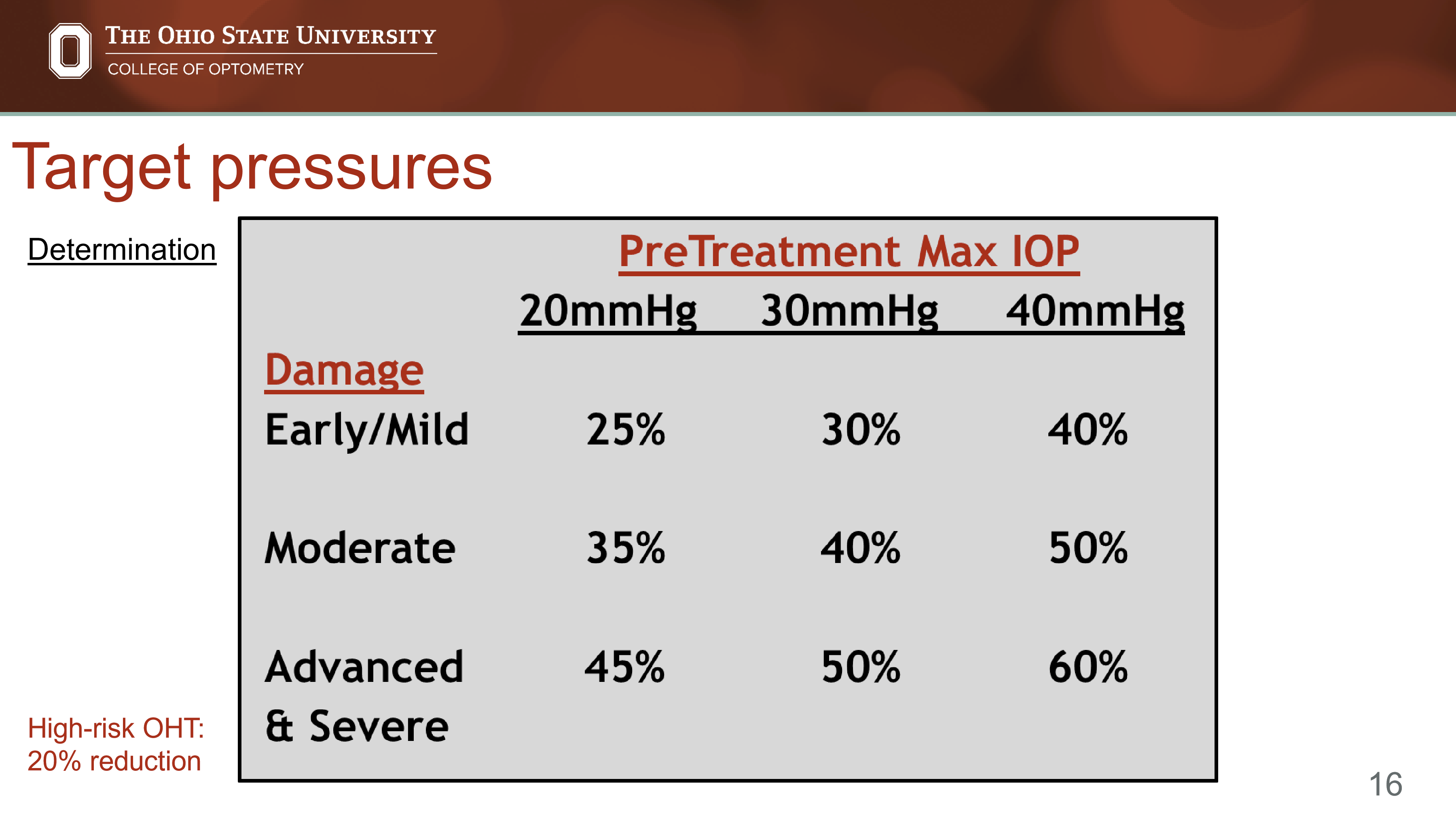
How do glaucoma severity and Tmax influence the recommended target IOP reduction?
The greater the severity and/or Tmax, the larger the recommended pressure reduction.
General guideline:
Early/Mild disease: ~25-40% reduction
Moderate disease: ~35-50% reduction
Advanced/Severe disease: ~45-60% reduction
What factors may warrant an additional 5% reduction beyond the initial target IOP?
Add ~5% additional IOP reduction for each of the following:
Long life expectancy at diagnosis (<60 years old)
Pseudoexfoliation glaucoma/syndrome (PXE)
Drance (optic disc) hemorrhage
Worsening β-peripapillary atrophy (β-PPA)
Once Tmax and target IOP are established, what should be done at each follow-up visit?
At every visit:
Measure current IOP
Compare it to Tmax
Compare it to the target IOP
Adjust therapy if necessary to achieve target pressure
How are OCT and visual field testing used after a target pressure is established?
OCT and visual field testing determine whether the patient is:
Stable → current target pressure may be appropriate
Progressing → target pressure likely needs to be lowered
How can you tell that a target pressure is too high?
If the patient continues to demonstrate:
Optic nerve deterioration
RNFL thinning
GCA loss
Visual field progression
then the target pressure was insufficient and should be lowered.
What is the overall goal when selecting glaucoma medications?
Reach the target IOP using:
The fewest drops
The fewest medications
The fewest bottles
What factors should be considered when selecting a glaucoma medication?
Efficacy
Magnitude of IOP reduction
Ability to flatten diurnal IOP fluctuations
Safety
Contraindications
Drug interactions
Side effects
Patient factors
Cost
Dosing schedule
Ease of use and acceptance
Why are prostaglandin analogs (PGAs) considered first-line therapy for most glaucomas?
Lower IOP by up to ~35%
Produce the flattest diurnal IOP curve
Have few systemic side effects
Are typically dosed once daily
Are widely available as generics
What is the mechanism/main clinical effect of prostaglandin analogs?
Increase uveoscleral outflow → significant reduction in IOP.
When are prostaglandin analogs (PGAs) NOT considered first-line therapy?
Inflammatory glaucoma
Patients with a history of recurrent uveitis
Patients with a history of cystoid macular edema (CME)
Situations requiring immediate large IOP reduction (e.g., acute angle-closure or other IOP emergencies)
Why should PGAs be used cautiously in patients with recurrent uveitis?
PGAs may potentially:
Worsen ocular inflammation
Trigger recurrence of uveitis in susceptible patients
Why are PGAs used cautiously in patients with a history of cystoid macular edema (CME)?
PGAs can increase the risk of:
New CME
Recurrent CME in predisposed eyes
When should prostaglandin analogues be used with caution due to cosmetic side effects?
Use PGAs with caution when cosmetic changes could affect the patient’s occupation or well-being. Key effects include:
Permanent iris pigmentation, especially in mixed-color irides
Periorbital pigmentation
Deepening of the upper eyelid sulcus
Lengthening of eyelashes/cilia
Why should clinicians “beware of monotherapy” with prostaglandin analogues?
PGA cosmetic effects can be asymmetric if used in only one eye, causing noticeable differences between eyes, such as unequal iris color, eyelash length, periocular pigmentation, or eyelid sulcus depth.
What are the major legacy prostaglandin analogues, and what are their high-yield distinguishing features?
Lumigan: bimatoprost 0.01%; generic bimatoprost 0.03%
High efficacy
Travatan Z: travoprost 0.004%
Longest duration of action
Branded version is BAK-free and preserved with SofZia
Xalatan: latanoprost 0.005%
Least likely to induce redness
What are the main non-BAK prostaglandin analogue options?
Zioptan: preservative-free tafluprost 0.0015%
Available as generic
Single-dose containers
Iyuzeh: preservative-free latanoprost 0.005%
Single-dose containers
Xelpros: BAK-free latanoprost 0.005%
Preserved with potassium sorbate
What are “next-generation” prostaglandin analogue medications designed to do differently from legacy PGAs?
Next-generation PGAs work slightly differently than legacy PGAs to either:
Increase IOP-lowering efficacy
Minimize side effects
Improve tolerability or adherence
How does Vyzulta differ from traditional latanoprost, and what is the clinical benefit?
It combines PGA activity with a nitric oxide-driven effect on the trabecular meshwork, which increases aqueous outflow through the conventional pathway. It averages 1.23 mmHg lower IOP than latanoprost and may increase perfusion to the optic nerve head.
What is Rocklatan, and why is it considered more potent than a standard PGA alone?
It combines:
Latanoprost: increases uveoscleral outflow
Netarsudil: ROCK/ROK inhibitor that increases trabecular meshwork outflow and lowers episcleral venous pressure
What is OMLONTI, and why may it have fewer cosmetic adverse effects than traditional PGAs?
It is an EP2 receptor agonist, not an FP receptor agonist.
Because many classic PGA cosmetic effects are associated with FP receptor activation, OMLONTI may reduce cosmetic adverse effects such as iris pigmentation, eyelash changes, and periorbital changes.
Is switching from one PGA to another effective for improving IOP control?
Yes. Many studies show that most patients have improved IOP response after switching to a second PGA. This may be due to:
Improved adherence when patients are told the first drug is not working
More time on therapy allowing IOP to reach target
Possible true pharmacologic differences between PGAs
If a patient has not reached target IOP on Xalatan or Travatan Z, what PGA switches are suggested?
If target pressure is not reached, switch from:
Xalatan or Travatan Z
to a stronger or next-generation option:
Lumigan
Vyzulta
Rocklatan
If a patient develops significant redness or hyperemia on Lumigan or Travatan Z, what switch may improve tolerability?
If redness is the issue, switch from:
Lumigan or Travatan Z
to options with potentially less redness or better tolerability:
Xalatan
Xelpros
Iyuzeh
Zioptan
How should PGA switching be guided clinically?
PGA switching depends on the problem:
Need more IOP lowering:
Switch to Lumigan, Vyzulta, or RocklatanNeed less redness or better tolerance:
Switch to Xalatan, Xelpros, Iyuzeh, or ZioptanNeed fewer preservative-related issues:
Consider Zioptan, Iyuzeh, or Xelpros
When are topical beta blockers used as an alternative first-line glaucoma medication?
Use topical beta blockers, up to about 30% IOP reduction, when prostaglandin analogues are contraindicated or ineffective. Preferred agents are the nonselective beta blockers:
Timolol
Levobunolol
What is the basic mechanism and typical starting dose of topical timolol?
Timolol lowers IOP by decreasing aqueous humor production from the ciliary body.
Typical first dose: timolol 0.25%, 1 drop QAM.
What are the major contraindications or cautions for topical beta blockers?
Avoid or use caution in patients with:
Asthma
COPD
Heart issues, especially bradycardia or heart block
Children
What is Alphagan P, and how effective is it for lowering IOP?
Alphagan P = brimonidine 0.1%; generic brimonidine is often 0.15%.
It produces about 25% IOP reduction.
Branded Alphagan P is preserved with Purite.
What is the dual mechanism of alpha-2 agonists like brimonidine?
Brimonidine lowers IOP by:
Decreasing aqueous humor production
Increasing uveoscleral outflow, especially after about 1 week
Possible additional benefit: possible neuroprotective effect.
How is brimonidine dosed as monotherapy versus add-on therapy?
TID as monotherapy
BID as additive therapy
What are the main topical carbonic anhydrase inhibitors, and how effective are they?
Main topical CAIs:
Trusopt = dorzolamide 2%
Azopt = brinzolamide 1%
Both are available generically and produce about 20% IOP reduction.
What is the mechanism of topical carbonic anhydrase inhibitors in glaucoma?
CAIs inhibit carbonic anhydrase in the ciliary body, reducing bicarbonate formation and therefore decreasing aqueous humor production.
What is Rhopressa, and what is its expected IOP-lowering effect?
Rhopressa = netarsudil 0.02%, a Rho kinase inhibitor.
It produces a consistent 4 to 5 mmHg drop in IOP.
Dosing: 1 drop QHS.
What are the major adverse effects of Rhopressa/netarsudil?
Key adverse effects include:
Conjunctival hyperemia, seen in about 53% of patients
Hyperemia leads to discontinuation in about 21%
Corneal verticillata, also called whorl or vortex keratopathy
Pain on instillation
Conjunctival hemorrhage
Side effects usually decrease dramatically after the first month of therapy.
What was the traditional purpose of a monocular trial when starting glaucoma medication?
A monocular trial involved treating one eye first to determine whether the medication:
Effectively lowers IOP before starting binocular therapy
Causes ocular side effects that may limit patient tolerance
Why are monocular trials no longer considered clinically useful for long-term IOP assessment?
Recent studies show monocular trials do not provide clinically relevant information about long-term IOP reduction because IOP varies throughout the day due to diurnal variation.
What is the current clinical philosophy for starting glaucoma medication in both eyes?
If the clinician intends to treat both eyes, therapy should be started in both eyes at the same time, rather than using a monocular trial first.
What two tests should every glaucoma suspect or glaucoma patient have at every visit?
Every visit should include:
IOP measurement
Optic nerve head evaluation, dilated or undilated
Why should IOP be measured before ultrasound pachymetry or gonioscopy?
IOP should be measured before ultrasound pachymetry or gonioscopy because those tests can artificially lower IOP, leading to an inaccurate pressure reading.
When should IOP be checked after initiating a new glaucoma medication?
Follow-up IOP check should occur 4 to 6 weeks after starting a new medication, roughly 30 days later.
What should be done if target IOP is reached after starting therapy?
If target IOP is reached:
Continue the medication
Measure IOP multiple times over the next 1 to 4 months
Monitor for diurnal IOP fluctuations, which may affect whether medications need to be added or changed
What should be done if target IOP is not reached after starting therapy?
If target IOP is not reached, determine whether to:
Switch medications, or
Add another medication
Why should clinicians be cautious about adding more glaucoma medications too quickly?
Medication adherence decreases as the number of medications increases.
Because adjunctive therapy can reduce compliance with the original medication, clinicians should consider switching before adding when target IOP is not reached.
What is the key question when deciding whether to switch medications or add another medication?
Ask whether the first medication produced its expected IOP reduction.
If the first medication did not give the expected drop, switch medications.
If the first medication did give the expected drop but target IOP is still not reached, add another medication.
When should a glaucoma medication be switched rather than adding another drop?
Switch if the first medication does not produce the expected IOP drop.
Example: If latanoprost is expected to lower IOP by about 25 to 30%, but the patient only gets a 16 to 17% reduction, the medication response is inadequate, so switching is appropriate.
When should a second glaucoma medication be added instead of switching the original medication?
Add a medication when the original medication gives the expected IOP reduction, but:
Target IOP is still not reached, or
Disease is still progressing
Why might an added medication not produce its full expected standalone IOP reduction?
Add-on therapy often has less efficacy than monotherapy with that same drug.
When are additive glaucoma agents used?
Additive agents are used when a single medication does not achieve target pressure, especially when a large IOP reduction is needed.
What factors guide the choice of an additive glaucoma medication?
Additive therapy should be chosen based on the clinical goal:
Reduce dosing / improve cost-effectiveness: use cost-effective first-line combinations
Avoid beta blocker: choose non-beta-blocker add-ons when beta blockers are contraindicated or undesirable
Maximize nocturnal IOP lowering: consider agents like topical CAIs
Get a consistent result: consider agents with reliable IOP-lowering effects
Why are combination glaucoma medications useful for adherence?
Combination medications reduce the number of bottles and dosing burden, which improves compliance. The goal is to prescribe the lowest number of medications/bottles necessary to reach target IOP.
What is maximal medical therapy (MMT) in glaucoma management?
MMT occurs when a patient is “maxed out” on topical medication mechanisms that can effectively lower IOP.
Clinically, this usually means the patient is already using multiple effective mechanisms and additional drops may add limited benefit or worsen adherence.
What is the typical medication structure of MMT?
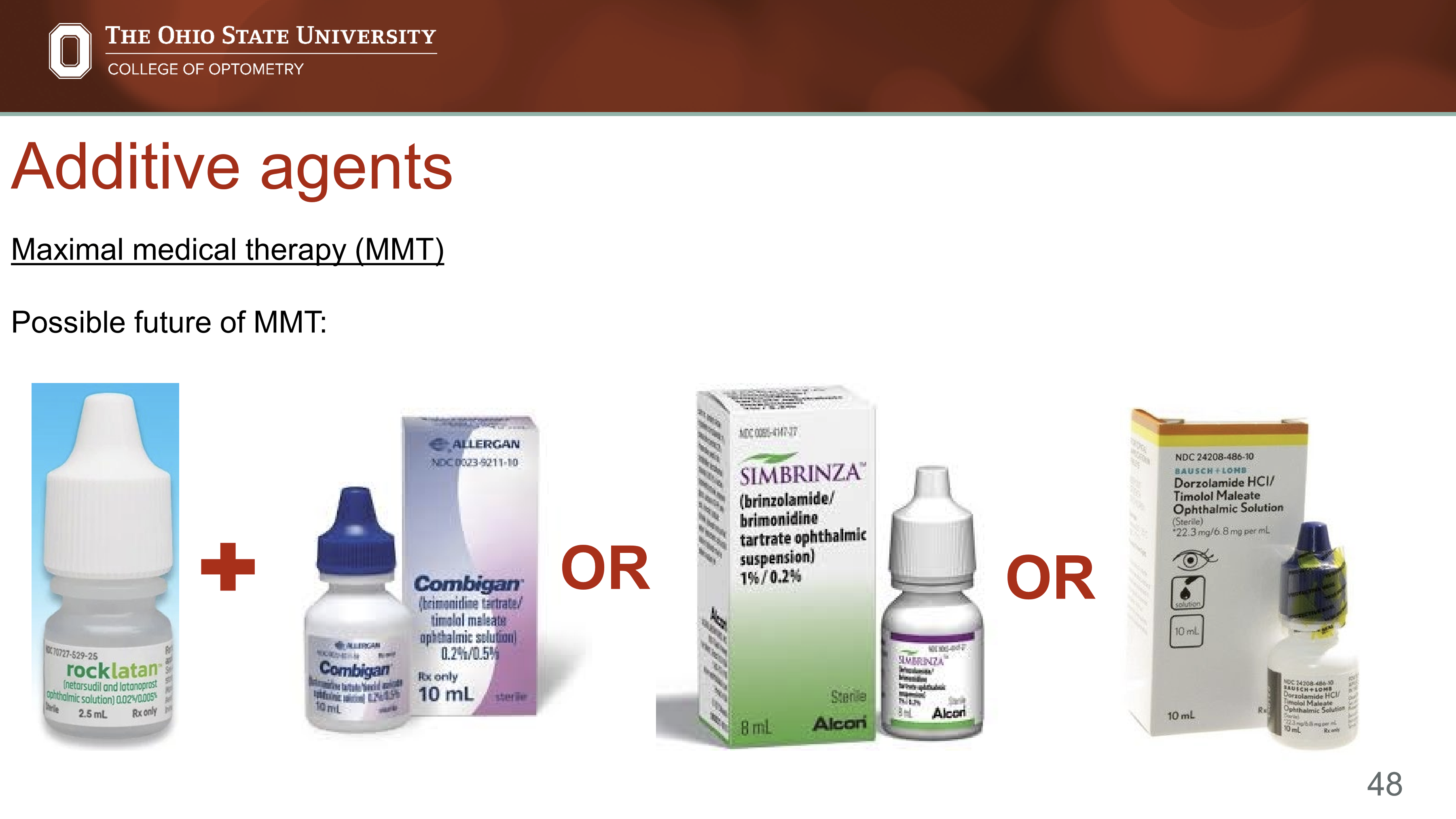
MMT typically occurs after three medications contained in two bottles, often:
PGA + combination drop, such as latanoprost + Combigan
orBeta blocker + combination drop, such as timolol + Simbrinza
Why are combination drops important when approaching MMT?
Combination drops help reduce the number of bottles, which can improve adherence. Since compliance decreases as medication burden increases, combination therapy allows multiple mechanisms while minimizing complexity.
What are examples of possible future MMT-style combinations?
Possible future combinations include:
Rocklatan + Combigan
Rocklatan + Simbrinza
Rocklatan + dorzolamide/timolol
Why can glaucoma patients have low motivation to use drops consistently?
Most glaucoma is asymptomatic, except acute angle closure and some intermittent angle closure. Patients often do not feel high IOP and early peripheral vision loss is usually subtle and gradual.
Treatment can also feel unrewarding because drops:
Cause side effects
Do not make vision better
Must be taken lifelong
What patient-centered challenge should clinicians remember when prescribing glaucoma drops?
Patients may not perceive the disease but may strongly perceive the burden of treatment. Drops can cause irritation, redness, allergy, or other side effects, while the benefit is preventing future vision loss rather than improving current vision.
Why is patient education a critical part of successful medical glaucoma therapy?
Patient understanding improves compliance with:
Follow-up appointments
Medication schedules
Long-term treatment plans
Because glaucoma therapy requires lifelong adherence, education should be a mainstay of every treatment plan.
How can pictures and brochures improve glaucoma patient education?
Visual aids can show advanced glaucomatous vision loss from the patient’s perspective, helping patients understand what treatment is trying to prevent. This can improve motivation and adherence.
What practical strategies help patients correctly instill glaucoma eye drops?
Patients should be taught drop instillation directly and clearly. Helpful strategies include:
Have the patient demonstrate drop instillation in-office using an artificial tear
Give large-print written instructions
Include pictures of the medication bottles
Specify the time of day each drop should be used
Refer to medications by cap color, since drug names are often confusing
Why should clinicians refer to glaucoma medications by cap color?
Patients are often confused by medication names, especially when there are multiple drops or generic substitutions. Cap colors provide a simpler visual cue that can improve medication identification and adherence.
What are the cap color associations?
Yellow or light blue: beta blockers
Yellow = timolol 0.5%
Light blue = timolol 0.25%
Navy blue: beta blocker combination agent
Light green: non-beta-blocker combination agent
Orange: carbonic anhydrase inhibitors
Teal: prostaglandin analogues
Purple: alpha-2 agonists
Red: mydriatics/cycloplegics
Green: miotics
Pink: steroids
Gray: NSAIDs
Brown/tan: anti-infectives
Besides asking about missed drops, how can clinicians assess adherence at follow-up visits
Clinicians can:
Monitor refill rates
Ask patients to bring medication bottles to appointments
Have patients explain how and when they use each drop
What are common clinical signs of ocular surface disease in glaucoma patients using topical medications?
OSD can present with inflammatory and surface findings such as:
Telangiectatic vessels at the lid margin
Matting of lashes
Meibomian gland disease
Diffuse conjunctival hyperemia
Diffuse punctate epithelial erosions with fluorescein staining
Diffuse conjunctival injection
Subepithelial haze
How does glaucoma medication burden affect ocular surface disease?
As the number of glaucoma medications increases, both the prevalence and severity of dry eye/ocular surface disease increase. OSD severity also increases with longer duration of therapy.
What inflammatory changes are associated with topical glaucoma medications?
Topical glaucoma medications can increase:
Surface macrophages
Mast cells
Lymphocytes
Surface inflammatory markers
What is the most common preservative in glaucoma medications, and why is it important?
Benzalkonium chloride (BAK) is the most common preservative. It is important because preservatives target bacterial cell walls but can also damage ocular epithelial cell membranes.
What are the major ocular surface effects of BAK?
BAK can cause:
Squamous metaplasia of conjunctival epithelium
Reduced goblet cell density
Irregular tear film
Increased keratocyte activation
More corneal damage and conjunctival infiltration, especially at higher concentrations
Mechanism → epithelial toxicity and inflammation → worse dry eye/OSD.
What are examples of non-BAK preservatives used in glaucoma medications?
Non-BAK preservative options include:
Purite: used in Alphagan P
SofZia: used in Travatan Z
Potassium sorbate: used in Xelpros
Purite and SofZia are oxidative preservatives.
What are examples of preservative-free glaucoma medications?
Tafluprost: Zioptan
Latanoprost: Iyuzeh
Dorzolamide/timolol: preservative-free Cosopt
Timolol: preservative-free Timoptic
How should ocular surface disease influence glaucoma medication choice?
If a glaucoma patient develops dry eye, irritation, conjunctival hyperemia, punctate epithelial erosions, or lid margin disease, consider medication-related OSD. Management may include reducing drop burden, using combination drops, switching away from BAK-preserved medications, or using BAK-free/preservative-free options.