FUNCTIONAL ANATOMY: LOWER LIMB & ANKLE

0.0(0)
Studied by 0 people
call kaiCall Kai
Locked
learnLearn
examPractice Test
spaced repetitionSpaced Repetition
heart puzzleMatch
flashcardsFlashcards
GameKnowt Play
Card Sorting

1/43

encourage image

There's no tags or description

Looks like no tags are added yet.

Last updated 2:01 AM on 9/20/26
Name
Mastery
Learn
Test
Matching
Spaced
Call with Kai
Chat

No analytics yet

Send a link to your students to track their progress

44 Terms
1
New cards
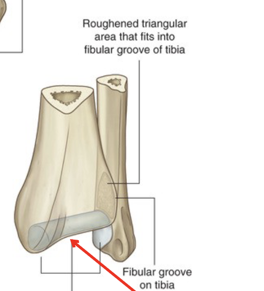
<p>What does this area of the Tibia sit on</p>

What does this area of the Tibia sit on

Articular surfaces for Talus

2
New cards

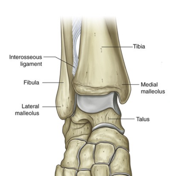
All about the Proximal Tibiofibular Joint

  • synovial planar joint

  • articulation between the head of the fibula and the lateral condyle of the tibia

  • held together by ASTFL and PSTFL and interosseous membrane


<ul><li><p><mark data-color="yellow" style="background-color: yellow; color: inherit;">synovial planar </mark>joint</p></li><li><p>articulation between the <mark data-color="yellow" style="background-color: yellow; color: inherit;">head of the fibula</mark> and the <mark data-color="yellow" style="background-color: yellow; color: inherit;">lateral condyle of the tibia</mark></p></li><li><p>held together by <mark data-color="yellow" style="background-color: yellow; color: inherit;">ASTFL</mark> and <mark data-color="yellow" style="background-color: yellow; color: inherit;">PSTFL</mark> and <mark data-color="yellow" style="background-color: yellow; color: inherit;">interosseous membrane</mark> </p></li></ul><p></p>
3
New cards
<p>All about the Distal Tibiofibular Joint</p>

All about the Distal Tibiofibular Joint

  • Fibrous syndesmoses joint

  • articulation between the distal fibula and tibia

  • held together by AITFL (anterior tibiofibular ligament) and PITFL (posterior tibiofibular ligament) and interosseous membrane


4
New cards
<p>What are these holes in the Interosseous Membrane?</p>

What are these holes in the Interosseous Membrane?

  • Apertures

    • they are for neurovascular structures


5
New cards
<p>Talocrural Joint</p>

Talocrural Joint

  • synovial hinge joint

    • facilitates DF and PF

  • articulation between fibula, tibia, surface of talus

  • held together by fibrous synovial and collateral ligaments


6
New cards

All about the Talus

  • has a wider body anteriorly than posteriorly

  • has a convex superior surface (body)

    • for DF and PF

  • has a convex anterior surface (head)

    • articulates with the navicular


<ul><li><p>has a wider body anteriorly than posteriorly</p></li><li><p>has a convex superior surface (body)</p><ul><li><p>for DF and PF</p></li></ul></li><li><p>has a convex anterior surface (head)</p><ul><li><p>articulates with the navicular</p></li></ul></li></ul><p></p>
7
New cards

Why is the Talar neck depressed?

Allows the joint to go into deeper DF

8
New cards

Medial Ligaments of the Talocrural Joint

Deltoid Ligamet: Anterior Tibiotalar, Tibionavicular, Tibiocalcaneal, Posterior Tibiotalar
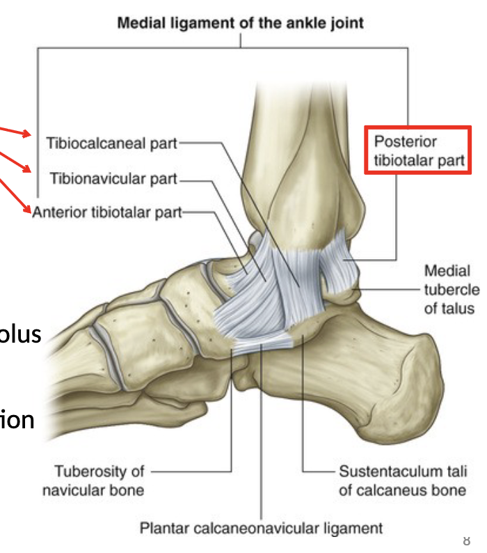
<p>Deltoid Ligamet: Anterior Tibiotalar, Tibionavicular, Tibiocalcaneal, Posterior Tibiotalar</p>
9
New cards

About the deltoid ligament

  • fans out from the medial malleolus

  • limits talar eversion, lateral translation, and rotation

  • Anterior fibres = limit PF

  • Posteiror fibres = limit DF


10
New cards

Lateral Ligaments of the Talocural joint

  1. anterior talofibular

  2. posterior talofibular

  3. calcaneofibular


<ol><li><p>anterior talofibular</p></li><li><p>posterior talofibular</p></li><li><p>calcaneofibular</p></li></ol><p></p>
11
New cards

Anterior talofibular

  • goes from lateral malleolus to talus

  • limits inversion and PF


12
New cards

Posterior talofibular

  • goes from lateral malleolus to talus

  • limits inversions, and DF


13
New cards

Calcaneofibular

  • goes from lateral malleolus to calcaneus

  • limits inversion


14
New cards

Subtalar Joint

  • synovial ellipsoid joint

  • inversion/eversion and some abduction/adduction


15
New cards

Popliteal Fossa

  • superior border: hamstrings

  • Inferior border: gastrocnemius

  • contents:

    • plantaris muscle

    • tibial and common fibular nerves

    • popliteal vein

    • popliteal artery


16
New cards

All about the Interosseous Membrane

  • links the fibula and tibia together

  • provides surface for muscular attachment

  • separates the anterior and posterior compartments


17
New cards

Anterior Compartment Muscles

Tibialis anterior

Extensor Hallucis Longus

Extensor Digitorum Longus

Fibularis (peroneus) Tertius


18
New cards

Tibialis Anterior

O: lateral tibial condyle, proximal shaft of tibia, interosseous membrane

I: medial coneiform and 1st MT

A: DF and inversion, support medial longitudinal arch
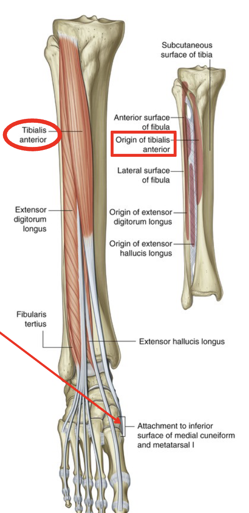
<p>O: lateral tibial condyle, proximal shaft of tibia, interosseous membrane</p><p>I: medial coneiform and 1st MT</p><p>A: DF and inversion, support medial longitudinal arch</p>
19
New cards

Extensor Hallucis Longus

O: mid-anterior surface of fibula, interosseous membrane

I: base of distal phalanx of big toe

A: extension of MTP and IP joints of big toe, DF of foot

<p>O: mid-anterior surface of fibula, interosseous membrane</p><p>I: base of distal phalanx of big toe</p><p>A: extension of MTP and IP joints of big toe, DF of foot</p>
20
New cards

Extensor Digitorum Longus

O: lateral tibial condyle, interosseous membrane, proximal 2/3 anterior fibula

I: base of middle and distal phalanges of lateral 4 toes via dorsal digital expansions

A: extension of MTP, PIP, DIP joints of lateral 4 toes, DF and eversion of foot
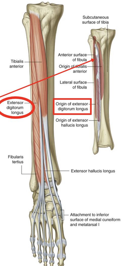
<p>O: lateral tibial condyle, interosseous membrane, proximal 2/3 anterior fibula</p><p>I: base of middle and distal phalanges of lateral 4 toes via dorsal digital expansions</p><p>A: extension of MTP, PIP, DIP joints of lateral 4 toes, DF and eversion of foot</p>
21
New cards

Fibularis (peroneus) tertius

O: distal 1/3 of anterior fibula

I: base of 5th metatarsal

A: DF and eversion of foot

<p>O: distal 1/3 of anterior fibula</p><p>I: base of 5th metatarsal</p><p>A: DF and eversion of foot</p>
22
New cards

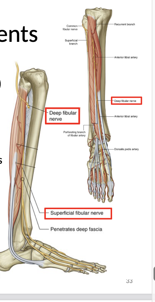
Nerve innervation of Anterior Compartment

Deep Peroneal Nerve

23
New cards

Extensor Retinaculum

  • There is a superior and inferior extensor retinaculum
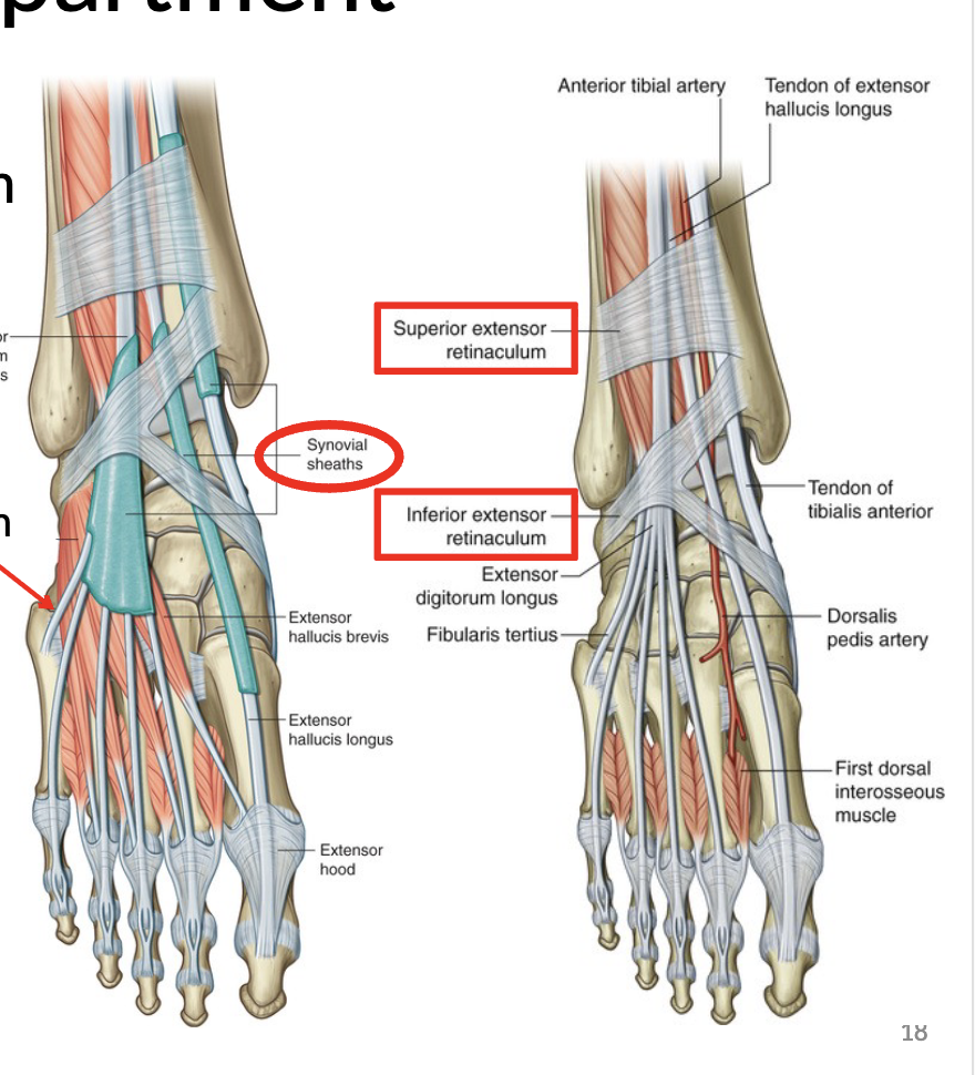

<ul><li><p>There is a superior and inferior extensor retinaculum </p></li></ul><p></p>
24
New cards

Lateral compartment muscles

Fibularis (peroneus) longus

Fibularis (peroneus) brevis

25
New cards

FIbularis (peroneus) longus

O: head and proximal 2/3 of lateral fibula

I: medial cuneiform and 1st metatarsal

A: eversion and plantar flexion of foot, support arches of foot
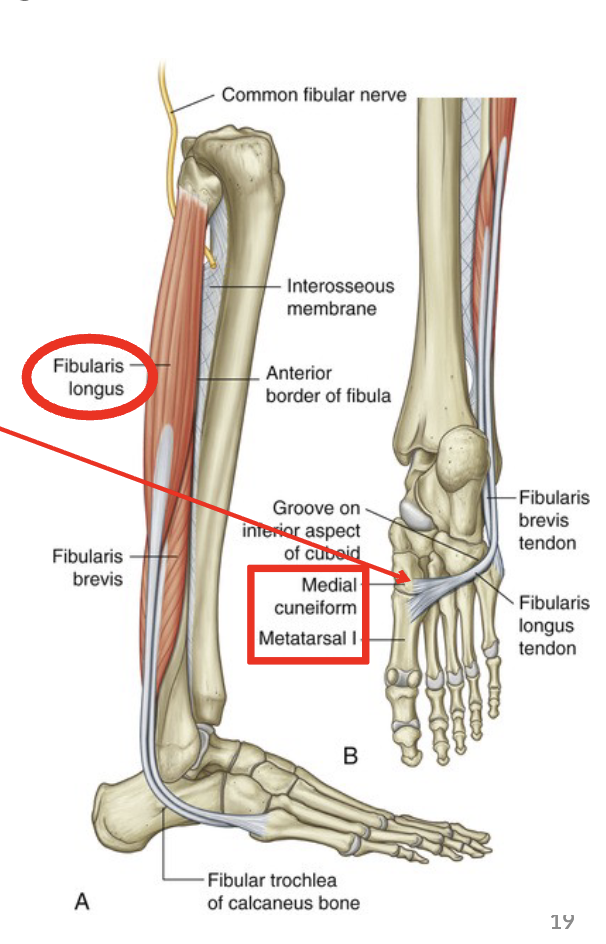
<p>O: head and proximal 2/3 of lateral fibula</p><p>I: medial cuneiform and 1st metatarsal</p><p>A: eversion and plantar flexion of foot, support arches of foot</p>
26
New cards

Fibularis (peroneus) brevis

O: distal 2/3 lateral fibula

I: 5th metatarsal

A: eversion and assistes in PF of foot
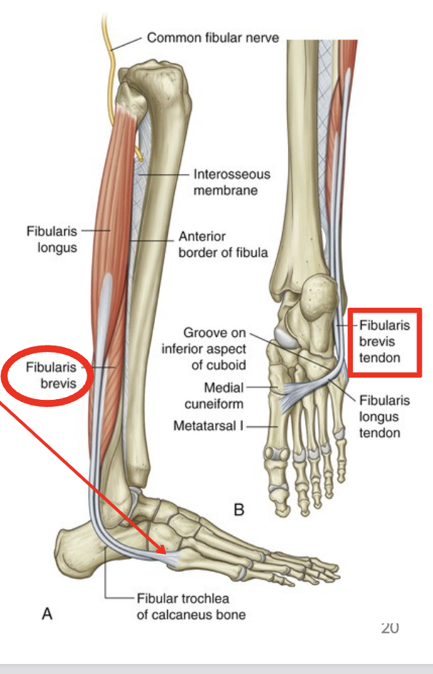
<p>O: distal 2/3 lateral fibula</p><p>I: 5th metatarsal</p><p>A: eversion and assistes in PF of foot</p>
27
New cards

Nerve innervation of lateral compartment

Superficial Peroneal Nerve

28
New cards

Posterior compartment muscles

Gastrocnemius

Plantaris

Soleus

Tibialis Posterior

Flexor digitorum longus

Flexor Hallucis Longus


29
New cards

Gastrocnemius

O: 2 heads originate from superior medial and lateral femoral condyles posteriorly

I: posterior calcaneus via calcaneal tendon

A: PF of foots, assist with knee flexion

<p>O: 2 heads originate from superior medial and lateral femoral condyles posteriorly</p><p>I: posterior calcaneus via calcaneal tendon</p><p>A: PF of foots, assist with knee flexion</p>
30
New cards

Plantaris

O: lateral supracondylar line, oblique popliteal ligament

I: posterior calcaneus via calcaneal tendon

A: weak PF of foot and flexion of knee

<p>O: lateral supracondylar line, oblique popliteal ligament</p><p>I: posterior calcaneus via calcaneal tendon</p><p>A: weak PF of foot and flexion of knee</p>
31
New cards

Soleus

O: proximal 2/3 of posterior tibia and fibula

I: posterior calcaneus via calcaneal tendon

A: plantar flexion of foot
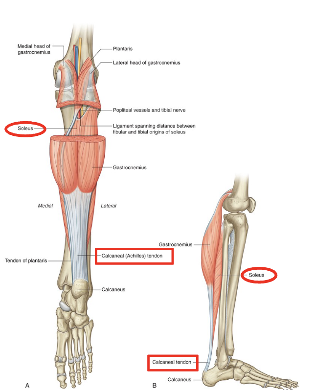
<p>O: proximal 2/3 of posterior tibia and fibula</p><p>I: posterior calcaneus via calcaneal tendon</p><p>A: plantar flexion of foot</p>
32
New cards

Tibialis posterior

O: proximal ½ of posterior tibia and fibula, interosseous membrane

I: navicular tuberosity and medial cuneiform

A: PF and inversion of foot, dynamic foot arch support
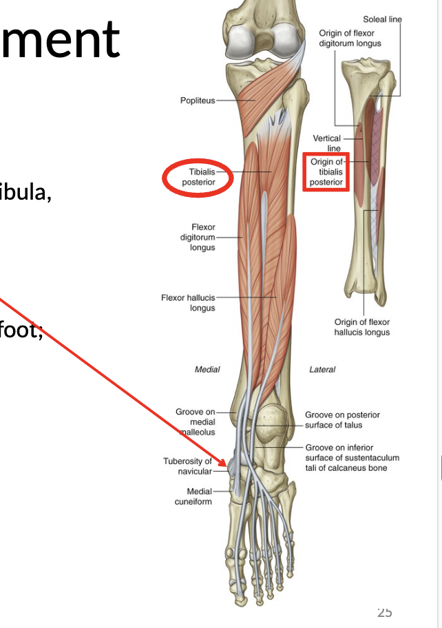
<p>O: proximal ½ of posterior tibia and fibula, interosseous membrane</p><p>I: navicular tuberosity and medial cuneiform</p><p>A: PF and inversion of foot, dynamic foot arch support</p>
33
New cards

Flexor digitorum longus

O: proximal middle 1/3 of posterior tibia

I: bases of distal phalanges of lateral 4 toes

A: flexion of MTP, PIP, DIP joints of lateral 4 toes, PF and inversion of foot

<p>O: proximal middle 1/3 of posterior tibia</p><p>I: bases of distal phalanges of lateral 4 toes</p><p>A: flexion of MTP, PIP, DIP joints of lateral 4 toes, PF and inversion of foot</p>
34
New cards

Flexor Hallucis longus

O: proximal middle 1/3 of fibula and interosseous membrane

I: base of distal phalange of the big toe

A: flexion of MTP and IP joints of big toe, PF and inversion of foot

<p>O: proximal middle 1/3 of fibula and interosseous membrane</p><p>I: base of distal phalange of the big toe</p><p>A: flexion of MTP and IP joints of big toe, PF and inversion of foot</p>
35
New cards

Innervation of Posterior Compartment

Tibial Nerve

36
New cards

Flexor Retinaculum

Tom, Dick, and Harry (anterior to posterior) (medial to lateral), tibial nerve pass posterior to the medial malleolus
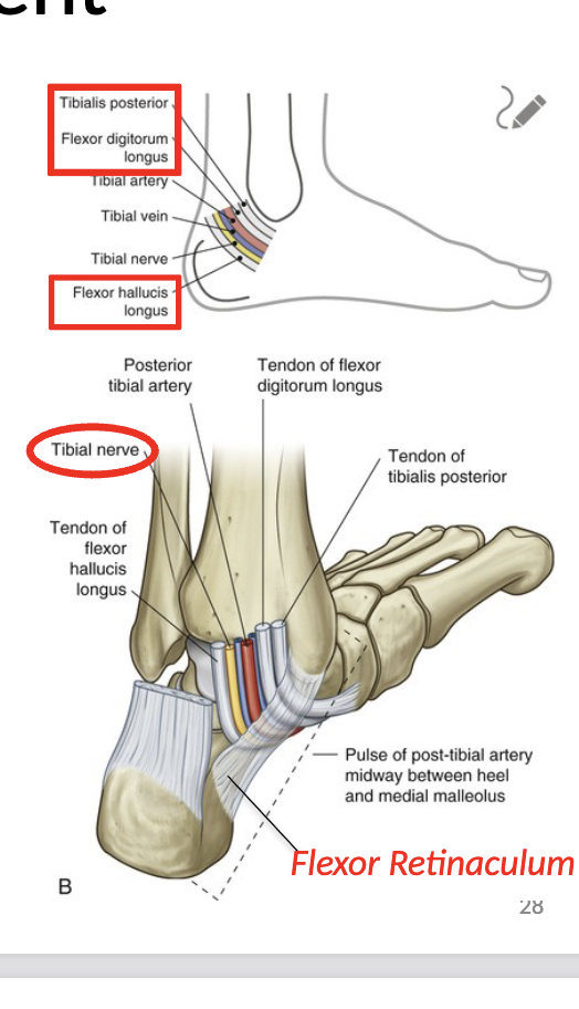
<p>Tom, Dick, and Harry (anterior to posterior) (medial to lateral), tibial nerve pass posterior to the medial malleolus</p>
37
New cards

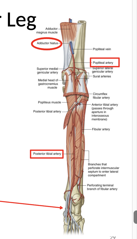
Blood supply to lower leg (posteriorly)

Femoral Artery

  • passes through the adductor hiatus and becomes the popliteal artery behind the knee

  • bifurcates and continues down as the posterior tibial artery superficial to TDH and deep to gastroc/soleus

  • passes through flexor retinaculum


<p>Femoral Artery</p><ul><li><p>passes through the adductor hiatus and becomes the <mark data-color="yellow" style="background-color: yellow; color: inherit;">popliteal artery</mark> behind the knee</p></li><li><p>bifurcates and continues down as the <mark data-color="yellow" style="background-color: yellow; color: inherit;">posterior tibial artery </mark>superficial to TDH and deep to gastroc/soleus</p></li><li><p>passes through flexor retinaculum</p></li></ul><p></p>
38
New cards

Blood supply to lower leg (anteriorly)

Femoral artery

  • passes through adductor hiatus to become the popliteal artery behind the kee

  • bifurcates and passes through superior hiatus and continues down anterior leg as the anterior tibial artery deep to the tib ant

  • continues as dorsalis pedis artery lateral to extensor hallucis longus


<p>Femoral artery</p><ul><li><p>passes through adductor hiatus to become the <mark data-color="yellow" style="background-color: yellow; color: inherit;">popliteal artery </mark>behind the kee</p></li><li><p>bifurcates and passes through superior hiatus and continues down anterior leg as the <mark data-color="yellow" style="background-color: yellow; color: inherit;">anterior tibial artery</mark> deep to the tib ant</p></li><li><p>continues as <mark data-color="yellow" style="background-color: yellow; color: inherit;">dorsalis pedis artery</mark> lateral to extensor hallucis longus</p></li></ul><p></p>
39
New cards

Posterior Thigh Nerve innervation

Sciatic Nerve (L4-S3)

  • enters gluteal region through greater sciatic foramen (inferior to piriformis)

  • runs posterior to adductor magnus muscle and splits as tibial and common fibular nerve

innvervates (motor):

  • bicerps femoris

  • semitendinosus

  • semimebranosus

  • adductor magnus


<p>Sciatic Nerve (L4-S3)</p><ul><li><p>enters gluteal region through greater sciatic foramen (inferior to piriformis)</p></li><li><p>runs posterior to adductor magnus muscle and splits as <mark data-color="yellow" style="background-color: yellow; color: inherit;">tibial and common fibular nerve</mark></p></li></ul><p>innvervates (motor):</p><ul><li><p>bicerps femoris</p></li><li><p>semitendinosus</p></li><li><p>semimebranosus</p></li><li><p>adductor magnus</p></li></ul><p></p>
40
New cards

Tibial nerve innervation

  • descends posterior leg, superficial to TDH and deep to gastroc/soleus

Innervates:

Motor:

  • popliteus, plantaris, gastrocs, soleus, tibialis posterior, flexor digitroum longus, flexor hallucis longus

sensory:

  • posterolateral lower leg, pinky toes, sole of foot via plantar nerves


<ul><li><p>descends posterior leg, superficial to TDH and deep to gastroc/soleus</p></li></ul><p>Innervates:</p><p>Motor:</p><ul><li><p>popliteus, plantaris, gastrocs, soleus, tibialis posterior, flexor digitroum longus, flexor hallucis longus</p></li></ul><p>sensory:</p><ul><li><p>posterolateral lower leg, pinky toes, sole of foot via plantar nerves</p></li></ul><p></p>
41
New cards

Common peroneal nerve innervation

  • bifurcates into the deep and superficial fibular nerves

motor:

  • short head of biceps femoris

sensory:

  • lateral aspect of proximal lower leg


<ul><li><p>bifurcates into the<mark data-color="yellow" style="background-color: yellow; color: inherit;"> deep and superficial fibular nerves</mark></p></li></ul><p>motor:</p><ul><li><p>short head of biceps femoris</p></li></ul><p>sensory:</p><ul><li><p>lateral aspect of proximal lower leg</p></li></ul><p></p>
42
New cards

Common peroneal nerve innervation

  • runs between peroneus longus and brevis


motor:

  • peroneus longus and brevis

sensory:

  • anterior aspect of distal lower leg and dorsum of foot
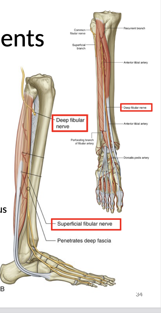

<ul><li><p>runs between peroneus longus and brevis</p></li></ul><p></p><p>motor:</p><ul><li><p>peroneus longus and brevis</p></li></ul><p>sensory:</p><ul><li><p>anterior aspect of distal lower leg and dorsum of foot</p></li></ul><p></p>
43
New cards

Deep Peroneal Nerve innervation

  • runs deep to extensor digitorum longus and tibialis anterior


motor:

  • tibialis anterior, EHL, EHB, EDL, EDB, peroneus tertius

sensory:

  • between 1st and 2nd toes


<ul><li><p>runs deep to extensor digitorum longus and tibialis anterior</p></li></ul><p></p><p>motor:</p><ul><li><p>tibialis anterior, EHL, EHB, EDL, EDB, peroneus tertius</p></li></ul><p>sensory:</p><ul><li><p>between 1st and 2nd toes</p></li></ul><p></p>
44
New cards

Innervation overview (photo)

knowt flashcard image