Anatomy Prelab 4
 Call Kai
 Learn
Learn Practice Test
 Spaced Repetition
 Match
Match Flashcards
 Knowt Play
1/56

There's no tags or description
Looks like no tags are added yet.
Name | Mastery | Learn | Test | Matching | Spaced | Call with Kai | Chat |
|---|
No analytics yet
Send a link to your students to track their progress
57 Terms
The deep matter of the spinal cord is considered
Gray matter

Gray matter description
Contains neuronal cell bodies, dendrites, and local circuitry; organizes into Doral, ventral, and lateral horns
The superficial matter in the spinal cord is the __
White matter

White matter description
Primarily myelinated axons organized into funiculi; carries ascending sensory and descending motor information
Which spinal cord horn is thinner and pointier
Dorsal horn

Dorsal horn function
Receives/ processes sensory information entering through dorsal roots
Which horn is larger, like butterfly wings
Ventral horn

Ventral horn function
Contains somatic motor neuron cell bodies whose axons leave through ventral roots
Lateral horn
Visceral motor/autonomic region, especially prominent in T1-L2 spinal cord segments for sympathetic outflow
Central canal
Small CSF containing channel within the spinal cord, lined by ependymal cells
Posterior, anterior, and lateral funiculi
White matter regions containing ascending and descending tracts; recognize organization rather than individual tract detail
Posterior rootlets
Sensory axons enter the spinal cord through posterior rootlets and diverge from the dorsal root
Posterior rootlets
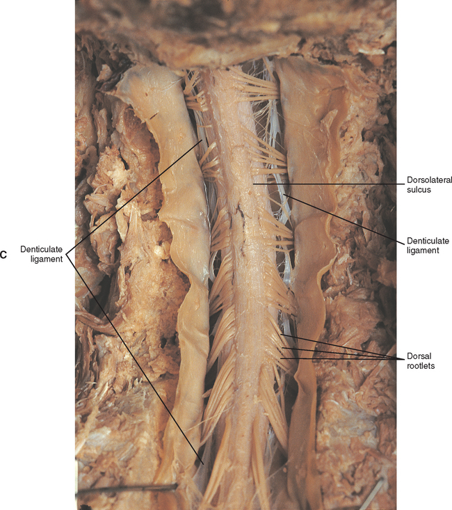
Posterior root
Carries afferent sensory fibers toward the spinal cord

Dorsal root ganglion
Contains sensory neuron cell bodies; enlargement on the dorsal root. A lesion may affect sensory input without directly interrupting ventral root motor neurons

Anterior rootlets
Motor axons leave the spinal cord through anterior rootlets and converge into the ventral root
Anterior root
Carries efferent motor fibers away from spinal cord
Mixed spinal nerve
formed by union of dorsal and ventral roots; contains sensory, somatic motor, and autonomic fibers that form just distal to DRG

Posterior ramus
Mixed branch supplying intrinsic back muscles, vertebral joints, and overlying skin

Anterior ramus
Mixed branch supplying anterolateral trunk and limbs, forms major nerve plexuses except in much of the thoracic region

Spinal cord
CNS structure connecting brain and peripheral nerveser
Cervical enlargement
Increased neural tissue associated with upper-limb innervation and the brachial plexus
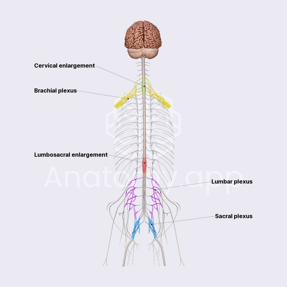
Lumbosacral enlargement
Increased neural tissue associated with lower limb innervation and the lumbar/sacral plexuses

Conus medullaris
Tapered inferior end of the adult spinal cord, typically near L1-L2

Cauda equina
Descending lumbar and sacral nerve roots inferior to the conus; roots travel within the lumbar cistern before exiting

Filum terminale
Slender continuation from the conus that contributes to longitudinal stabilization of the spinal cord

Lumbar cistern
CSF-containing subarachnoid region containing cauda equine: lumbar puncture is performed below the adult cord to reduce risk of cord injury

Meningeal branch
Re-enters the vertebral canal to supply structures within/around the canal
Intervertebral foramen
Passage through which spinal roots and associated vessels enter or leave the vertebral canal; clinically relevant to nerve-root compression
Peripheral nerve
Bundle of PNS axons carrying sensory, motor, and autonomic signals between the CNS and peripheral targets
Nerve fascicle
Bundle of axons within a peripheral nerve
Endoneurium
Connective tissue surrounding individual peripheral nerve fibers eriP
Perineurium
Connective tissue sheath surrounding a nerve fascicle; contributes to the protected internal environment of the fascicle
Epineurium
Outer connective tissue sheath surrounding the entire peripheral nerve and its fascicles
Cervical plexus
Network formed primarily by anterior rami of upper cervical spinal nerves; recognize as a ventral-ramus plexus

Brachial plexus
Anterior-ramus plexus distributing spinal nerve fibers to the upper limb

Lumbar plexus
Anterior ramus plexus contributing peripheral nerves to the lower trunk and limb

Sacral plexus
Anterior-ramus plexus contributing major peripheral nerves to the pelvis and lower limb

Neuron cell body
Metabolic/integrative region containing the nucleus; motor neuron somata may be recognized in spinal gray matter
Dendrites
Receive synaptic input and conduct graded signals toward the cell body
Axon
Conducts action potentials away from the cell body toward another neuron or effector
Myelin sheath
Electrical insulation around selected axons that increases conduction efficiency
Node of ranvier
Gap between myelin segments where action potentials are regenerated during saltatory conduction
Schwann cell
PNS glial cell responsible for myelination/ensheathment of peripheral axons
Oligodendrocyte
CNS glial cell responsible for myelinating CNS axons
Satellite cell
PNS glial cell surrounding neuronal cell bodies in peripheral ganglia, including the dorsal root ganglion
Astrocyte
CNS support cell involved in structural/ metabolic support and regulation of the neural environment
Microglia
Resident immune/phagocytic cells of the CNS
Ependymal cells
Line the ventricles and central canal; specialized ependymal cells contribute to choroid plexus CSF production tL
Lateral horn/intermediolateral cell column
Contains preganglionic sympathetic neuron cell bodies in T1-L2

White ramus communicans
Carries myelinated preganglionic sympathetic fibers from a spinal nerve into the sympathetic trunk; present at only T1-L2
Sympathetic trunk
Paired longitudinal chains alongside the vertebral column that allow sympathetic fibers to synapse, ascend, or descend
Sympathetic trunk ganglion
Contains sympathetic postganglionic neuron cell bodies
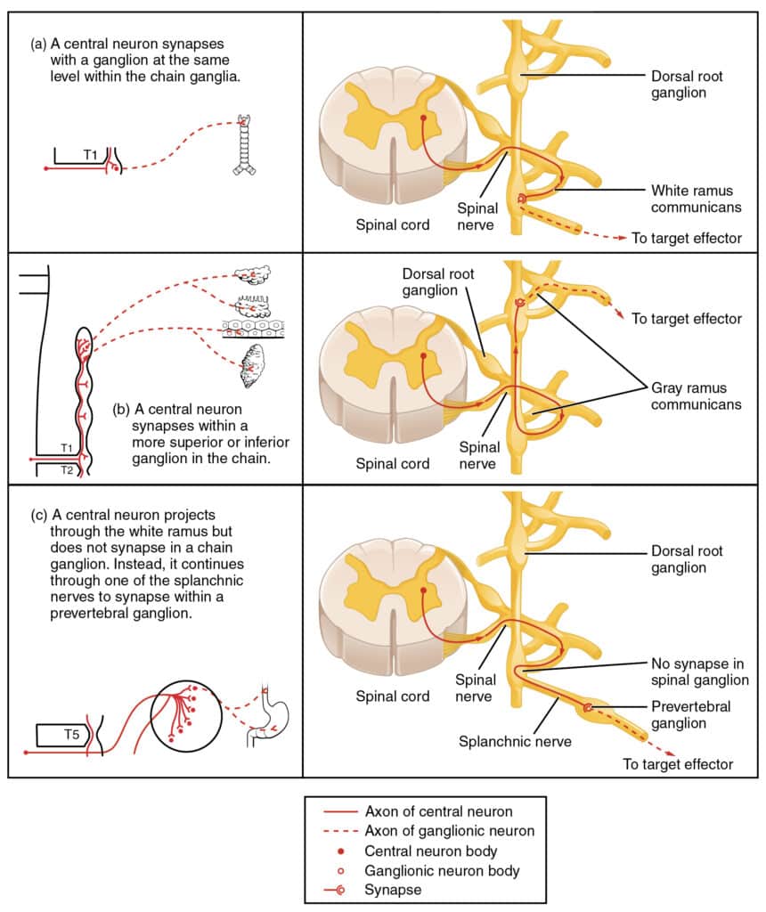
Gray ramus communicans
Carries unmyelinated postganglionic sympathetic fibers from the sympathetic trunk back to a spinal nerve; gray rami occur at all spinal nerve levels
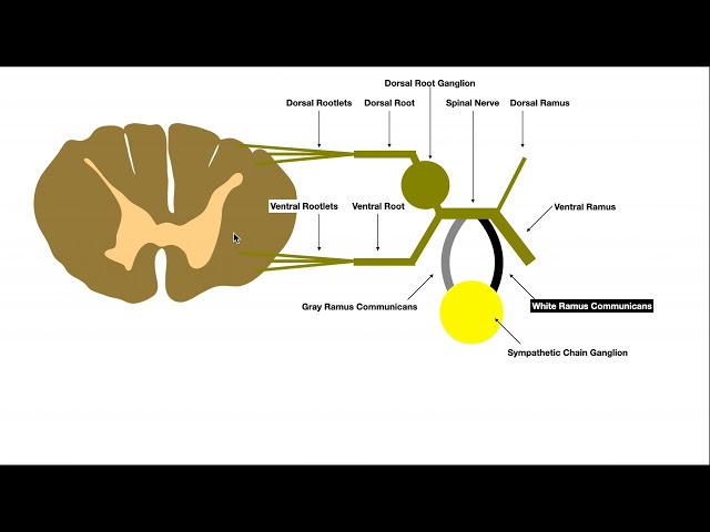
Thoracic splanchnic nerve
Carries primarily preganglionic sympathetic fibers toward pre vertebral ganglia; distinguish from gray rami returning fibers toward spinal nerve
Prevertebral ganglion
Sympathetic ganglion anterior to the vertebral column/aorta where many abdominopelvic preganglionic fibers synapse

Adrenal medulla
Modified sympathetic ganglion receiving preganglionic sympathetic fibers directly; releases catecholamines into bloodstream
