MSK TBL 1: Amino Acid & Skeletal Muscle Metabolism
 Call Kai
 Learn
Learn Practice Test
 Spaced Repetition
 Match
Match Flashcards
 Knowt Play
1/45

There's no tags or description
Looks like no tags are added yet.
Name | Mastery | Learn | Test | Matching | Spaced | Call with Kai | Chat |
|---|
No analytics yet
Send a link to your students to track their progress
46 Terms
nitrogen balance
amount of nitrogen in body
*most healthy people have 0 nitrogen balance
transaminases
shuttle nitrogen
[amino acid catabolism in peripheral tissues produces ammonia, esp in muscle]
glutamine synthetase
glutamate [1 N] + NH3 → glutamine [2 N]
fate of glutamine if energy is needed
converted into a ketoacid for catabolism
free ammonia converted to urea & intmd can make ATP
negative nitrogen balance
illness
starvation
proteins converted to aa’s
diabetes
positive nitrogen balance
growth
childhood
pregancy
anabolic state
body needs more proteins & nitrogen, so less is removed & hunger functions activate
Hyperammonemia
occurs when excess nitrogen is NOT converted to urea
→ significant cognitive & movement issues
asterixus
flapping tremor when pt holds palms up
indicates hyperammonemia
Markers of skeletal muscle damage
AST [aspartate aminotransferase]
CK [creatine kinase]
Markers of liver disease
AST
ALT [alanine aminotransferase]
alkaline phosphatase
glutathione
Arginine catabolism
Arg enters TCA cycle & N is donated to urea cycle
Arginine anabolism
Creatine & skeletal muscle metabolism
Nitric oxide signaling [cGMP]
muscles use what as a storage fuel?
creatine phosphate
creatine’s starting material is
Arg & Gly
made in kidney
creatine kinase
catalyzes:
creatine reversibly donating its phosphate to ADP to make ATP
allows muscles to contract for a longer period of time
creatinine
waste product
amount of creatinine excreted each day is constant
used as indicator of normal excretory function of kidneys
Sources of Energy/ATP for Muscle Contraction ranked shortest to longest
ATP concentration in muscle fiber [1-2 sec]
Phosphocreatine making ATP via CK enzyme
Glycolysis & glycogen hydrolysis [1 min]
Oxidative metabolism [hrs]
requires O2
uses fatty acids, glucose, and aa’s as substrates
nitric oxide signaling
paracrine
NO is a signaling molecule that uses cGMP signaling
clinically important cGMP signals
“BAD GraMPa”
BNP, ANP [cardiac hormones
EDRF [NO]
Endothelium-derived relaxing factor
EDRF/NO
smooth muscle relaxant
stops myosin activity
vasodilation
decreases calcium to decrease BP
NT
other
decreases blood coagulation & phagocytosis
NO signaling mechanism: local secretion & NT
NO synthase catalyzes conversion of Arg → NO
NO is gas that readily crosses cell membranes
activates guanylate cyclase, which makes cGMP
As a NT:
NO released into NMJ as vesicles
to stop signal, phosphodiesterase hydrolyzes cGMP
nitroglycerin
NO variant
used for symptom control in acute heart conditions
BCAA metabolism
connected to developmental delays
predominantly catabolized in skeletal muscle
BCAA have a branch in incorrect location that doesn’t allow them to be a direct substrate for beta-oxidation
BCAA metabolism pathway: Ile or Val or alloisoleucine
amine removed by transamination & carried to urea cycle by glutamine
C is removed by oxidative decarboxylation [branched-chain ketoacid dehydrogenase]
uses thiamine [B1] as cofactor
remaining aa skeleton shortened to make Propionyl-CoA
Propionyl-CoA converted to Methylmalonyl-CoA by propionyl-CoA carboxylase
uses biotin [B7] as a cofactor
Methylmalonyl-CoA can enter TCA as Succinyl-CoA by enzyme methylmalonyl-CoA mutase
uses cobalamin [B12] as a cofactor
BCAA metabolism pathway: Leu
*converted exclusively to ketogenic substrates
amine removed by transamination & carried to urea cycle by glutamine
C is removed by oxidative decarboxylation [branched-chain ketoacid dehydrogenase] → product Isovaleric-CoA
uses thiamine [B1] as cofactor
remaining aa skeleton shortened
shorter skeleton converted into acetoacetate
uses biotin [B7] as a cofactor
Maple Syrup Urine Disease [MSUD]
caused by decreased activity of branched-chain ketoacid dehydrogenase
manifests v. early [few days]
Findings
poor feeding, vomiting, lethargy
if untreated → irreversible neurological damage → cerebral edema at high levels, coma, & metabolic catastrophes
intellectual disability
Leu prevents uptake of other aa’s → loss of NTs
poor muscle tone
characteristic odor: maple syrup smell in urine or ear wax
Labs
ketosis: ketones in blood & urine
accumulation of 3 BCAAs & alloisoleucine
in some cases, symptoms can be decreased by high levels of thiamine [vit. B1]
assuming some residual enzyme activity
Methionine catabolic pathway
Met makes S-adenosylmethionine [SAM], which transfers methyl groups to make products
SAM → homocysteine [has free SH group needed to make Cys]
cystathione beta synthase converts homocysteine → cystathione thru process called transsulfuration using Ser
occurs in liver & kidney
requires Vitamin B6
cystathione directly makes Cys
remaining C skeleton converted into Propionyl-CoA
SAM required to make
phospholipids, DNA, & NTs
normally, homocysteine levels are
v. low in serum b/c its either remethylated to methionine or catabolism
B12 cycle
B12 carries methyl groups
in Met Synth, methyl group comes from folate
in catabolism its donated to folate
in generation of succinyl-CoA, it moves the 1 carbon
source of B12
animal products
enzymes that require vitamin B12 [2]
methionine synthase
remethylation of homocysteine
methylmalonyl-CoA mutase
makes TCA cycle intmd, succinyl-CoA during catabolism
B12 cycle is connected to which other vitamin
B9
both are catalytic cycles termed 1-carbon metabolism
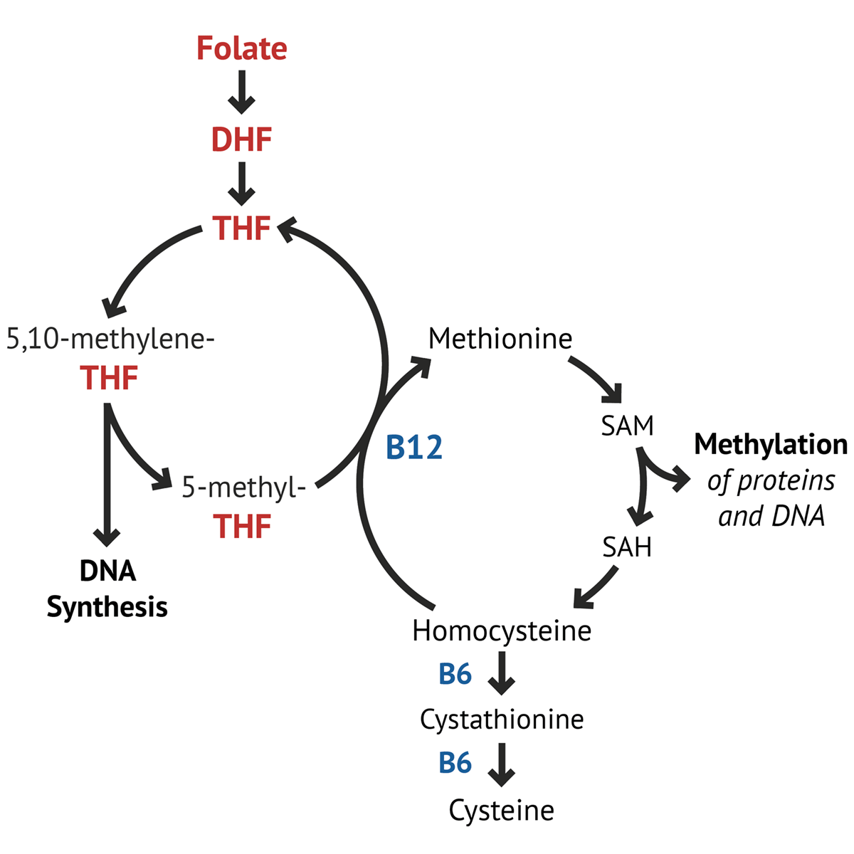
B12 deficiency assessed by
accumulation of methylmalonyl-CoA & urinary excretion of methylmalonic acid
→ anemia
elevated homocysteine
Met makes SAM
B12 is methylated by
5-methyltetrahydrofolate [FH4-CH3]
active folate
dihydrofolate reductase [FH2 reductase]
catalyzes synth of FH4 from folate
methylenetetrahydrofolate reductase [FH4 reductase]
converts FH4-methylene [also an active folate] to FH4-CH3
2nd pathway in liver to remethylate homocysteine to methionine
uses betaine as methyl donor and enzyme betaine methyltransferase
why is folate important in pregancy
allows RNA → DNA
which develops faster: B12 or B9 deficiency?
B9
B12 takes a long time to develop because liver has ~1-2 yr supply of B12
B12 deficiency leads to what kind of anemia?
pernicious, macrocytic anemia
low folate → poor DNA synth → immature red cells released from marrow [large bc have nucleus & organelles]
neutrophils will also have >5 nuclear lobes
prolonged B12 deficiency →
symmetric tingling
proprioception loss
weakened vasculature
*once neurological symptoms take place, its IRREVERSIBLE
methylamalonyl-CoA acidemia
occur from either B12 deficiency or enzymatic mutation to methylmalonyl-CoA mutase
methylmalonyl-CoA converts to methylmalonic acid in blood → blood pH imbalance
appears in early infancy
autosomal recessive
findings
hypotonia
hepatomegaly
hyperammonemia
ketoacidosis
thromnopenia
heart problems can also occur
high levels of methylmalonic acid & other ketones in urine
cystathionine beta synthase [CBS] deficiency
AKA homocystinuria
CBS mutations → elevated homocysteine in blood & urine
more common in people from Germany, Norway, Ireland, & Qatar
usually appears at 3-5 yrs old
findings
connective & fiber tissue problems
limited joint mobility
dislocation of ocular lens in eye
skeletal abnormalities
excessive blood clotting → BV obstruction → atherosclerosis, MI, or stroke
sight issues seeing far away [myopia = nearsighted]
*can be confused with Marfan Syndrome
HIGH homocysteine & Met
LOW Cys
isovaleric acidemia
caused by dysfunction in leu catabolism
less severe version of propionyl-CoA carboxylase deficiency
urine odor that smells like feet
propionyl-CoA carboxylase deficiency
caused by mutation to either gene that codes the 2 subunits of propionyl-CoA carboxylase
imp in the following macronutrients: VOMIT
Val
Odd-chain fatty acid
Met
Ile
Thr
some correlation w/ Saudia Arabia, Amish, & Inuit populations
Findings
1st, is severe vomiting in the 1st weeks after birth
encephalopathy & bone marrow suppression
manifests as failure to thrive, recurrent infections, cardiomyopathy, & pancreatic infections
high levels of propionic acid & other ketones in urine