BME 311 Lecture 3
 Call Kai
 Learn
Learn Practice Test
 Spaced Repetition
 Match
Match Flashcards
 Knowt Play
1/46

There's no tags or description
Looks like no tags are added yet.
Name | Mastery | Learn | Test | Matching | Spaced | Call with Kai | Chat |
|---|
No analytics yet
Send a link to your students to track their progress
47 Terms
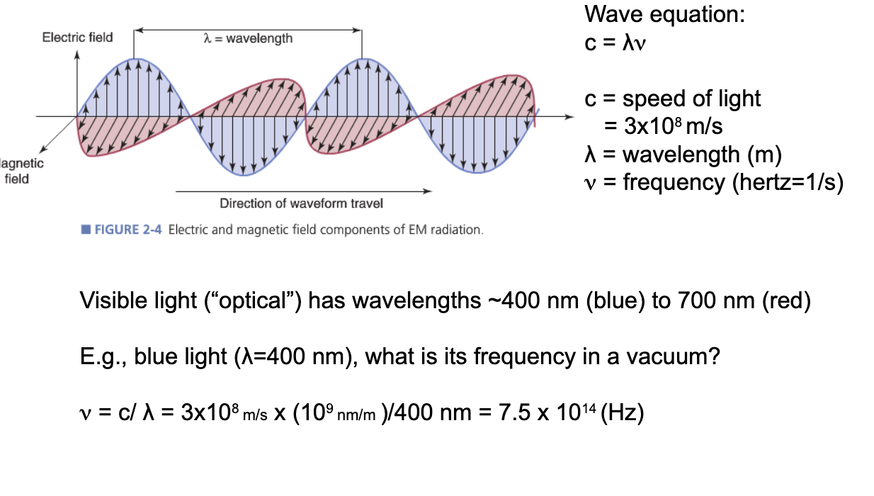
electromagnetic spectrum wave characteristics
visible light has wavelengths ~400 nm (blue) to 700 nm (red)
v=c/y=3×108m/s x (109 nm/m)/400nm=7.5×1014(Hz)
electromagnetic spectrum particle characteristics
-the discrete (particle-like) packets (or quanta) of EM energy are called photons
-the energy of a photon is: E(keV)=1.24/y (nm)
1 eV
energy acquired by an electron as it traverses an electrical potential difference (voltage) of one volt in vacuum = 1.6 × 10⁻¹⁹J
in most medical imaging (except MRI), we think of electromagnetic energy as photons, not waves
-point-like particle with speed and direction
-no rest mass, speed is constant, independent of frame of reference
energy of any given photon is represented by its temperature
-cold is low energy
-hot is high energy
think of a beam of photons as hot and cold marbles moving at constant speed in one direction
intensity of beam is represented by the number of marbles
interactions with matter
atoms/molecules
absorption
just removes marbles/photons = reduces intensity but not energy of remaining photons
scattering
changes direction, can lower energy but not necessarily the intensity (number of marbles is the same)
probability
of interaction can be low - penetration of cm or more into tissue with no interaction at all
energy conservation
-in an interaction, total energy cannot change
-sum of kinetic, potential (eg. gravity), etc
-and rest energy using E=mc2 (mass not conserved because some interactions convert m to E)
the quantum world
-bound states in atoms and nuclei
-only certain energies are allowed
-complicated to predict but we know what these energies are for all the elements
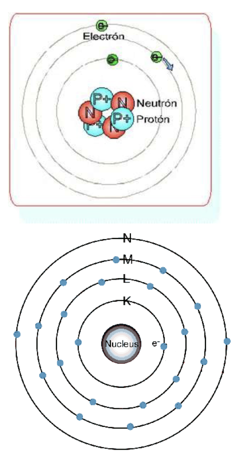
atom composed of
-proton-positively charged
-neutron-neutral
-electrons-negatively charged
-atomic size~10-10m, neutron size~10-14m
-maximum electron capacity of orbital = 2 (n)2
n=shell quantum number also represented by letters
-K means n=1
-L means n=2
atoms “prefer” lowest energy state = all lower electron orbitals filled to capacity with electrons
-will spontaneously change configuration to achieve lowest energy
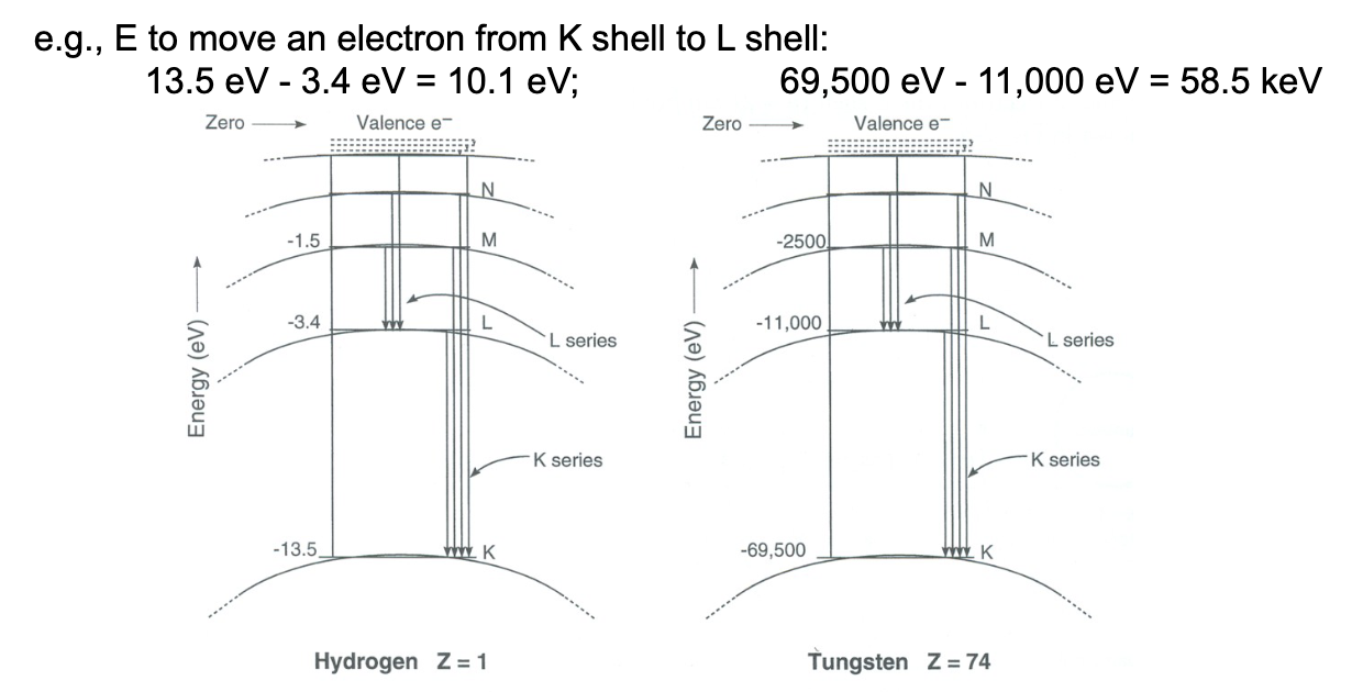
-energy levels are given negative sign, so zero is “free”, and >0 is kinetic energy
binding energy
-energy required to completely remove an electron from the atom
-to ionize an atom: energy from incoming photon or particle must equal to exceed the magnitude of electron’s binding energy
-to excite an atom: enough energy to push from lower shell to higher (unoccupied) shell
EM radiation of frequency greater than the UV-region carries sufficient energy per photon to remove from atomic shells-ionized atoms and molecules
-EM radiation of frequency below than the UV-region-non ionizing radiation
-threshold energy for ionization depends on the type of matter
-remove an electron from calcium and liquid water

radiation after ionization
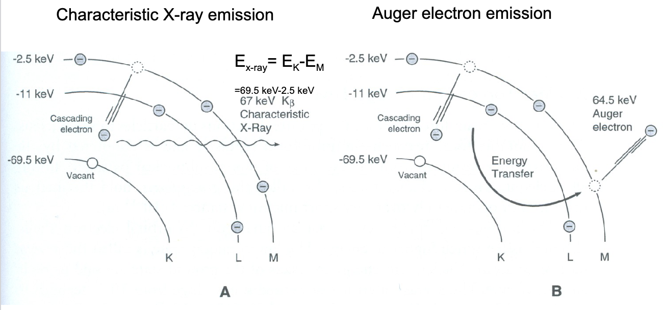
-energy released as characteristic x-rays or auger electrons
-electron cascade-series of electronic transition from outer to inner orbitals
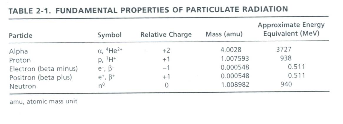
particle “radiation”
-not electromagnetic radiation
-but can have similar effects on atoms and nuclei (excitation)
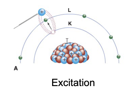
excitation vs de-excitation (emission)
ex. phosphorescence

how often/under what conditions do interactions between radiation and matter happen
-all about probabilities
-not easy to calculate exactly
-but there are trends, almost always with density, Z
excitation
transfer the incident particles’ energy to electrons in the absorbing material, promoting them to electron orbits farther from the nucleus (higher energy level)
de-excitation (emission)
release the absorbed energy, the electron returns to a lower energy level

Bremsstrahlung (to brake) and Strahlung (radiation)
x ray radiation (yellow) emitted by charged particles, such as electrons (blue) which are braking around other charged particles, such as an atomic nucleus (red). it forms the continuum component of the x-ray spectrum generated by an x-ray tube
-no excitation of target or projectile
-nothing to do with orbitals, just direct electron-nucleus interaction
x-rays given off when electrons lose kinetic energy (KE) due to deflection by nuclei
probability of interaction is proportional to Z2 of absorber/target

x-ray interactions
photons penetrate, scatter or get absorbed by matter
for a given photon, there is a probability for each of these, depending on
-energy of photon
-properties of target (density, Z, …)
four types of x-ray interactions
-rayleigh scattering
-compton scattering
-photoelectric absorption
-pair production

rayleigh scattering (coherent scattering)
-incident photon interacts with the entire atom without exciting it
-basically elastic = like billiard ball bouncing off wall of pool table
-likely at low x-ray energies (few keV)
-photon energy does not change but angle does change
-probability of interaction (ie amount of scatter) depends strongly on energy
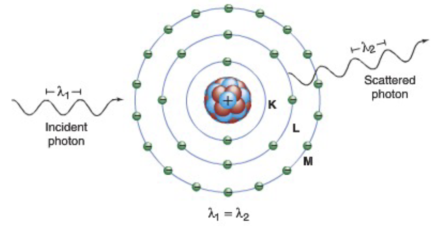
rayleigh scattering is the elastic scattering of light from particles less than approximately one-tenth the wavelength of the light
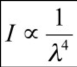
-the intensity of the light scattered is inversely proportional to the fourth power of the wavelength of light
-answer to why is the sky blue as the blue light with smallest wavelength is sunlight scattered more intensely than the remaining colors
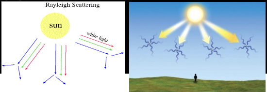
rayleigh scattering energy
x-ray 15-30 keV-used in mammography
-deleterious effect on image quality-random change of direction is bad
-in soft tissue, this accounts for 5% x-ray interactions above 70 keV
-~12% at 30 keV
compton scattering
-results in the ionization of the atom
-dominates above ~30 keV in soft tissue
Eo=ESC+Ee- (assuming binding energy is low and can be neglected)
ESC=Eo/1+(Eo/511 KeV)(1-cosθ)
at θ=0 degrees (forward or no scatter), Esc=Eo
at θ=180 degrees (backward), Esc is minimized

as Eo increases, scattered photons and electrons “prefer” forward direction which is received by image detector, reducing image contrast
probability also depends on electron density of target (and thus Z)
photoelectric effect (what you prefer to happen in x-ray imaging)
-photo “disappears” rather than changes direction
-probability of PE absorption per unit mass ~Z3/E3
Z=atomic number, E=Energy of incident photon eg PE of iodine (Z=53) greater than calcium (Z=20)
-interaction increases above absorption edges
PE process dominates when lower energy photons interact with high Z materials
-primary mode of interaction with screen phosphors, radiographic contrast materials and bone

photoelectric effect results in
-photoelectron
-positive ion (ionized atom)
-characteristic x-rays or Auger electrons (cascade)

attenuation of x-rays
-removal of photons from a beam of x-rays as it passes through matter
-caused by absorption and scattering
-low photon energies (<~20keV), PE effect dominates
-higher photon energies or in low Z (soft tissues), compton scattering dominates
-rayleigh scattering low-10% of interactions in mammography, 5% chest
-high photon energy (>1.02 MeV), beyond the range of x-ray or nuclear radiology, “pair production” contributes to attenuation
-photon can convert into electron-positron pair
-not relevant for medical imaging
linear attenuation coefficient
-fraction of photons removed from a monoenergetic beam of x-rays per unit thickness
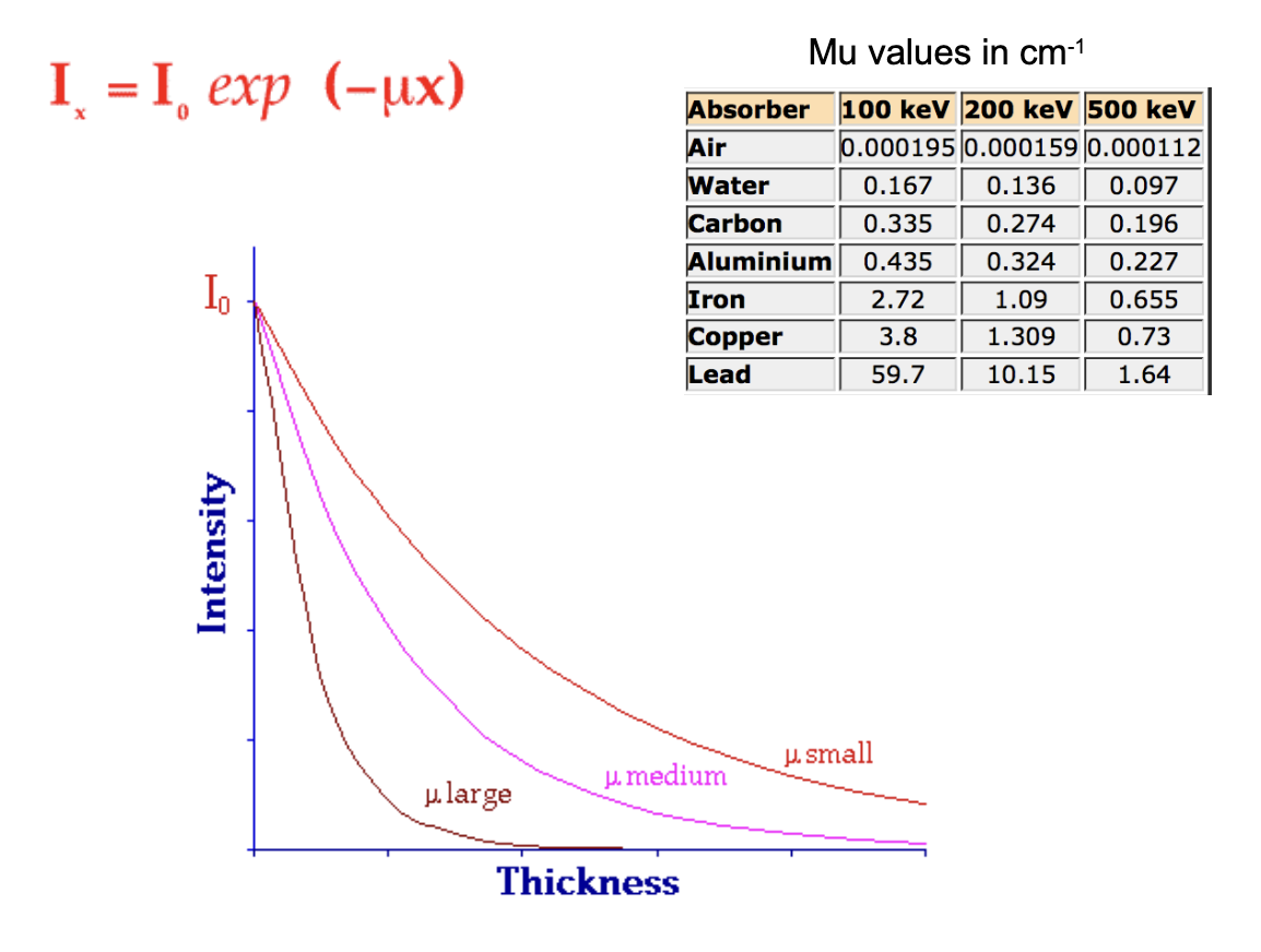
-as thickness increases, relationship exponential Nout=Ninexp[-udeltax]
-u = linear attenuation coefficient
=deltax=distance (x out - x in) or tissue thickness
![<p>-fraction of photons removed from a monoenergetic beam of x-rays per unit thickness</p><p>-as thickness increases, relationship exponential N<sub>out</sub>=N<sub>in</sub>exp[-udeltax]</p><p>-u = linear attenuation coefficient</p><p>=deltax=distance (x out - x in) or tissue thickness</p>](https://assets.knowt.com/user-attachments/d8b11e39-7cdd-4ca2-ad3f-9ee0d4aa32b5.png)
linear attenuation units
-dN=uNdx
-dN=number of photons removed
-N=incident photons
-dx=very small thickness
u=linear attenuation coefficient
-u=u(rayleigh)+u(compton)+u(photoelectric)+u(pair production)
-decreases with increasing energy except at absorption edges
units=1/cm
-soft tissue-0.35-0.16cm-1 for photon energies 30-100keV
mass attenuation coefficient
-for given thickness probability of interaction depends on number of atoms per unit volume and thus the density
-this dependency can be removed by dividing the density out of the linear attenuation coefficient of the material

attenuation
-depends on material, density, photon energy
-measured in artificial geometry (narrow beam)
-real world
multiple photon energies, broad beam geometry
want easy quantity to work with like thickness
especially for shielding (target=worker!)

half value layer
-used to calculate attenuation for shielding purposes for radiation workers
-thickness of material required to reduce the intensity to ½ initial value
-HVL~indirect measure of photon energies
-broad beam geometry -lower attenuation, safer shielding estimates
-most practical applications of attenuation occur under broad beam conditions
-tenth value layer (TVL)-reduce the intensity of beam by 1/10th
-often used in x-ray room shielding design calculations
reduction in beam intensity = (1/2)n, where n=number of half layers
-fraction transmitted through 3 HVLs=1/8=0.125
-easy to calculate in your head
-relation between HVL and u for single energy photons in narrow beam geometry: HVL=0.693/u
effective energy
average energy of photons in the beam
x-ray beam-polyenergetic
not just a single energy, but a distribution of energies
characterized the “hardness” or “quality” of a beam
ability to pass through tissue
HVL is a surrogate measure of average energy of photons in the beam
-mean free path
-average distance travelled by photon before interaction with matter
-MFP=1/u=1.44 HVL