Venous Insufficiency/PPG/APG/Non-Atherosclerotic Pathologies
 Call Kai
 Learn
Learn Practice Test
 Spaced Repetition
 Match
Match Flashcards
 Knowt Play
1/34

There's no tags or description
Looks like no tags are added yet.
Name | Mastery | Learn | Test | Matching | Spaced | Call with Kai | Chat |
|---|
No analytics yet
Send a link to your students to track their progress
35 Terms
Anterior Accessory Vein
Runs parallel and anterior to femoral vein
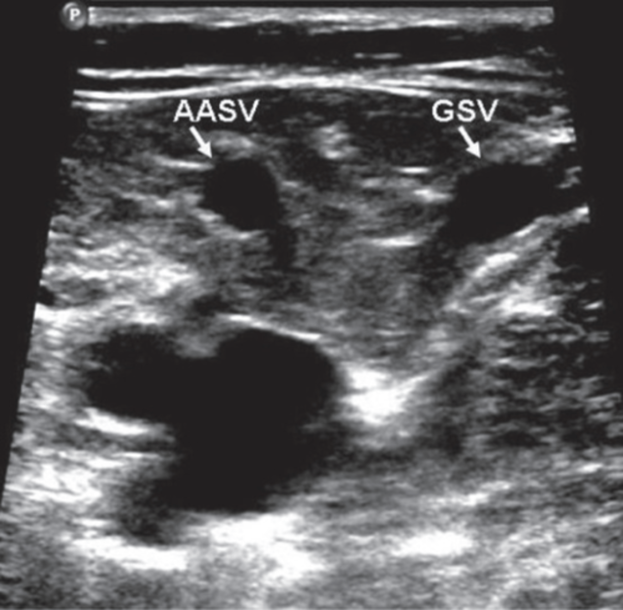
GSV
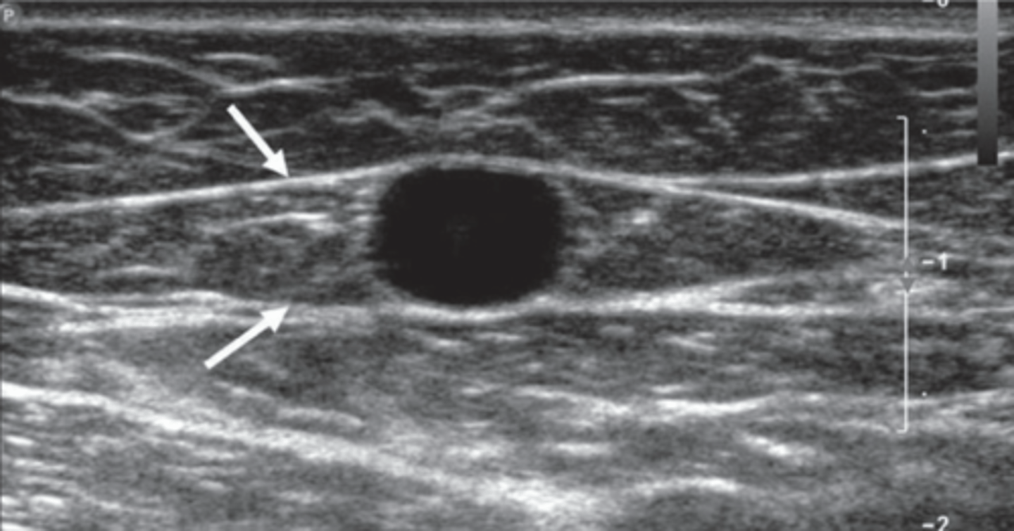
Saphenous Eye
Between facial layers
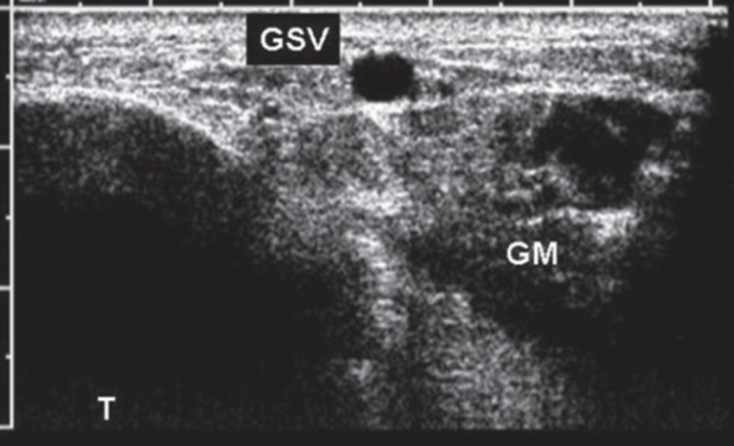
GSV in Calf
Angle Sign
Triangular form between gastrocnemius muscle and tibial bone
Tributary
Outside fascial plane
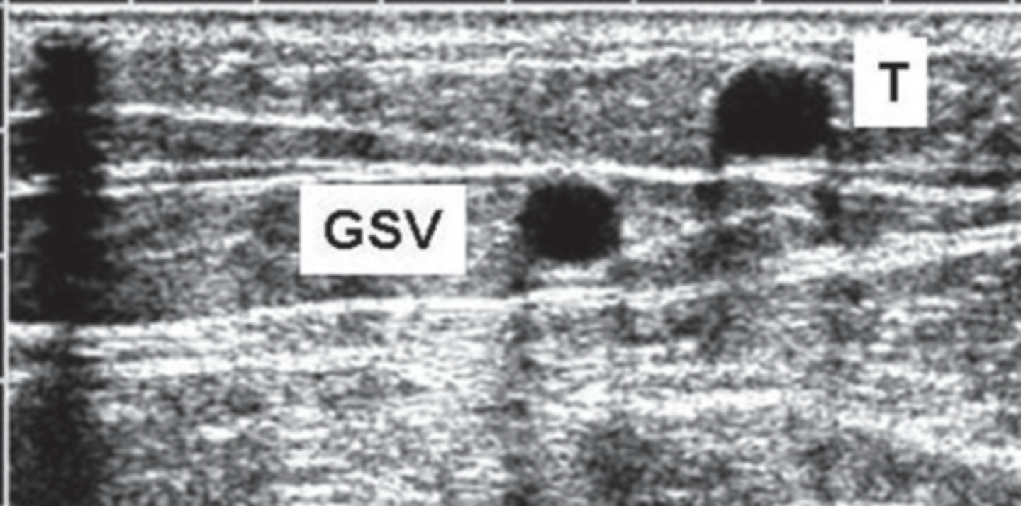
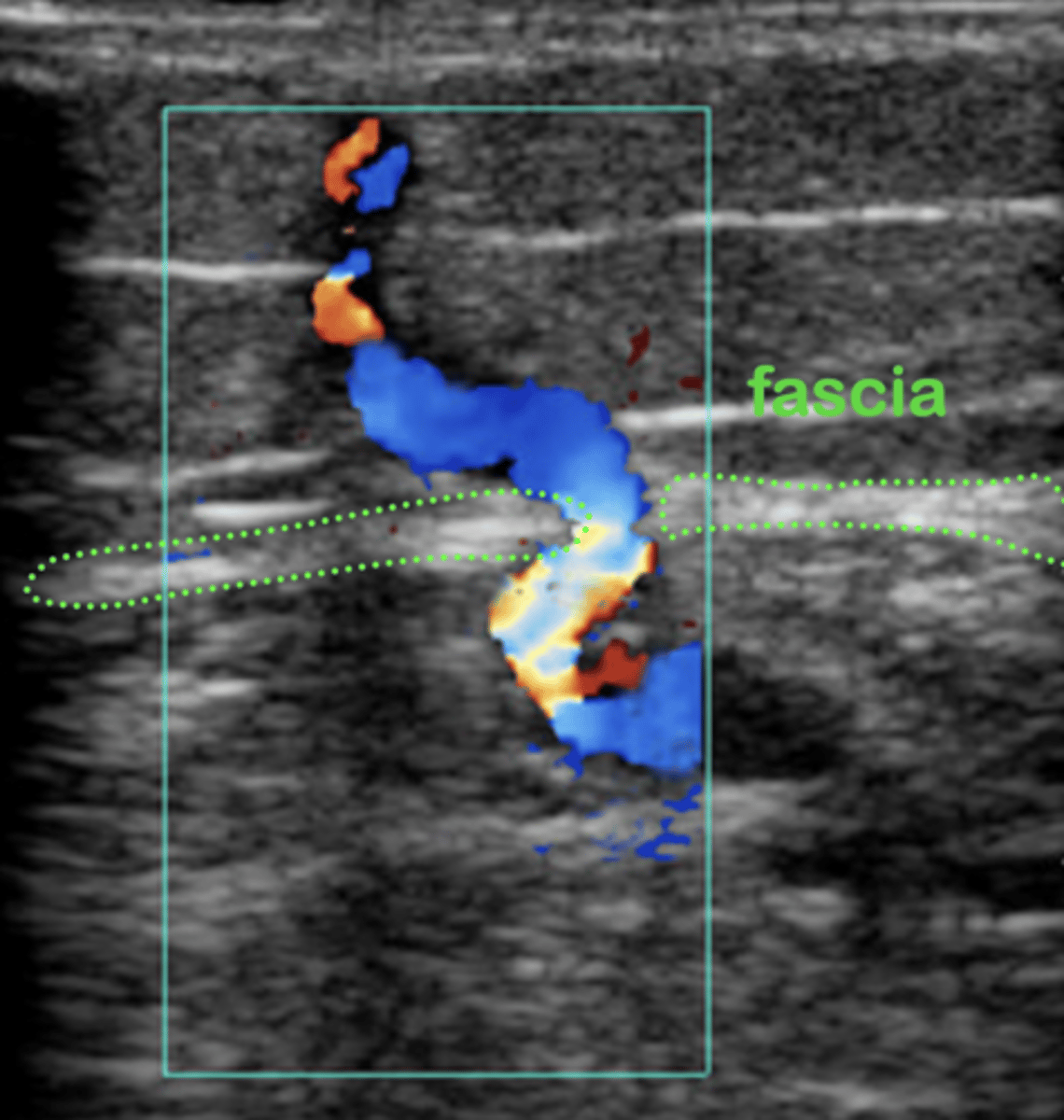
Perforator
Pass through muscular fascia layers - connect deep system to superficial system
Primary Valvular Insufficiency
Congenital absence of veins
Secondary Valvular Insufficiency
Damaged valves caused by thrombotic events
Chronic Venous Insufficiency
Obstruction & valve insufficiency
Arterial Ulcers
Found in tibial area/bony areas
Severe pain
Deep, regular shape
Little bleeding
Shiny skin, hair loss, thickened toe nails
Venous Ulcers
Found near medial malleolus
Minimal pain
Shallow, irregular shape
Oozing/Odor
Stasis dermatitis/brown discoloration/varicosities
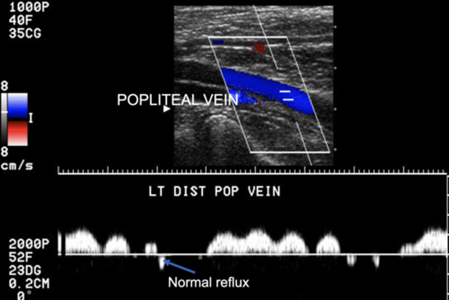
Normal Physiologic Reflux
Small amount of flow in the opposite channel is normal
Deep System Reflux
≥ 1 sec
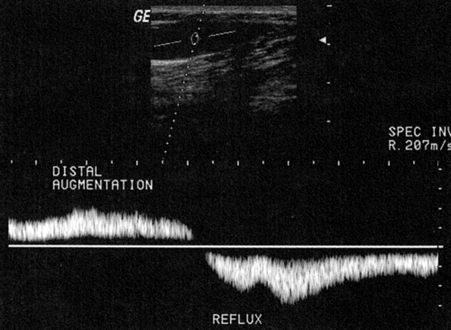
Superficial System Reflux
≥ 0.5 sec
Reflux Photoplethysmography (PPG)
Evaluates venous valve competency
Easy & reliable results
Positive findings must be confirmed with other studies
PPG Testing Protocol
Relaxed limb - normal baseline
Dorsiflex foot 5-6x - to empty calf veins
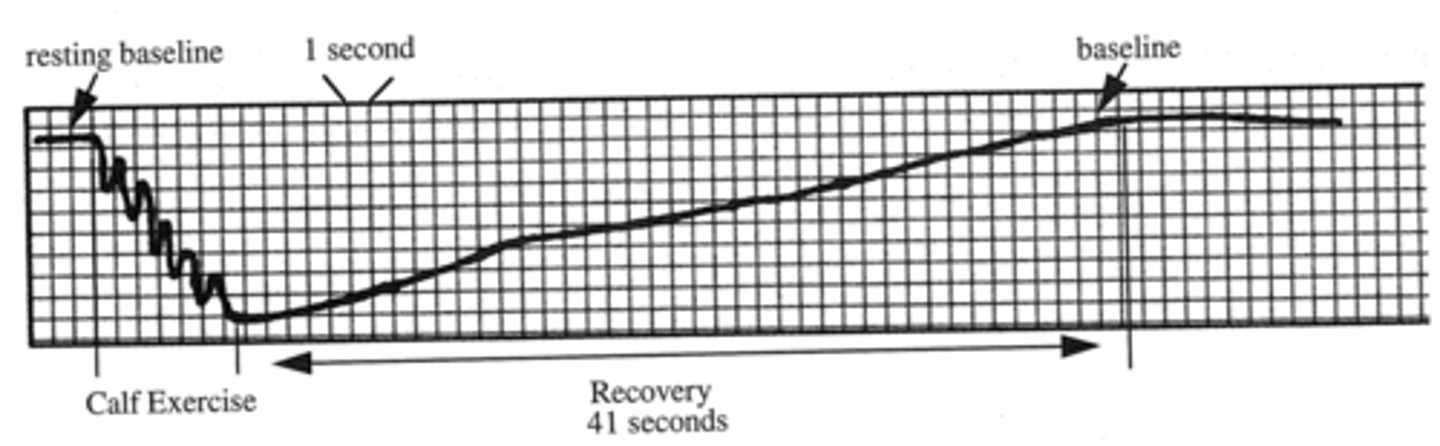
Venous refilling time - time from when exercise stops to stabilization period of ≥ 5 seconds
Normal PPG
Refill time/time to baseline > 20 seconds
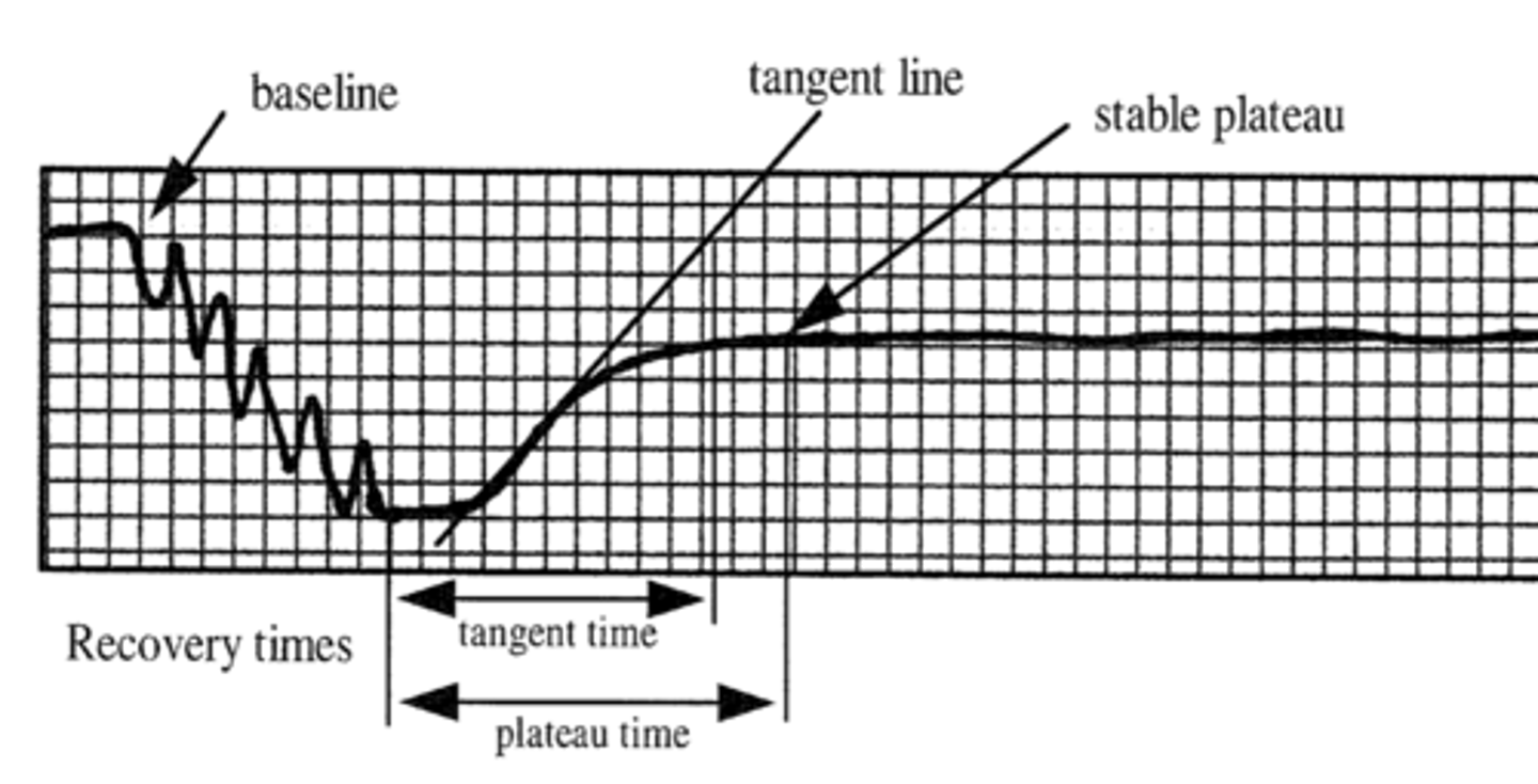
Abnormal PPG
Does not return to baseline
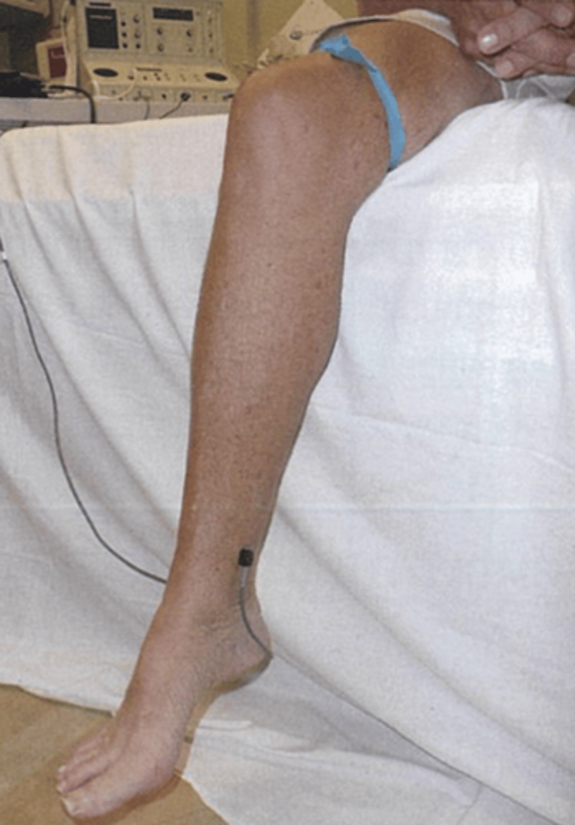
PPG - Tourniquet Test
Differentiates superficial from deep system
GSV Reflux PPG
Refill time < 20 sec
Normalizes with tourniquet above knee
SSV Reflux PPG
Refill time < 20 sec
Normalizes with tourniquet below knee
Incompetent Deep Valves PPG
Consistent reflux < 20 sec
Air Plethysmography (APG)
Recommended for chronic venous insufficiency
Cuff inflated to 10 mmHg
Leg is elevated
Patient quickly stands - bearing weight on non-tested leg
Distribute weight evenly
1 toe raise
10 toe raises
Return to supine and elevate tested leg
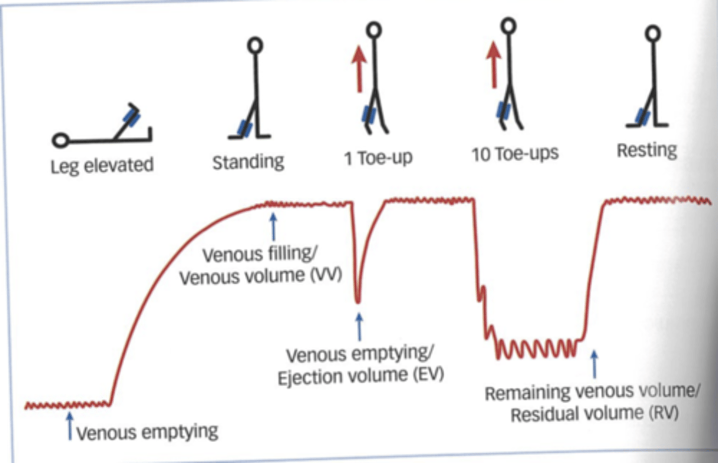
Filling Time
How long to accumulate blood in calf to 90% venous volume
NL = > 25 seconds
Ejection Fraction
After one toe
> 60% = normal
Venous Filling Index
Blood accumulated per unit time
NL = < 2 mL/s
Residual Volume
Percentage of venous volume
How much volume is pumped from calf after 10 toe raises
NL = < 20 - 35%
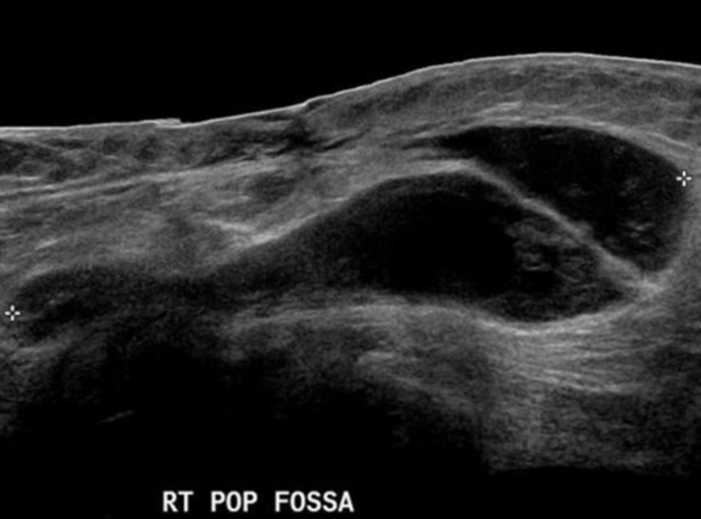
Baker's Cyst
Leaking synovial fluid into joint space
Connect to knee joint in medial popliteal fossa
Rupture looks like rat tail
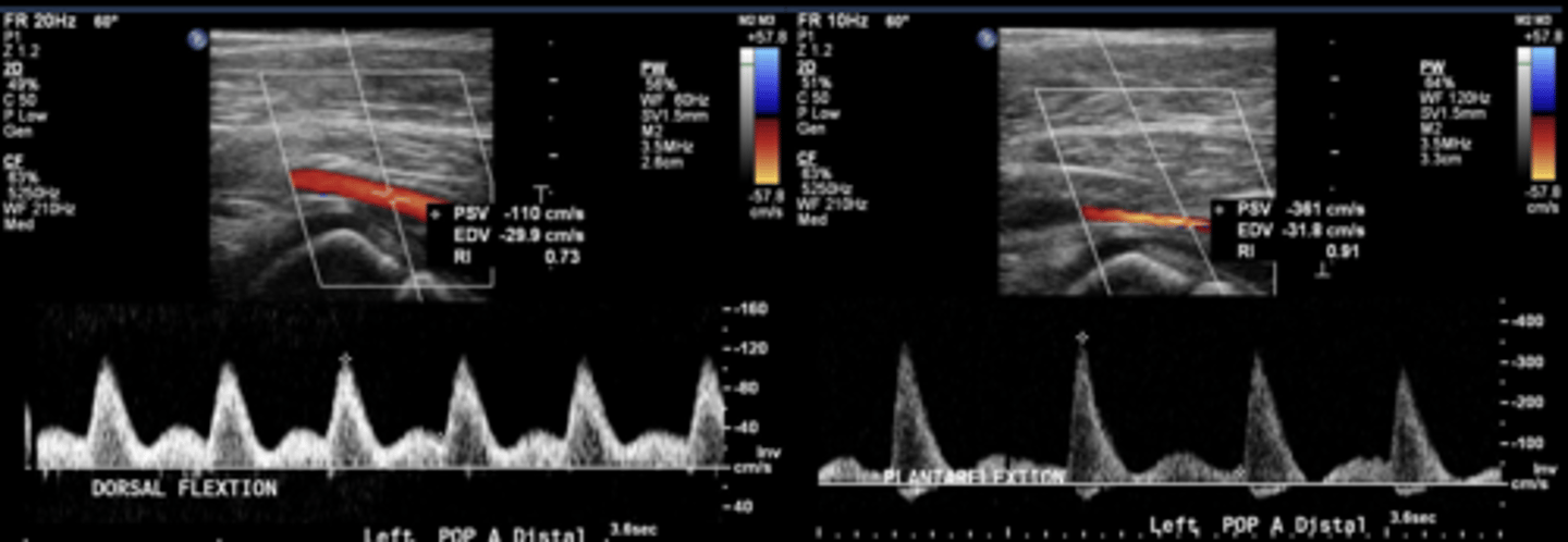
Popliteal Entrapment
Exercise ABI decreases 30-50%
Pain or cramping in calf during exercise
Discoloration
Swelling
Numbness
Untreated = ischemia or arterial damage
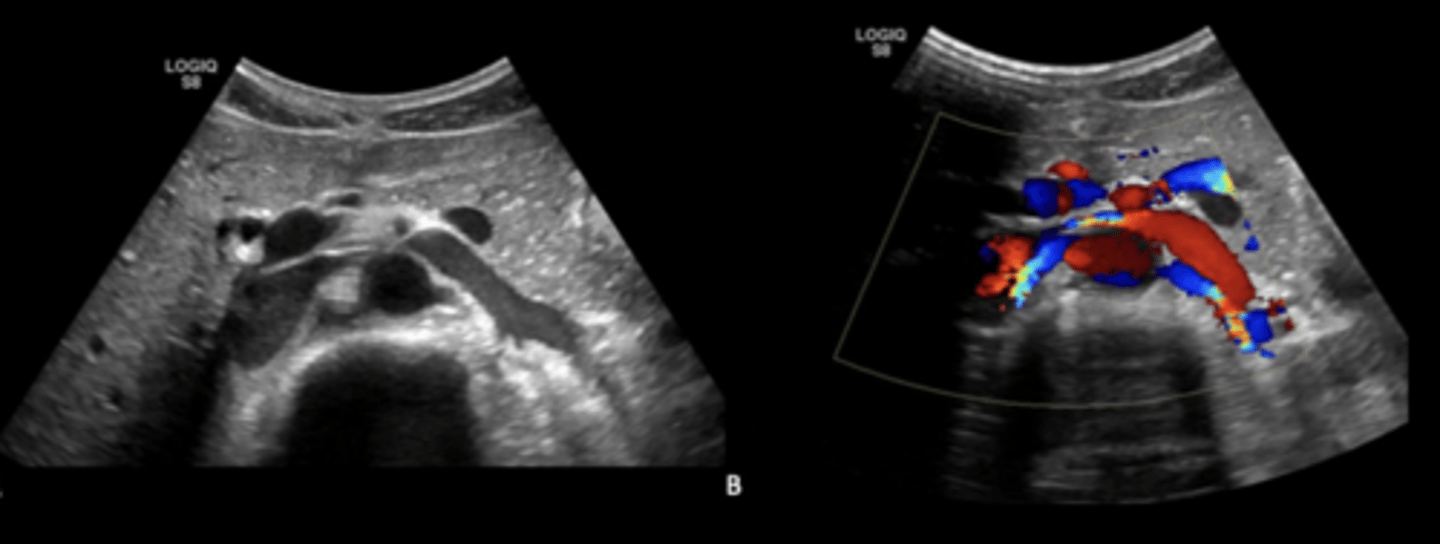
Nutcracker Syndrome
Left renal vein compressed by SMA
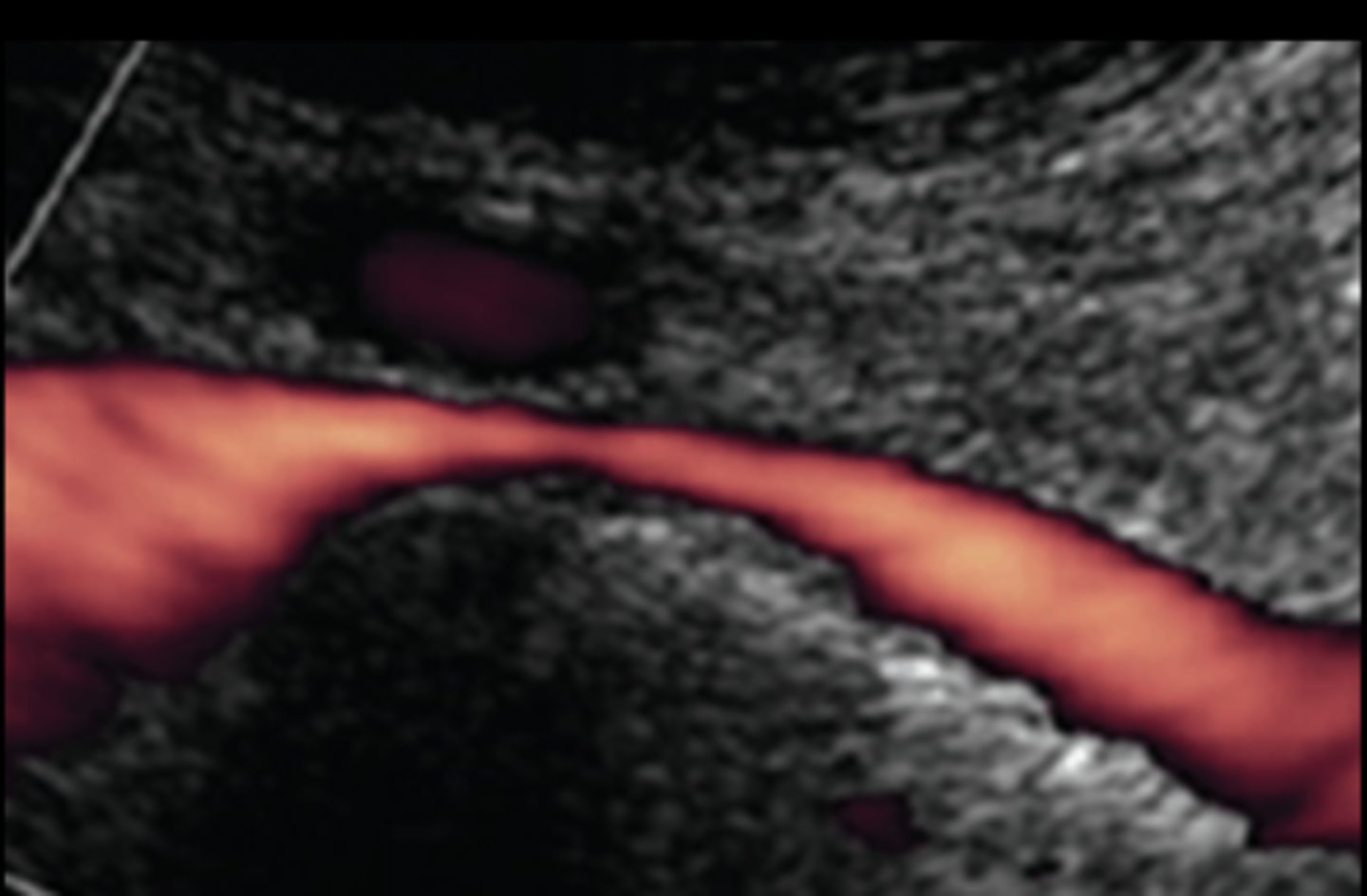
May-Thurner/Iliac Vein Compression Syndrome
Iliac vein is compressed between iliac artery and vertebral body
More common on left side
Can present as iliofemoral DVT or lower extremity edema/insufficiency
Treated by stent/balloon
Acute Compartment Syndrome
Increased intracompartmental pressure - > 30 mmHg
Diminishes vascular flow and tissue viability
Most commonly caused by fractures
Pain
Pareshthesia
Pallor
Paralysis
Pulselessness
Poikothermia (cold)
Giant Cell Arteritis
Inflammatory immune system disorder
Affects temporal & carotid arteries
Visual & neurological symptoms
Polymyalgia Rheumuatica
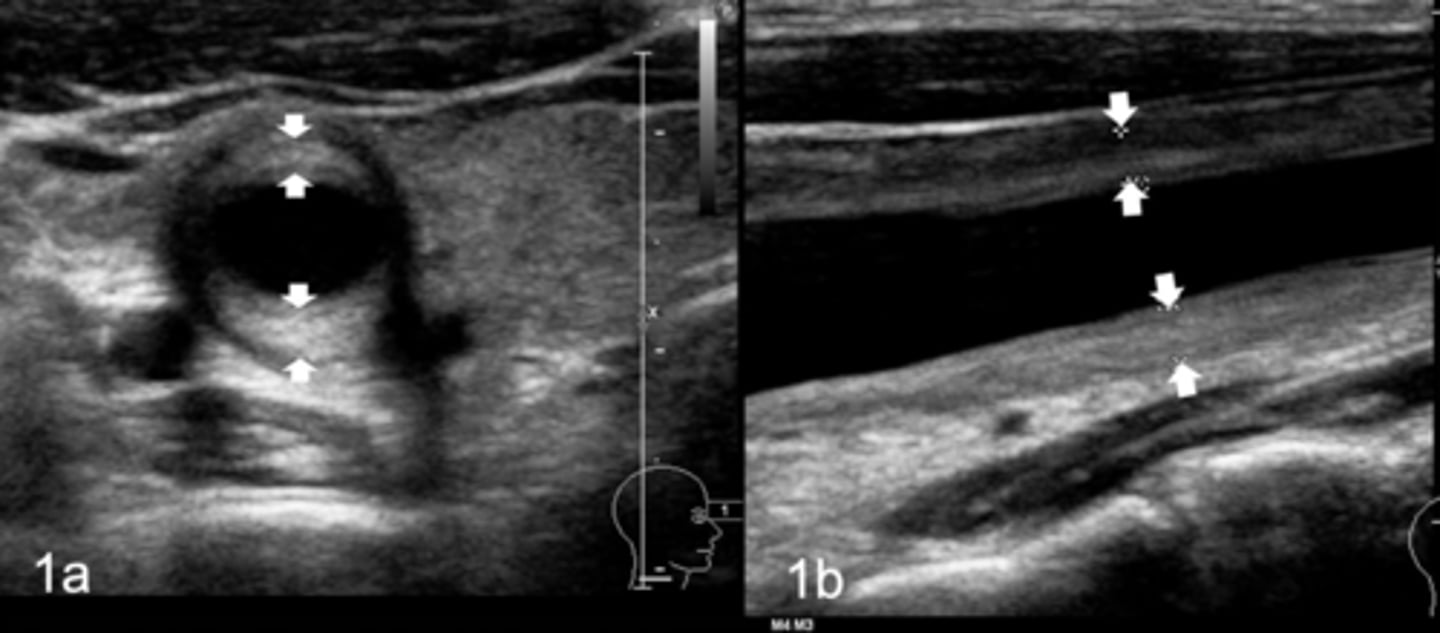
Takayasu's Arteritis
Inflammatory disease
Pulselessness of aorta, carotid, & branches
Narrowing & blockages
Macaroni sign
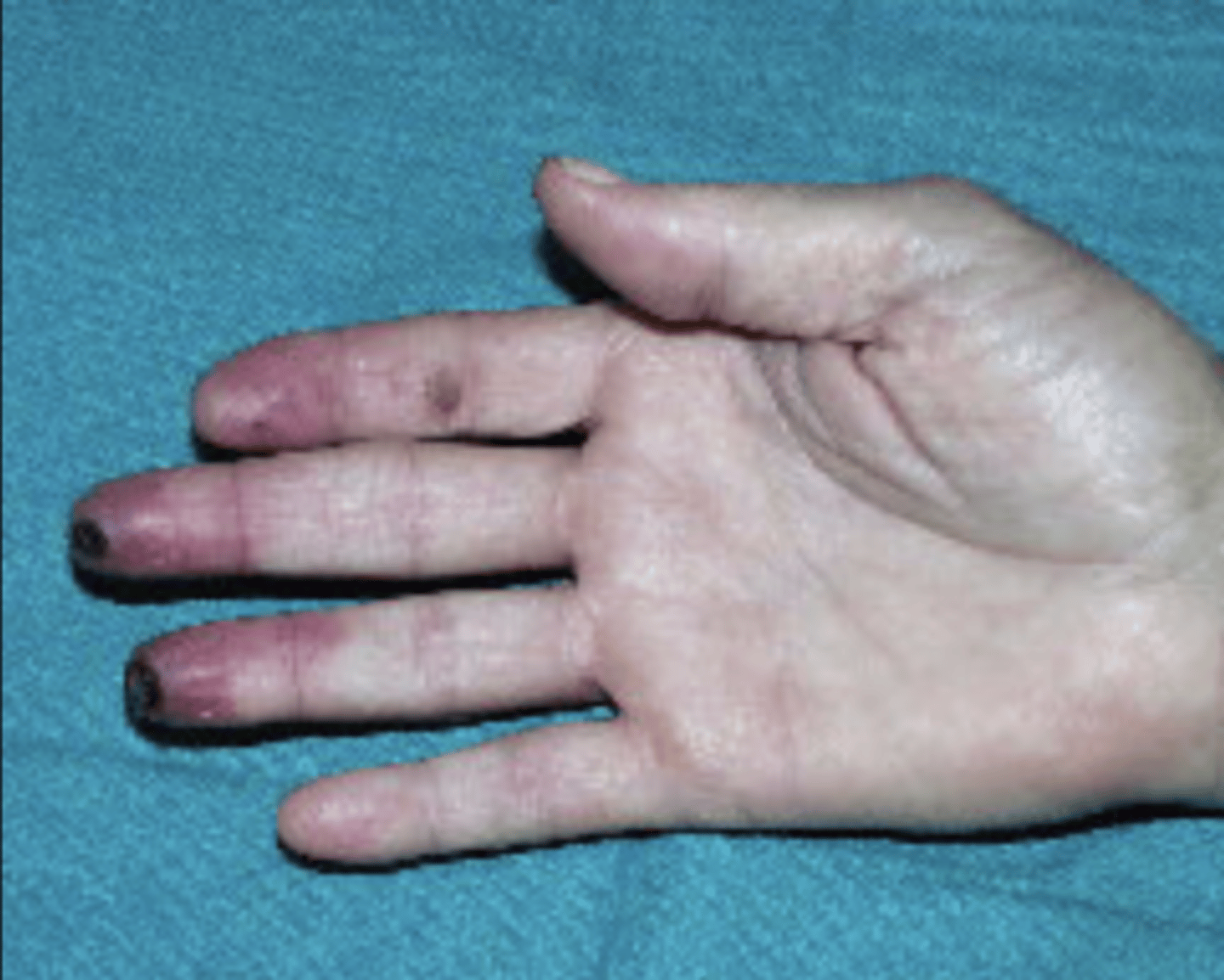
Buerger's Disease (Thromboangitis Obliterans)
Bilateral inflammatory disease of digital arteries in hands and feet
Always related to tobacco use
Fixed occlusive disease process
Ulcerations/gangrene - ¾ limb involvement common
Decreased distal pulses
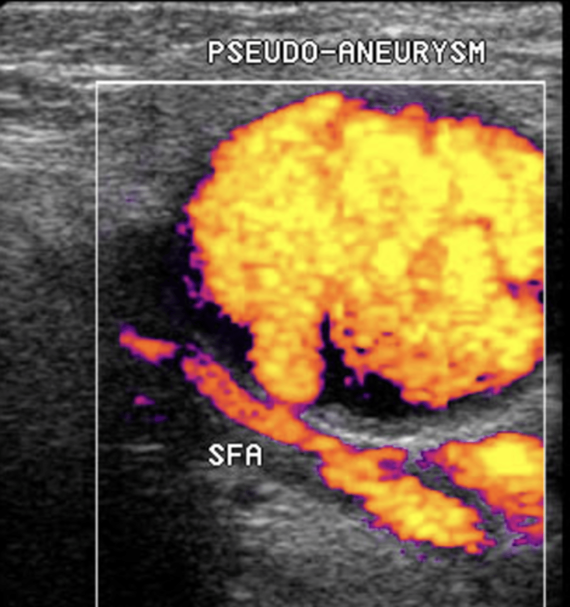
Iatrogenic Injury/Pseudoaneurysm
Due to percutaneous arterial catheterization, penetrating trauma, or graft anastomosis "blowout"
Treated by compression, surgical repair, or thrombin injection