Seasons Structure Week 1: NTK
 Call Kai
 Learn
Learn Practice Test
 Spaced Repetition
 Match
Match Flashcards
 Knowt Play
1/45

Earn XP
Description and Tags
Normal Anatomy of Chest Wall and Rib Cage
Name | Mastery | Learn | Test | Matching | Spaced | Call with Kai | Chat |
|---|
No analytics yet
Send a link to your students to track their progress
46 Terms
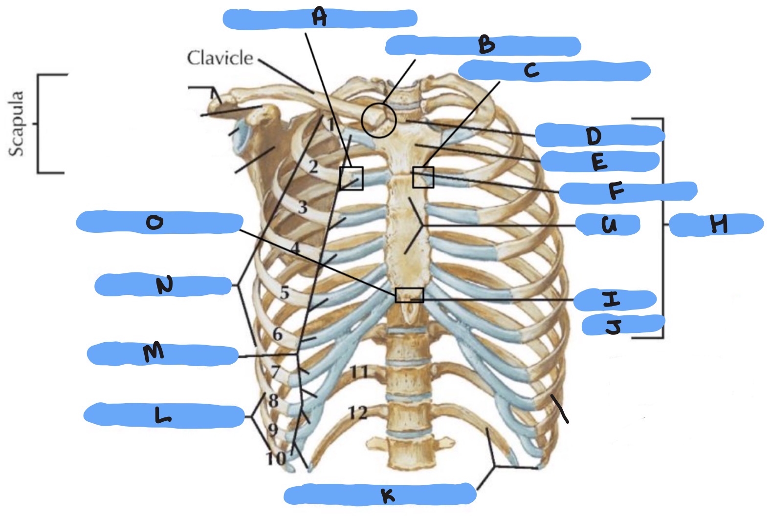
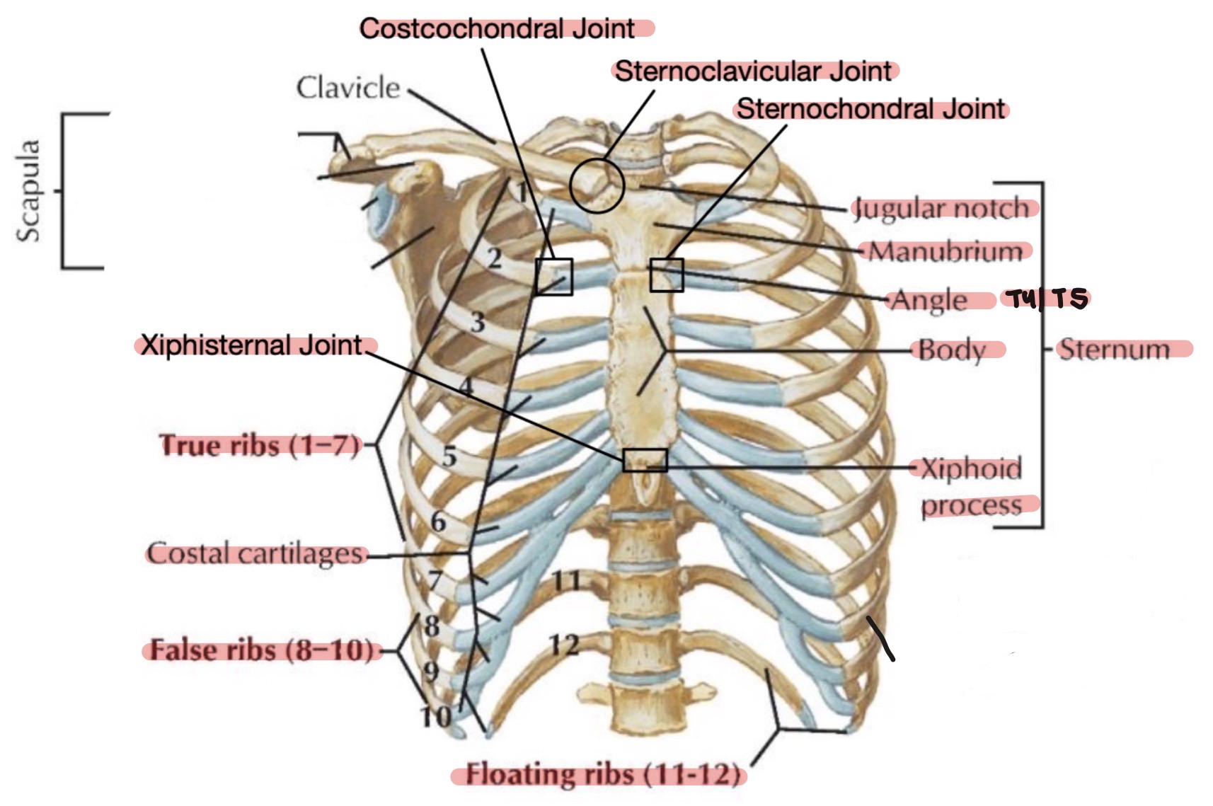
NTK
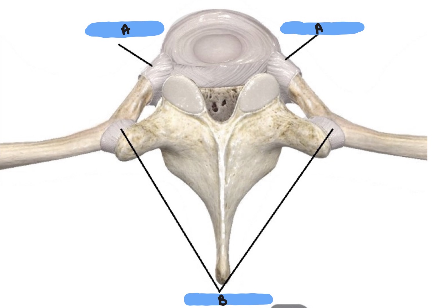
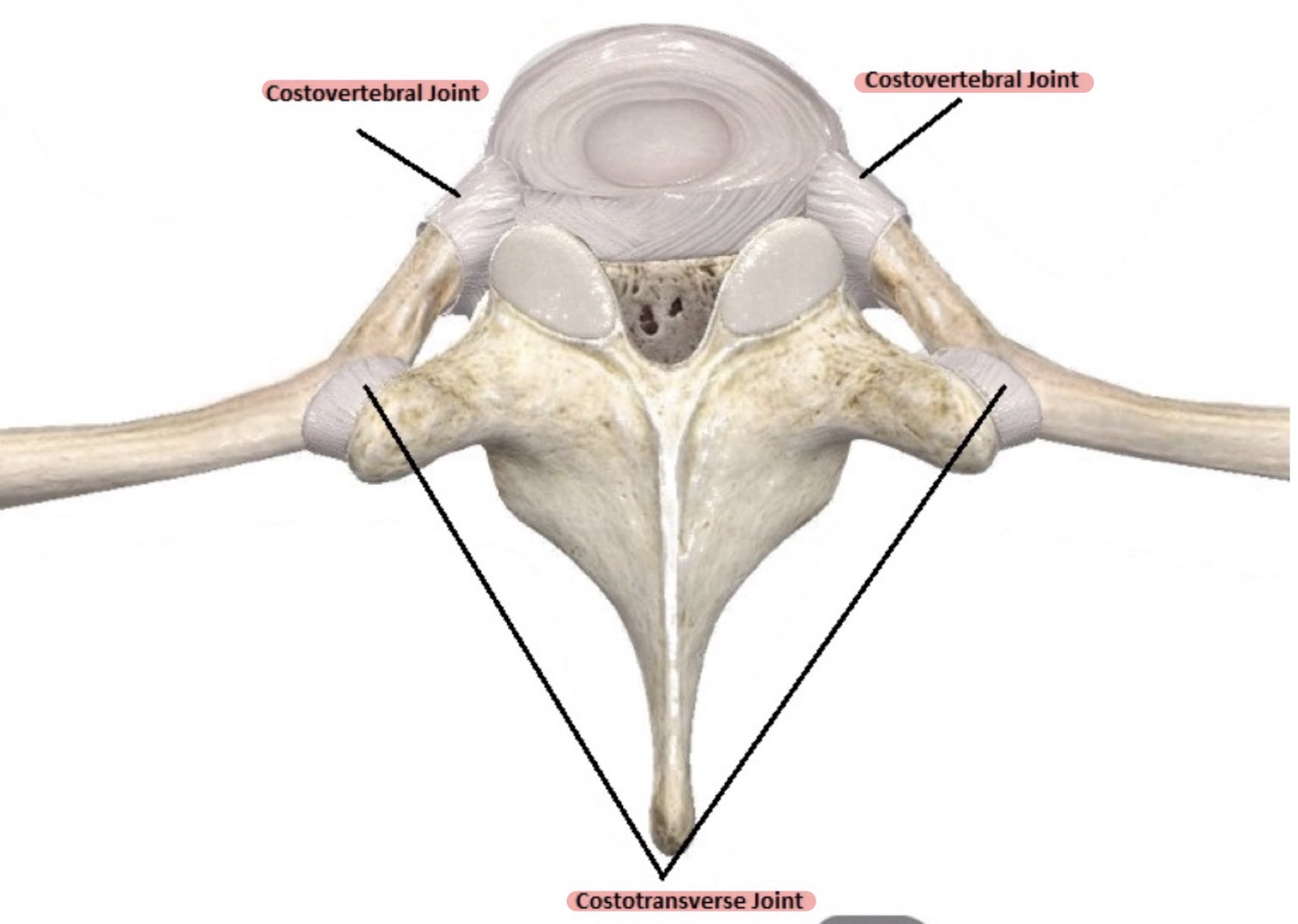
NTK
NTK
NTK
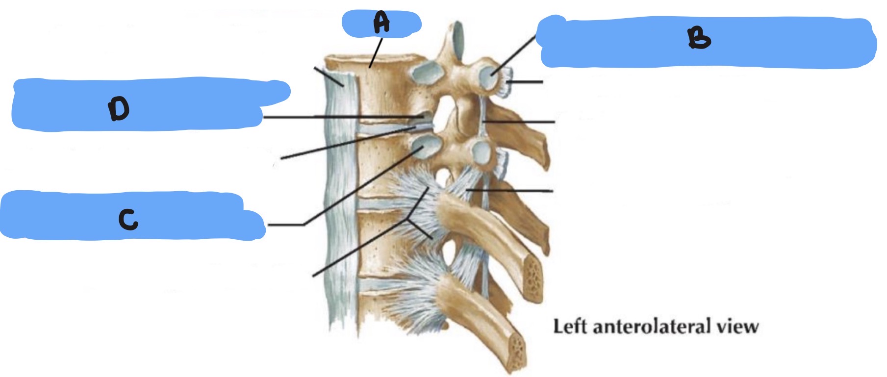
NTK
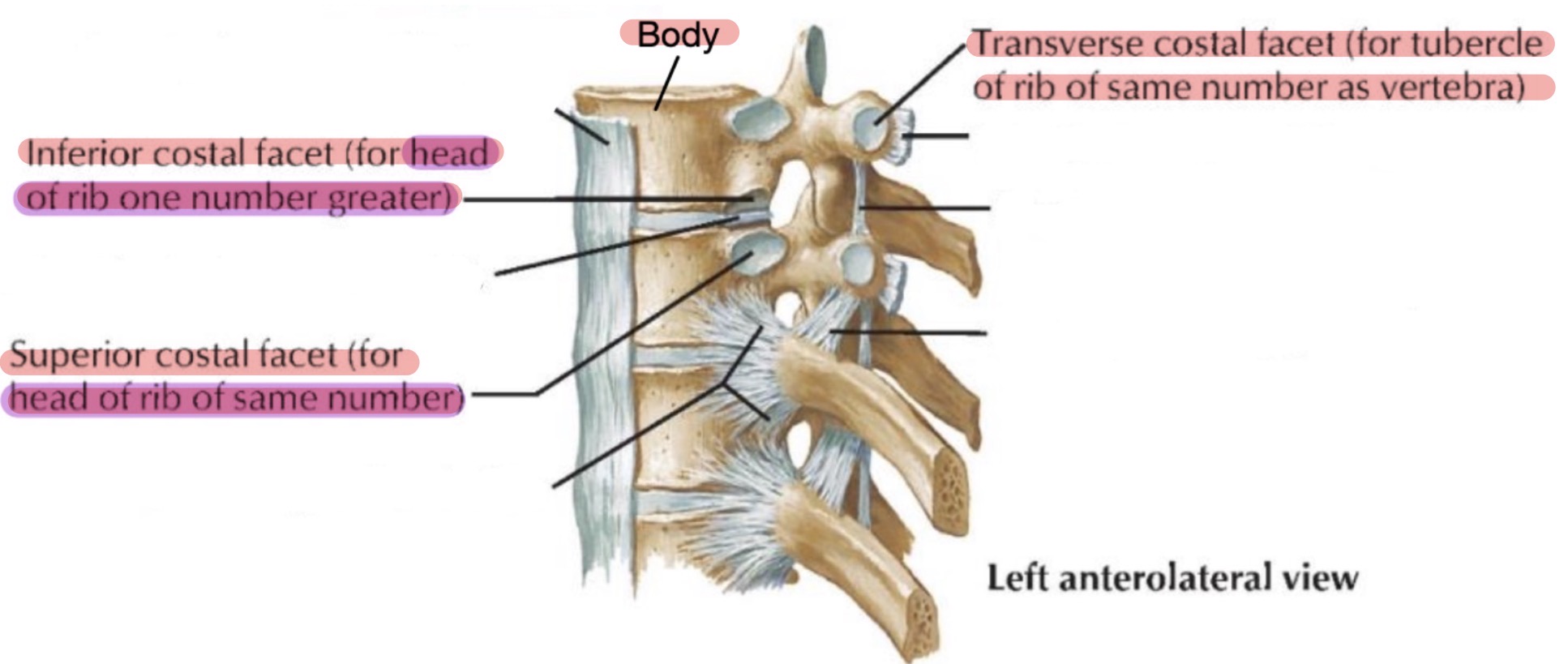
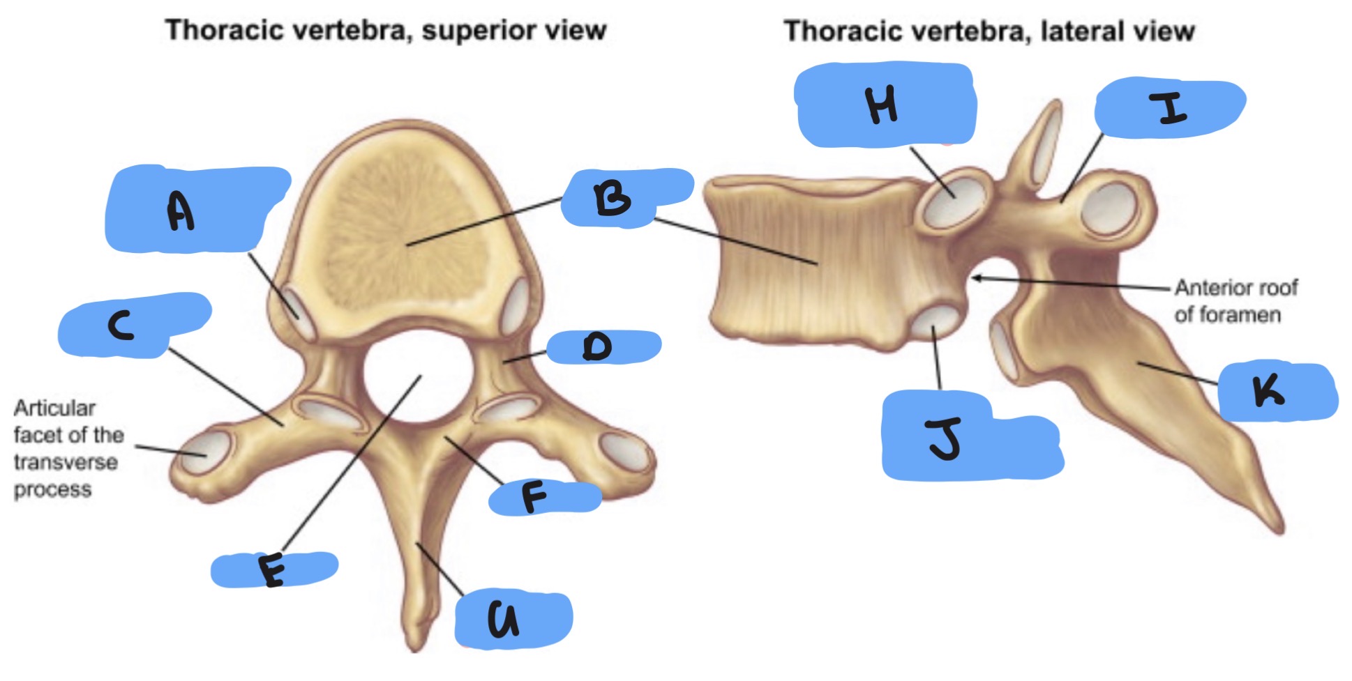
NTK
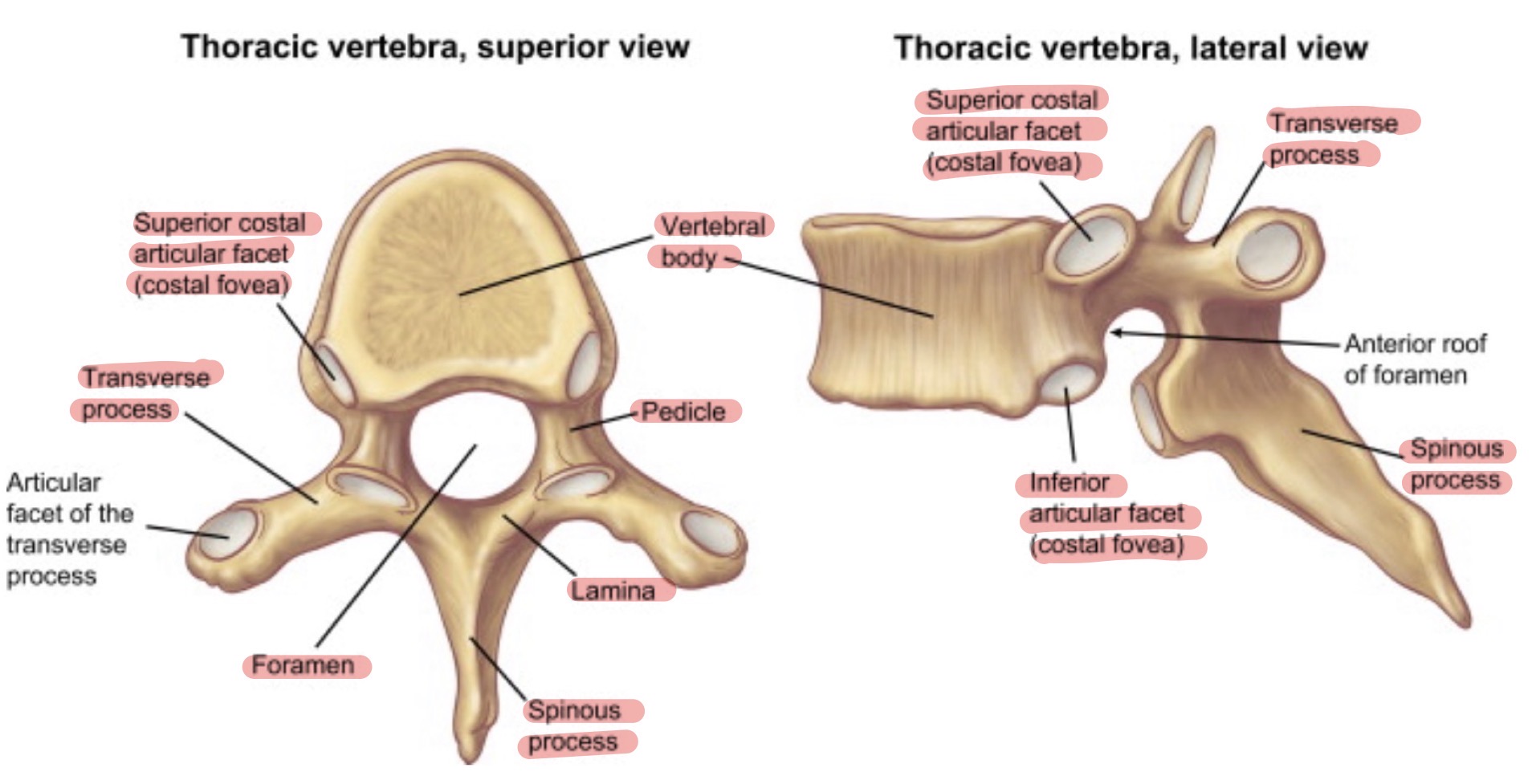
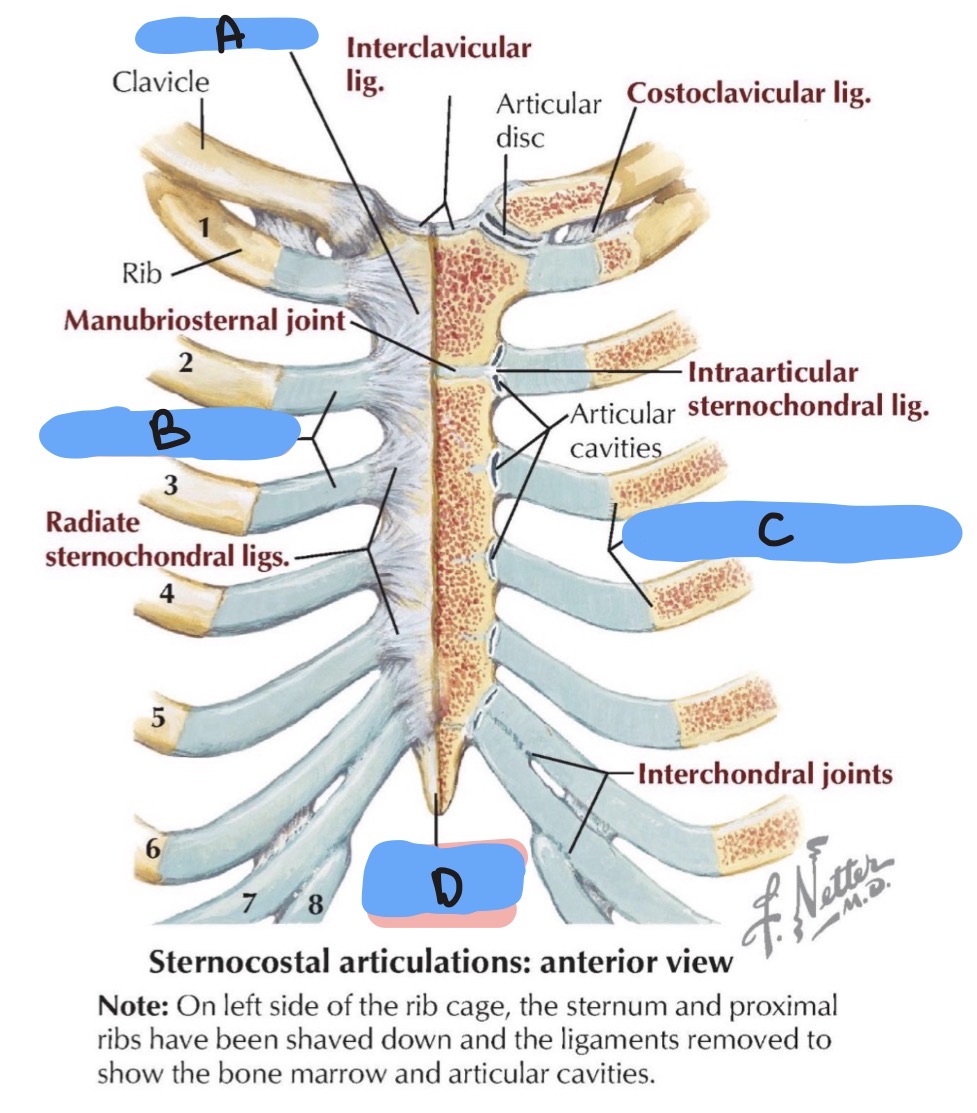
NTK
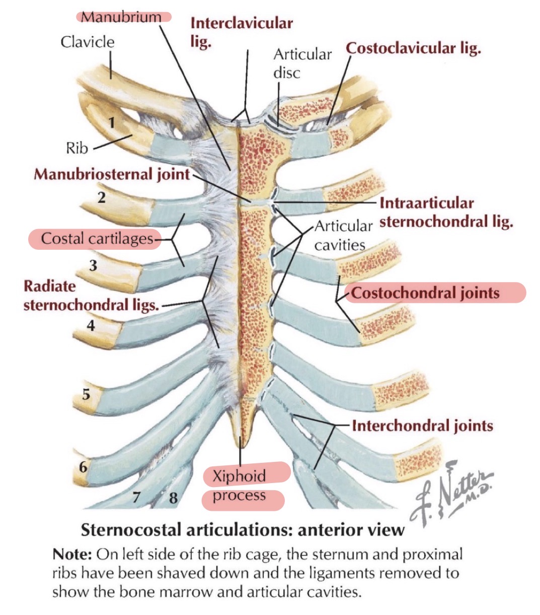
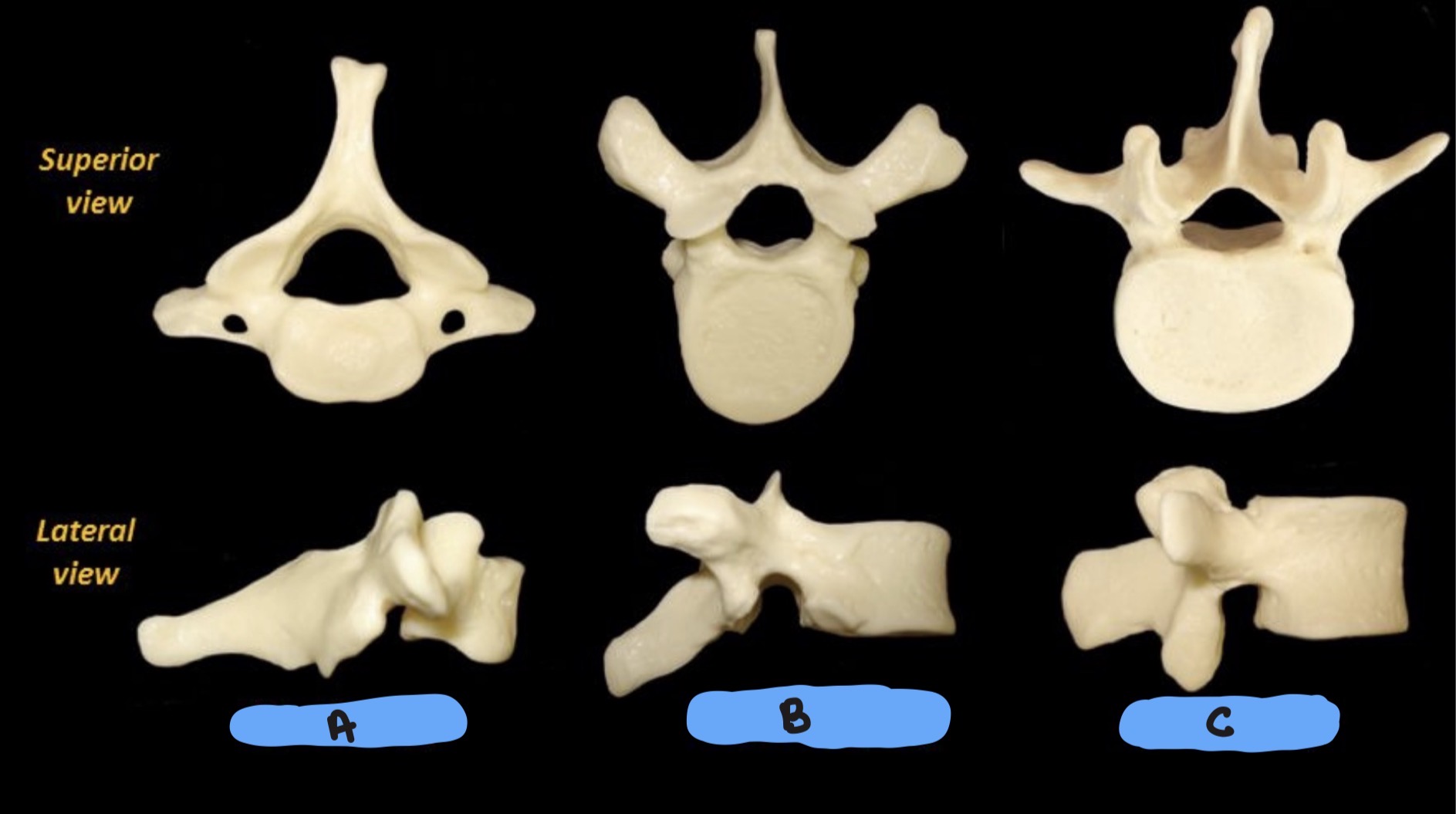
NTK
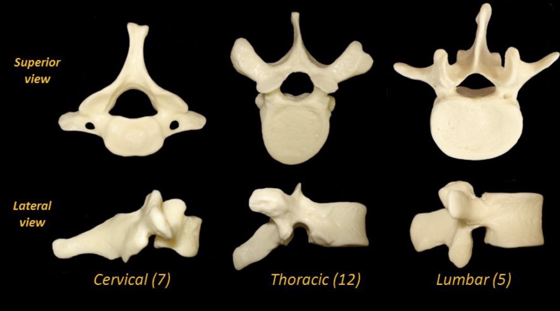
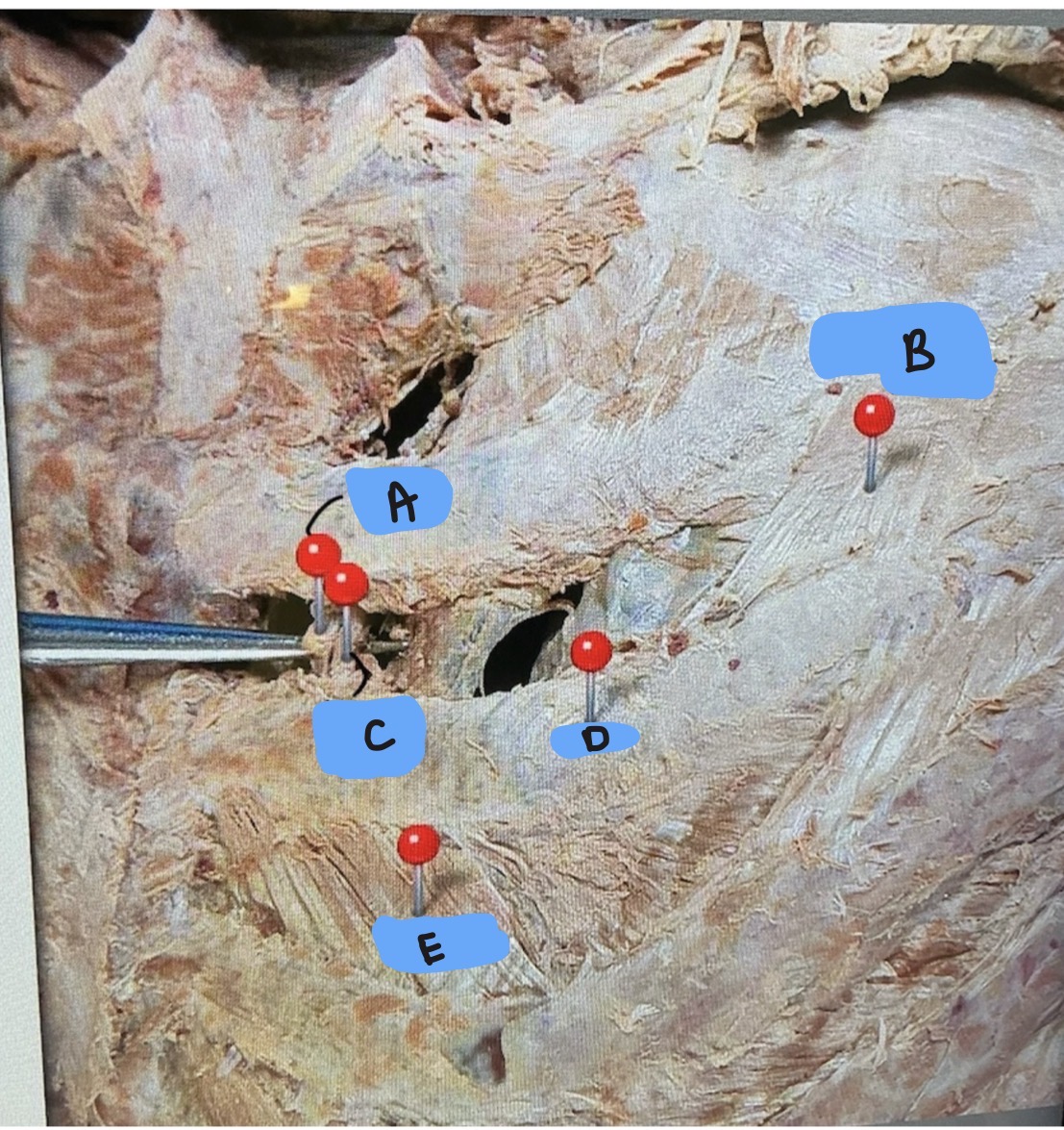
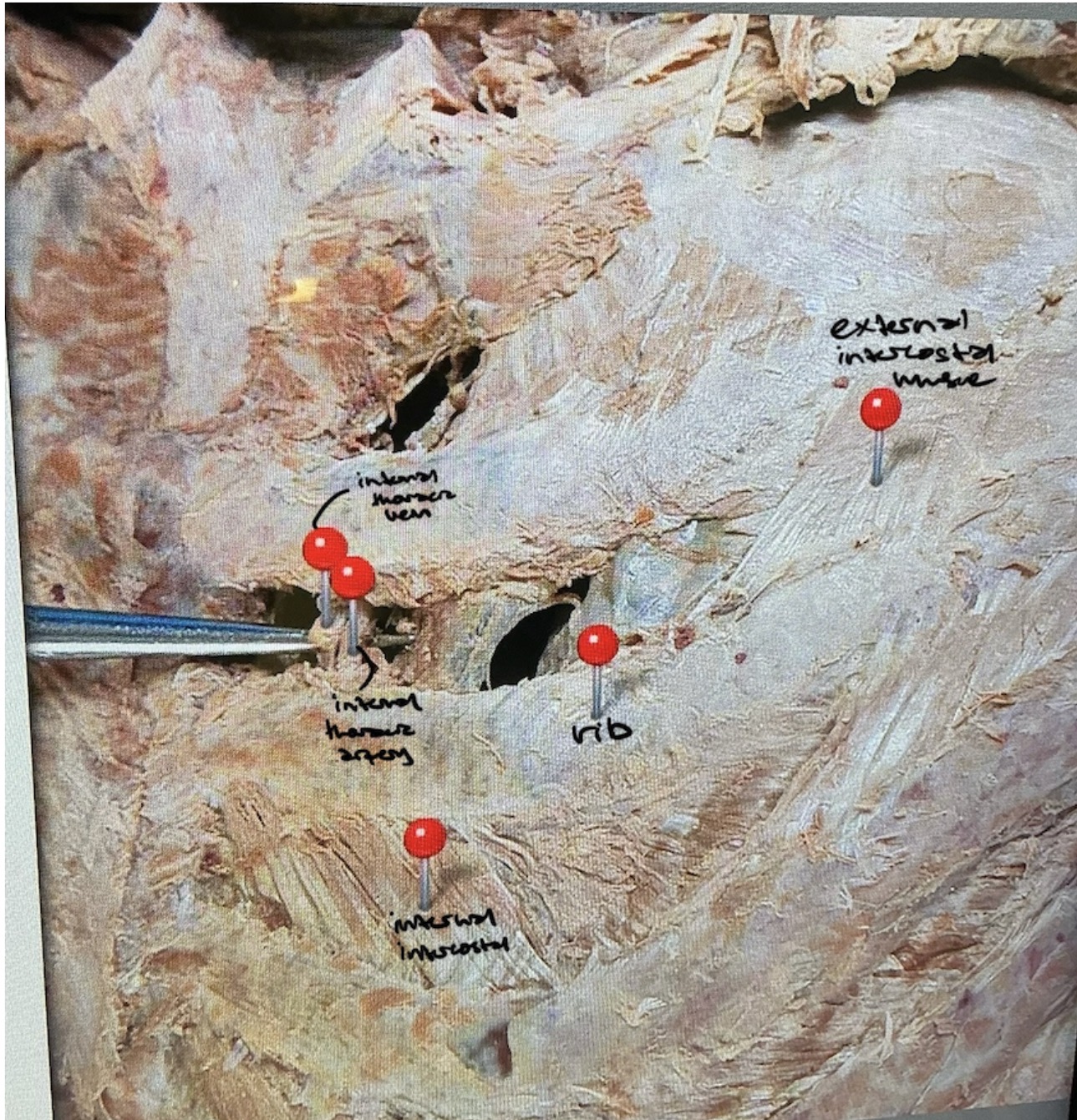
NTK
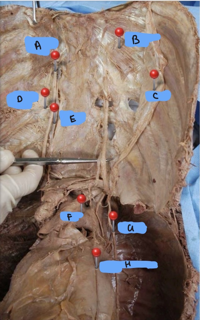
NTK
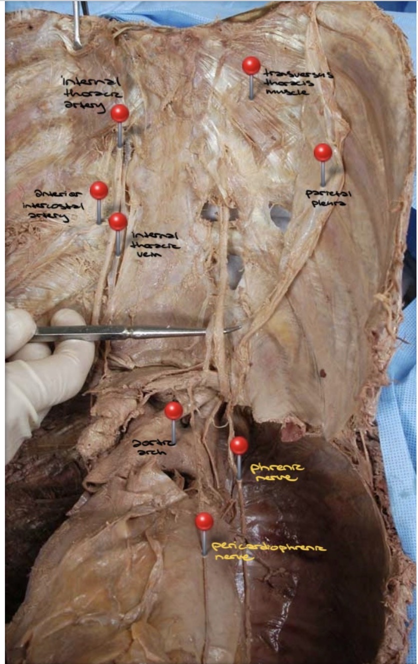
NTK
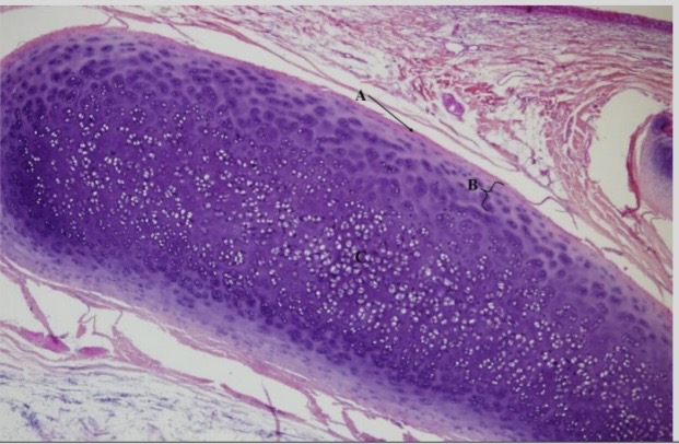
NTK: what type of cartilage is this
Hyaline
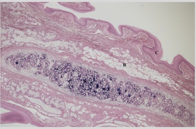
NTK: what type of cartilage is this
Elastic
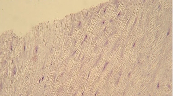
NTK: what type of cartilage is this
Elastic
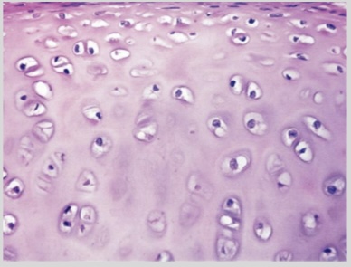
NTK: what type of cartilage is this
Hyaline
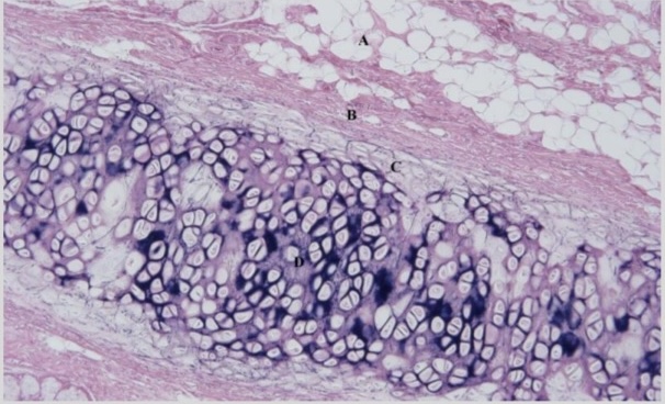
NTK: what type of cartilage is this
Elastic
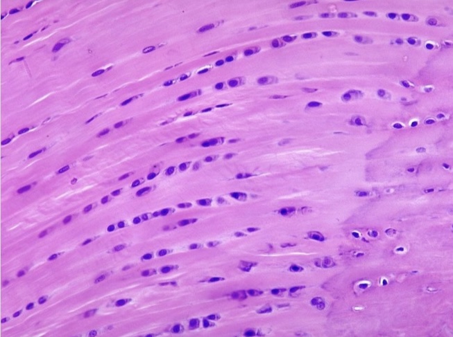
NTK: what type of cartilage is this
Elastic
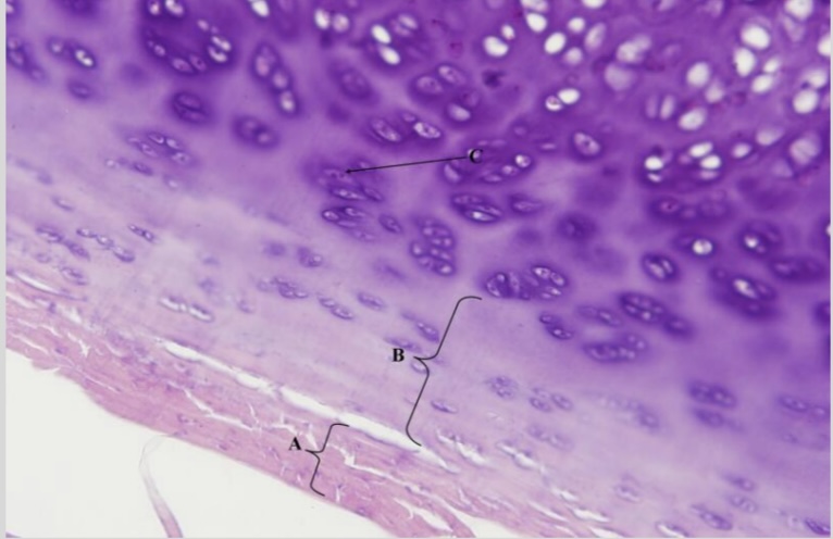
NTK: what type of cartilage is this
Hyaline
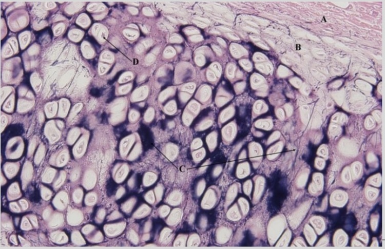
NTK: what type of cartilage is this
Elastic
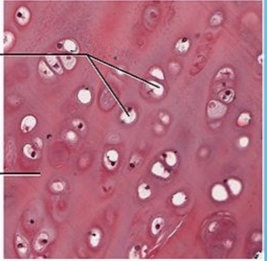
NTK: what type of cartilage is this
Hyaline
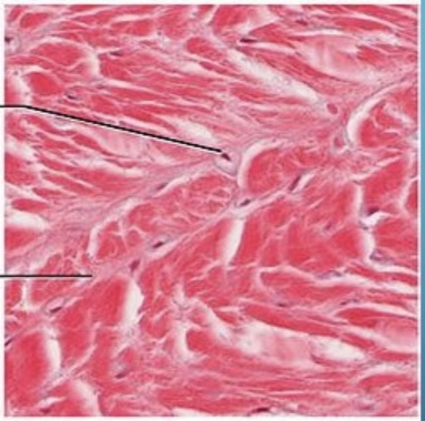
NTK: what type of cartilage is this
Fibrocartilage
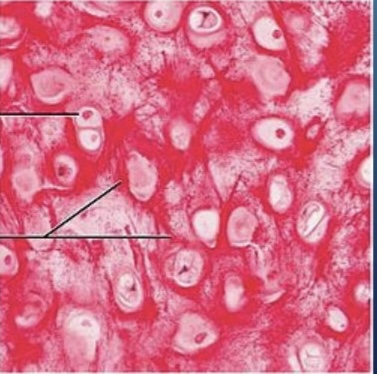
NTK: what type of cartilage is this
Elastic
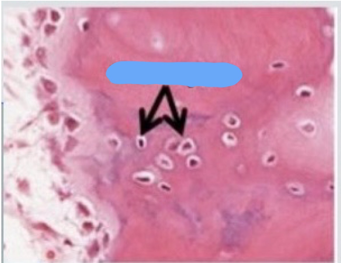
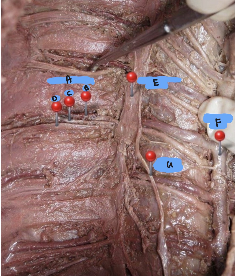
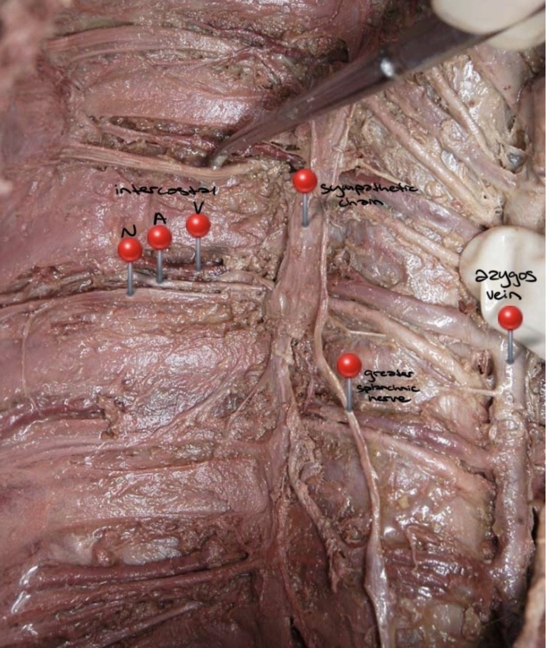
NTK: what is this
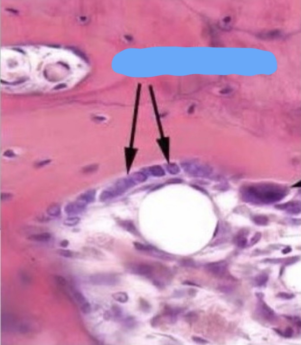
NTK: what is this
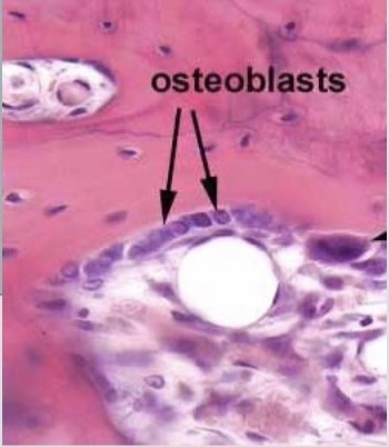
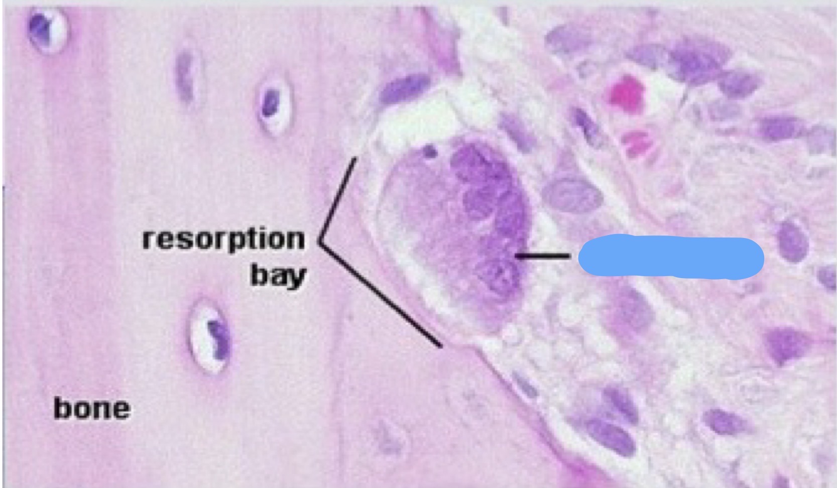
NTK: what is this
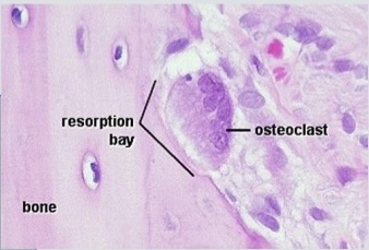
ICG: Is an intercostal nerve a dorsal or ventral ramus?
ventral
ICG: If an intercostal nerve is severed, what deficits would the patient exhibit?
Intercostal muscle weakness/paralysis (myotome), loss of somatic sensation in that dermatome, loss of sympathetic innervation
ICG: What is the difference between a true rib, false rib, and floating rib?
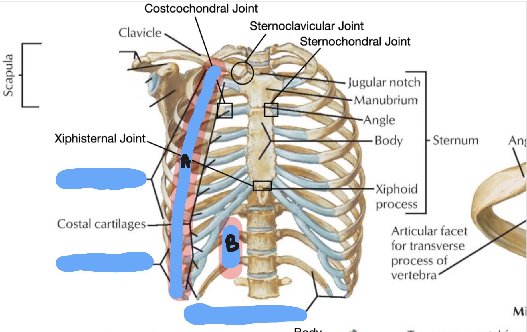
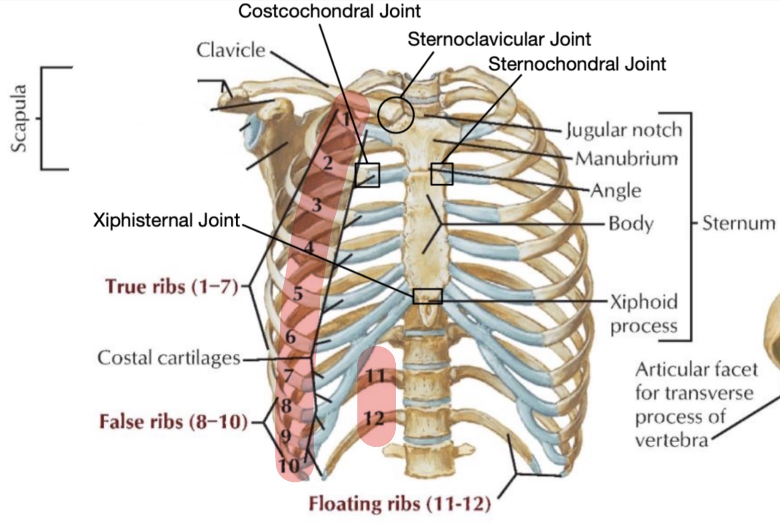
The difference between true ribs, false ribs, and floating ribs lies in their attachment to the sternum:
True ribs: These are the first seven ribs and are directly attached to the sternum.
False ribs: These are the next three pairs of ribs and attach indirectly to the sternum via the lowest true rib.
Floating ribs: The last two pairs of ribs do not attach to the sternum at all and only connect to the spine
ICG: What are some potential consequences of significant chest trauma with broken ribs?
Pulmonary contusion, flail chest, pneumothorax, hemothorax
ICG: How might fracture of several ribs at two or more places on the right-side effect respiratory function?
The patient has a flailed chest and a pulmonary contusion. The flail chest inhibits proper negative pressure ventilation.
The pulmonary contusion results in lung tissue that is not efficient at gas exchange creating a shunt and hypoxia.
ICG: How might a pulmonary contusion look like on CXR? How would this compare to a pneumothorax or hemothorax?
The opacity of contusion is similar to pneumonia.
Pneumothorax is dark (radiolucent due to just air present). Hemothorax very dense (opacity) with meniscus perhaps and does not follow lobar patterns.
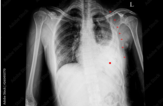
ICG: is this a pulmonary contusion, pneomothorax, or hemothorax
hemothorax
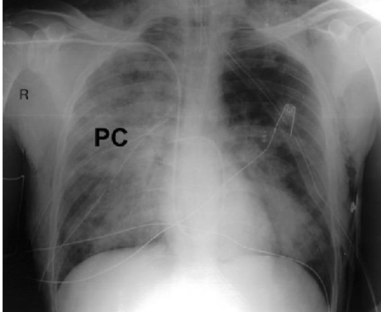
ICG: is this a pulmonary contusion, pneomothorax, or hemothorax
PC
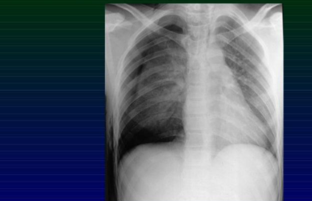
ICG: is this a pulmonary contusion, pneomothorax, or hemothorax
pneumothorax with rib fractures
ICG: The trauma team places metal plates to stabilize the rib fractures and the patient can be weaned from the ventilator after 4 days. While on the ventilator he is receiving narcotic analgesic IV and is switched to oral narcotics. He complains of significant chest wall pain.
4. What nerves are relaying this pain? What kind of sensory system is this? Trace the path of the rib pain back to the sensory cortex of the brain. How many neurons are involved?
Somatosensory from intercostal nerves mostly.
spinothalamic tract- 1st cell body in DRG, 2nd cell body in spinal cord-lateral or dorsal horns, third cell body in thalamus in brain, 4th cell body in cerebral cortex
IGC: The patient is diagnosed with neuropathic pain and started on Gabapentin. He has some relief from this, but he experiences some undesirable side effects and stops it, He starts taking his oral narcotics again. He is referred to pain management and undergoes an intercostal nerve block with significant improvement.
5. How would an intercostal block be done? Where should the anesthetic be injected?
Rib space at the affected level as well as the level immediately superior and immediately inferior. Inject on underside of each rib ideally.
ICG: Vignette 2: A 45-year-old male presents with severe chest pain following a motor vehicle accident. On physical examination, there is tenderness and swelling over the left lateral chest wall.
1. What are the main bones that form the chest wall?
The main bones forming the chest wall include the sternum, 12 pairs of ribs, and the thoracic vertebrae. (added protection form muscles, scapula)
ICG: 2. Describe the muscle layers of the chest wall.
The muscle layers of the chest wall include:
• Superficial layer: Pectoralis major and minor muscles.
• Intermediate layer: External intercostal muscles.
• Deep layer: Internal intercostal muscles, innermost intercostal muscles, subcostal muscles, and transversus thoracis muscles.
ICG: How might an injury to the chest wall affect the underlying structures?
An injury to the chest wall can lead to fractures of the ribs or sternum, which may puncture the pleura and lungs causing pneumothorax, hemothorax, or damage to the intercostal nerves and vessels.
Sternal fractures can also result in cardiac damage
Vignette 3: A 60-year-old female presents with a persistent cough and localized pain in the right upper chest. A chest X-ray reveals a lesion on the 3rd rib.
1. Compare and contrast the different types of ribs and their landmarks.
True ribs (1-7): Attach directly to the sternum via costal cartilages.
False ribs (8-10): Attach indirectly to the sternum via the costal cartilage of the rib above.
Floating ribs (11-12): Do not attach to the sternum at all.
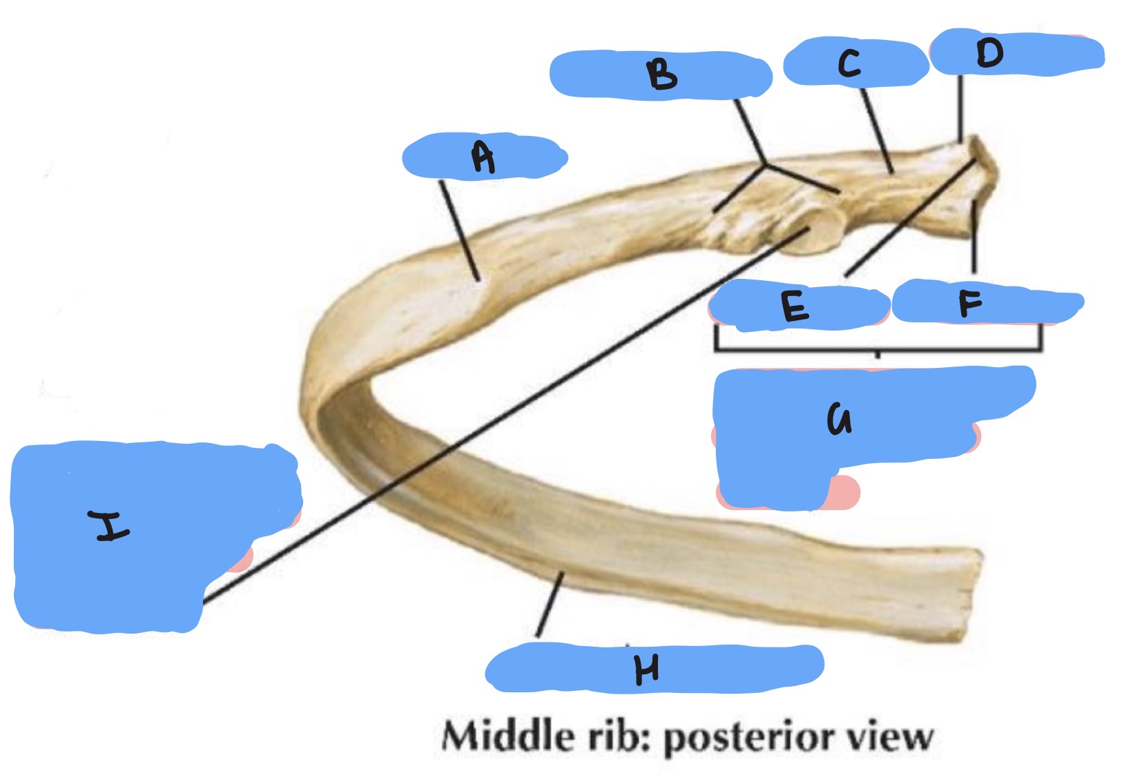
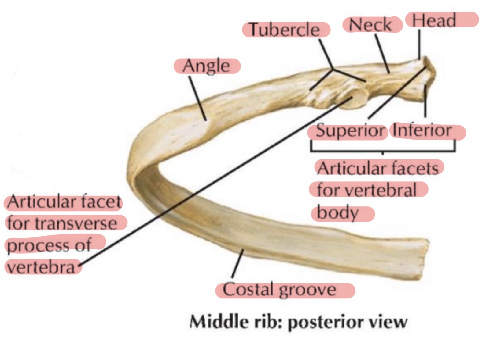
Landmarks: Head, neck, tubercle, angle, and body of the rib
ICG: What are the attachment points for the 4th rib?
The 4th rib attaches posteriorly to the thoracic vertebrae bodies at T3 and T4 and the transverse process (3 articular spots posteriorly) and anteriorly to the sternum via its costal cartilage
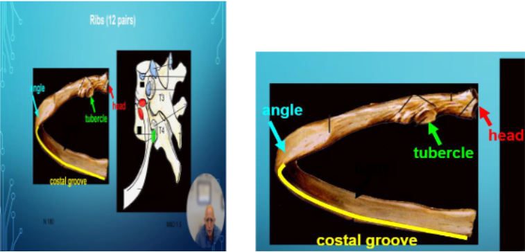
ICG: How can rib lesions impact the surrounding structures?
Rib lesions can impinge on the intercostal nerves and vessels, cause pleuritic pain, and may compromise respiratory movements if the integrity of the rib cage is affected
Vignette 4: A 30-year-old male presents with shortness of breath and difficulty breathing. On examination, there is limited movement of the diaphragm on the left side.
1. What are the primary muscles of respiration?
The primary muscles of respiration include the diaphragm, intercostal muscles (external, internal, innermost), and accessory muscles such as the scalene and sternocleidomastoid during forced respiration.
ICG: Describe the innervation of these muscles.
Diaphragm: Innervated by the phrenic nerve (C3-C5).
Intercostal muscles: Innervated by the intercostal nerves (T1-T11).
Accessory muscles: Innervated by the cervical spinal nerves and cranial nerve XI (sternocleidomastoid)
ICG: What might cause limited diaphragm movement
Limited diaphragm movement can be caused by phrenic nerve injury, paralysis, or conditions such as diaphragmatic hernia or elevated intra-abdominal pressure.
ICG: Vignette 5: A 50-year-old female is undergoing surgery for a mediastinal mass. Understanding the borders of the thoracic cavity is crucial for the surgical team.
1. What are the borders of the thoracic cavity?
Superior border: Thoracic inlet (bounded by the first rib, manubrium, and T1 vertebra).
Inferior border: Thoracic diaphragm.
Anterior border: Sternum and costal cartilages.
Posterior border: Thoracic vertebrae.
Lateral borders: Ribs and intercostal muscles.