CHAPTER 28: MANAGEMENT OF PX WITH OROFACIAL CLEFT
 Call Kai
 Learn
Learn Practice Test
 Spaced Repetition
 Match
Match Flashcards
 Knowt Play
1/48

Earn XP
Description and Tags
ppt + book
Name | Mastery | Learn | Test | Matching | Spaced | Call with Kai | Chat |
|---|
No analytics yet
Send a link to your students to track their progress
49 Terms
cleft
colloquial term is ”harelip”
occurs in 1 in 700 births
a congenital abnormal space or gap in the upper lip, alveolus or palate
the most common serious congenital anomalies to affect the orofacial region
cleft lip is more common in boys
cleft palate is more common in girls
¾ of the clefts are unilateral, ¼ bilateral
4 weeks after conception
development of the face:
frontonasal process
paired mx-mn processes
5th-6th weeks
maxillary processes and the medial nasal process merge to form the upper lip, alveolus, primary palate
lateral nasal process → ala of the nose
mandibular processes → lower lip and jaw
6th-8th weeks
palatal shelves fuse to form the secondary palate
primary palate
structures anterior to the incisive foramen (lip and alveolus)
secondary palate
structures posterior to the incisive foramen (hard and soft palates)
maxillary processes and medial nasal process
give rise to: upper lip, alveolus, primary palate
lateral nasal process
give rise to: ala of the nose
mandibular processes
give rise to: lower lip and jaw
palatal shelves
give rise to: secondary palate
cleft lip
aka: cheiloschisis
4-6 weeks in utero
incomplete fusion of maxillary process and medial nasal process
cleft palate
6-8 weeks in utero
palatal shelves
if soft palate = staphyloschisis
if hard palate = uranoschisis
class 1 cleft palate
cleft of the soft palate
class 2 cleft palate
cleft of the hard and soft palate
Class 3 cleft palate
complete unilateral cleft lip and palate
class 4 cleft palate
complete bilateral cleft lip and palate
causative factors of cleft
multifactorial; exact cause is UNKNOWN
nonsyndromic
isolated or no other health problem
genetic predisposition
syndromic
due to birth disorders or syndromes, such as:
van der woude, treacher-collins, stickler’s
pierre-robin’s syndrome, down syndrome
environmental factors
nutritional deficiencies vitamin excess or deficiencies
radiation, drugs, hypoxia, viruses
van der woude syndrome
around 3% of patients with syndromic cleft lip and palate
problems with IRF6 gene (interferon regulating factor 6)
a genetic autosomal dominant condition that affects embryologic facial development
pathognomonic:
bilateral pitting
paramedial pits of the lower lip

treacher collins syndrome
aka: mandibulofacial dysostosis
an autosomal dominant
clinical findings:
malformed middle ear ossicles
eye abnormalities (coloboma of the lower eyelids)
hypoplasia of the mandible and zygomatic complex

pierre robin syndrome
congenital birth defects that occur during fetal developments
clinical findings:
airway obstruction
glossoptosis → backward positioned tongue
micrognathia → underdevelopment of lower jaw
causes:
not fully understood
isolated genetic mutation
syndrome (stickler syndrome)

stickler syndrome
a genetic condition affecting connective tissues leading to problems with vision, hearing and movement
facial abnormalities like a cleft palate
clinical findings:
abnormal functioning & physiology of the eyes
recessed small lower jaw, flat face, small nose
common symptoms:
bone and joint
ear and hearing problems
eye and sight problems

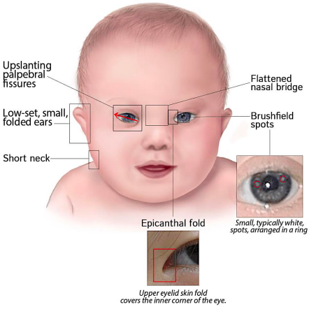
down syndrome
aka: trisomy 21
more of periodontal problems than dental caries
clinical findings:
low-set small folded ears
upslanting palpebral fissures
brushfield spots, epicathal fold
short neck, flattened nasal bridge
problems of individuals with clefts
nasal deformity esp if cleft extends to the nasal floor
associated anomalies 20x to have another congenital anomalies
speech difficulties retardation of consonant sounds (P,B,T,D,K,G)
ear problems predisposition to middle ear infections (otitis media)
pseudoprognathism → malocclusion class III MO (retrusion of the maxilla not protrusion of the mandible)
congenital absence of teeth (lateral and canine) or supernumerary teeth
feeding underdeveloped musculature → no negative pressure to allow sucking breast milk or bottle milk
types of cleft
cleft lip
alveolar cleft
submucosal cleft
cleft of the soft palate
cleft of the hard palate
cheilorrhaphy
surgical correction of the cleft lip
usually the earliest surgical procedure for cleft deformities.
derived from:
cheilo = lip
rhaphy = junction by a seam or suture
considers “rule of 10”
10 weeks old
10 lb body weight
10 g/dL hemoglobin
10k WBC
benefits:
restoring the orbicularis oris muscle:
improves muscular function
favorable molding effect on the developing alveolar segments
limitations:
ideal results are difficult to achieve despite surgical skill.
main problems include:
poor quality of tissues at the cleft margins
distortion of normal structures before surgery
surgical technique
unilateral cleft
the unaffected side serves as a guide for: lip length, symmetry
scar design
the scar lines should be broken up rather than forming one straight line, why?
fibrosis and scar contracture can distort the lip
linear closure may result in characteristic notching of the upper lip
techniques for cheilorrhaphy
tennison operation
wynn operation
LeMesurier technique for incomplete unilateral cleft
millard operation (rotation advancement technique) → most common technique
2 main objectives of cheilorrhaphy
functional objective
reestablish normal upper lip function
restore the normal arrangement and continuity of the orbicularis oris musculature
if muscle continuity is not restored
an aesthetically undesirable depression can appear when the lip is functioning.
aesthetic objective
create a lip with normal anatomic features, including:
vermilion tubercle
cupid's bow
philtrum
the repaired lip should be:
symmetric, soft, supple
well contoured
have inconspicuous scars
nasal deformity
another aesthetic goal is to partially correct the associated nasal deformity
palatorrhaphy
surgical repair of the cleft palate, usually completed in one operation.
sometimes performed in 2 operations:
soft palate closure → staphylorrhaphy (8-18 months of age)
hard palate closure → uranorrhaphy (4-5 y/o)
objectives:
create a palate that allows:
normal speech, deglutition
minimal interference with future maxillary growth
2 important requirements
competent velopharyngeal mechanism
complete separation of the nasal and oral cavities
surgical technique:
techniques vary greatly because each cleft is unique.
clefts differ in:
width, completeness, palatal length
amount of hard and soft tissue available
therefore, the technique may differ:
between surgeons, from one patient to another
techniques for palatorrhaphy
vomer flap technique
von langenbeck operation
von langenbeck operation
this technique is a one-layer closure
for closure of the hard palate using lateral releasing incision
nasal aspect of palatal flaps will epithelialize, as will denuded areas of the palatal bone

vomer flap technique
a one-layer closure
indication:
the vomer is long
attached to the palatal shelf opposite the cleft
cleft is not too wide
technique:
raise a mucosal flap from the vomer.
suture it to the palatal tissues on the cleft side.
advantages:
requires little stripping of palatal mucoperiosteum
produces minimal scar contraction
therefore, less potential restriction of maxillary growth
healing:
denuded areas of the vomer and flap reepithelialize.

advantages of early closure
easier feeding
better auditory tube function
better development of phonation/speech skills
better palatal and pharyngeal muscle development
improved psychological state of the parents and baby
better oral hygiene because the oral and nasal cavities are separated
2 major disadvantages of early closure
surgical scarring may cause restriction of maxillary growth.
surgery is more difficult in younger children because the structures are smaller.
advantages of delaying hard palate closure
allows more maxillary growth
reduces the effect of surgical scarring on growth
makes the use of orthodontic appliances easier
soft palate
usually closed between 8–18 months
functions as an important valve to control the distribution of escaping air between the oropharynx and nasopharynx
a muscular "valve" that separates the oral and nasal cavities during speech and swallowing
hard palate
may be left open as long as possible to allow unrestricted maxillary growth
may not be closed at the same time as the soft palate, esp when the cleft is wide
removable palatal obturators → may be used during the waiting period (it separates the oral cavity and nasal cavity)
how long can it be delayed?
→ closure may be postponed until at least all deciduous teeth have erupted
→ since a significant amount of maxillary growth has already occurred by 4–5 years, hard palate closure is usually performed around this time (completed before the child enters school)
compensatory velopharyngeal mechanism
passavant's ridge a temporary bulge on the posterior pharyngeal wall that forms during speech and swallowing
pharyngeal walls narrow the passageway between the oropharynx and the nasopharynx
tongue postures to help valve the air
superficial muscles around the nose limit the amount of air escaping from the nasal cavity
submucosal cleft
aka: occult cleft
a defect due to lack of continuity in the musculature of the soft palate
not readily seen on clinical exam since oronasal mucosa covers the muscular defect
how to diagnose?
let the patient say “ah”
this action lifts the soft palate → a furrow in the midline is seen
hypernasal speech without obvious soft palate cleft
alveolar cleft
a cleft of the alveolus
usually not repaired during the initial cleft lip or palate surgery
residual oronasal fistulas may also remain; maxillary alveolus remains discontinuous.
problems caused if unrepaired:
oral fluids can enter the nasal cavity.
nasal secretions can enter the oral cavity.
teeth may erupt into the cleft.
alveolar segments may collapse.
a large cleft may negatively affect speech.
benefits if repaired:
unites the alveolar segments
helps prevent collapse, constriction of the dental arch
provides bone support for teeth
closes oronasal fistula
separates: oral cavity, nasal cavity
prevents fluid from passing between them.
augments the alveolar ridge
provides a better supporting base for dental prostheses.
supports the lip and nasal alar base
creates a solid foundation for: lip, alar base of the nose
improves nasal structure because the tissues receive osseous support.
timing of alveolar bone grafting
performed between 6-10 y.o, why?
much of maxillary growth has already occurred
the graft is less likely to interfere with future maxillary growth.
ideally, the graft should be in place before permanent canine eruption.
ideal timing relative to canine
when ½ -⅔ of the unerupted canine root has formed.
some surgeons prefer grafting closer to the time when the maxillary central incisors are erupting.
[alveolar bone grafting should be performed before nasal revisions]
surgical procedure for alveolar cleft graft
soft tissue coverage
bone grafts must be covered by intact mucoperiosteal flaps
flaps are developed from:
nasal, palatal, labial mucosa
closure
sutured → prevents infection of the bone graft.
tension-free, watertight
bone graft material
ilium, cranium
other materials:
allogeneic bone → bone from another individual
bone morphogenetic proteins
graft preparation
graft is made into a particulate form:
packed into the defect after:
nasal / palatal mucosa closure
labial mucosa is then closed over the graft
orthodontic expansion
maxillary arch expansion can be done:
before / after grafting
both can be effective.
[some surgeons prefer expansion before bone grafting because it provides better surgical access to the cleft]
palatal obturator
latin word 'obturare' which means to close or shut off
a prosthesis that can be used to close defects such as an opening in the roof of the mouth
staphylorrhaphy
soft palate closure
considered the most technically difficult procedure
always closed in 3 layers and in this order: nasal, muscular, oral
why is it difficult?
it is located deep in the back of the mouth.
problems include:
limited access
difficulty with lighting & retraction
working from the oral side while repairing both oral and nasal surfaces
thin or atrophic tissues → closure must remain intact during function and healing
uranorrhaphy (hard palate)
it is closed using soft tissue only
usually, an osseous partition is not created between the oral and nasal cavities.
two-layer closure: nasal layer, oral/palatal layer
nasal mucosa is mobilized from:
nasal floor, septal region
lateral nasal wall
the nasal mucosa is sutured together before the oral closure
basic technique
incise the soft tissues along the cleft margins.
dissect the tissues away from the palatal shelves.
bring the tissues together over the cleft.
lateral relaxing incisions near the teeth are often necessary
closure
soft tissues are sutured watertight over the cleft.
the exposed bone from the lateral relaxing incisions heals by secondary intention.
the superior surface of the palatal flaps becomes the nasal floor and develops respiratory epithelium.
common procedures for palatal lengthening
W-Y push-back procedure (Wardill)
U-shaped push-back procedure (Dorrance and Brown)
common skeletal problems of cleft px
maxillary retrusion
transverse maxillary constriction
pharyngeal flap procedure
most commonly used secondary procedure for improving velopharyngeal competence
a wide vertical strip is raised from the posterior pharyngeal wall of:
pharyngeal mucosa, pharyngeal muscle
the flap is inserted into the superior aspect of the soft palate
result:
the flap connects the pharynx and soft palate.
this leaves two lateral openings/ports between the:
oropharynx, nasopharynx
this reduces airflow between the two spaces.