IMED1001 - Nervous System Anatomy (I and II)
1/30
There's no tags or description
Looks like no tags are added yet.
Name | Mastery | Learn | Test | Matching | Spaced |
|---|
No study sessions yet.
31 Terms
Central Nervous System (CNS)
- collect information
- process and evaluate information
- initiate response to information
- consists of the brain and spinal cord. Everything else is PNS
Sensory Nervous System
afferent; sends messages to the CNS. GO THROUGH HBIO NERVOUS SYSTEM FOR THIS CHAPTER
Motor Nervous System
efferent; sends messages to muscles. GO THROUGH HBIO NERVOUS SYSTEM FOR THIS CHAPTER
Rostral
towards the nose/towards to forehead (rmbr the saying: rostral towards nostril)
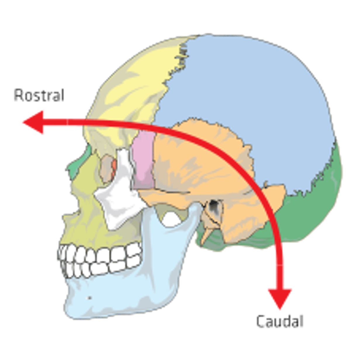
Caudal
towards the tail/towards the spinal cord (rmbr the saying caudal towards the cord)
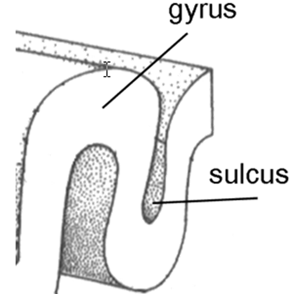
Gyri and Sulci
- lumps and grooves of the surface of the brain greatly increase surface area and thus cognition
- Gyri: 'hills' on the surface of the brain, often correlate with important functional areas
- Sulci: 'valleys' on the surface of the brain. Topographical borders of lobes and gyri. Long, deep sulci = fissure
Grey Matter in the Brain
consists of unmyelinated cell bodies and dendrites
White Matter in the Brain
myelinated axons
Structure of a Neuron
- Grey matter = cell bodies (cell body or soma
- White matter = myelinated neurons
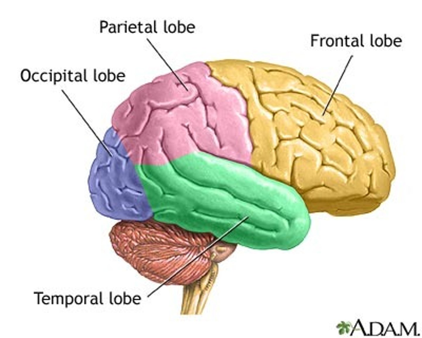
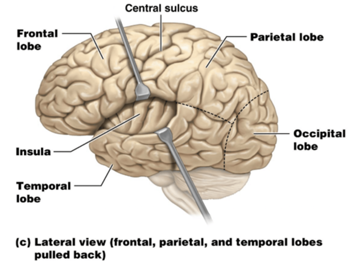
Lobes of the Brain
- Frontal
- Parietal
- Temporal
- Occipital
- named after the bones they sit under
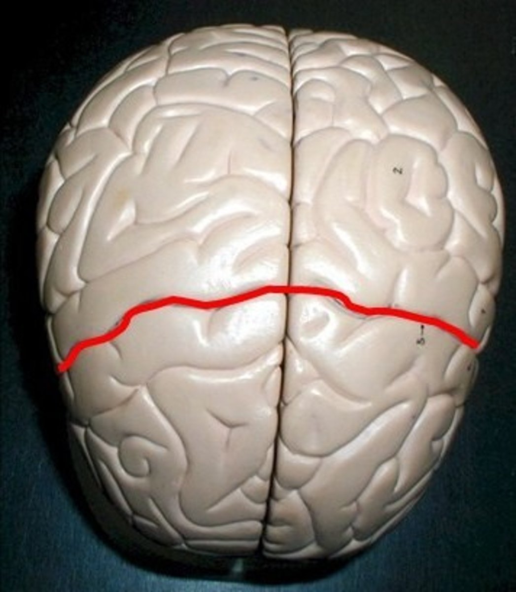
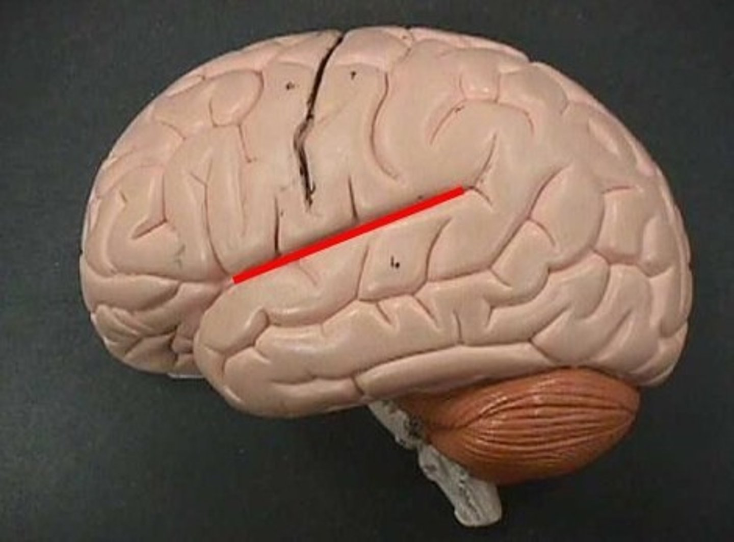
Central Sulcus
separates frontal and parietal lobes
Lateral Sulcus
Separates temporal lobe from parietal and frontal lobes
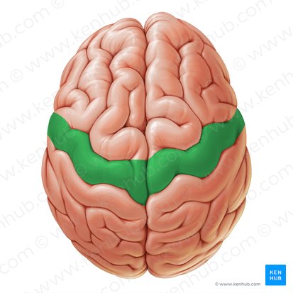
Precentral Gyrus
the strip of frontal cortex, just in front of the central sulcus, that is crucial for motor control
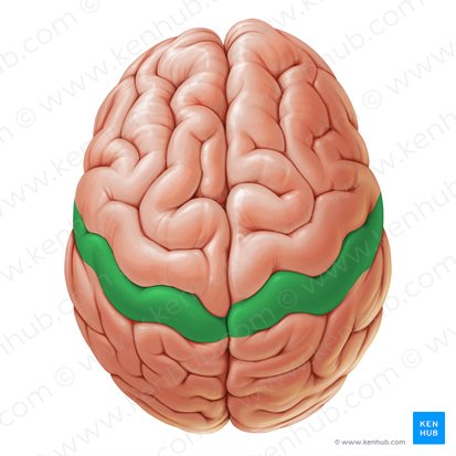
Postcentral Gyrus
the strip of parietal cortex, just behind the central sulcus, that receives somatosensory information from the entire body
5 lobes of the cerebrum
- learning outcome says know at least one function of each lobe:
- Frontal Lobe: abstract thought, mood, motivation, emotional control, decision making
- Insula (taste): taste, pain, consciousness, visceral sensation
- Parietal Lobe (touch and proprioception): taste, visual processing, language processing
- Occipital Lobe (vision): Visual awareness, visual processing
- Temporal Lobe (hearing and smell): hearing, smell, emotion, learning, verbal memory
Functional Regions of Cerebral Cortex (3 of them)
- Primary Motor Cortex: final command centre for voluntary movement. Left primary motor cortex controls right side of the body and vice versa
- Primary somatosensory cortex: conscious sensory awareness. Left primary somastosensory cortex controls right side and vice versa
Corticospinal Tract
connections between brain and spine (chain of two neurons)
Corpus Callosum
- facilitates communication between hemispheres, ensuring coordination of cognitive motor functions (synchronises hemispheric activity, prevents excessive competition between hemispheres)
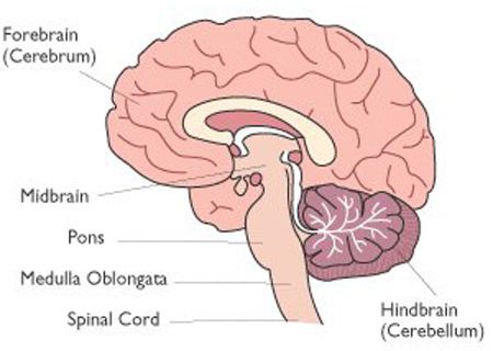
Cerebellum
- a lot of convolutions means high level of cognition
- fine tunes and coordinates skeletal muscle actions (does not initiate movement - that is a role of the cerebrum)
- stores memories for movement patterns. e.g riding a bike
- maintains balance and posture: regulates equilibrium, adjusts posture, controls muscle tone
- Integrates esnsory and motor input from the spinal cord, brainstem and cortex to refine movement and enhance proprioception (body position awareness)
- plays a role in mood regulation, emotions
The Brainstem (contains 3 secftions)
3 SECTIONS:
- Midbrain: vision, hearing, motor control, sleep/wake cycle regulation, arousal, temperature regulation
- Pons (= bridge): connects cerebellum to the brainstem, relays signals between cerebrum, cerebellum and spinal cord. It contains sleep and respiratory centres and regulates breathing rhythms and sleep cycles
- Medulla Oblongata: cardiac regulation (heart rate and blood pressure), respiratory controls (breathing rate and rhythm), vomiting, coughing, sneezing and swallowing reflexes
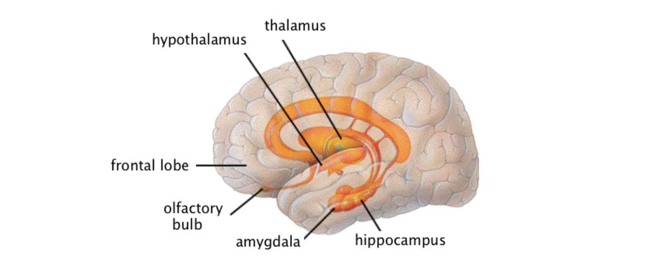
Thalamus
- gateway to the cerebral cortex. Filters and directs nearly all sensory input (except smell) to the appropriate cortical area
- Relays information related to taste, hearing, equilibrium, vision, touch, pain, pressure and temperature
- Plays a role in coordinating movement by relaying signals between the cerebellum and cerebrum
- involved in memory formation and the emotional regulation as part of the limbic system
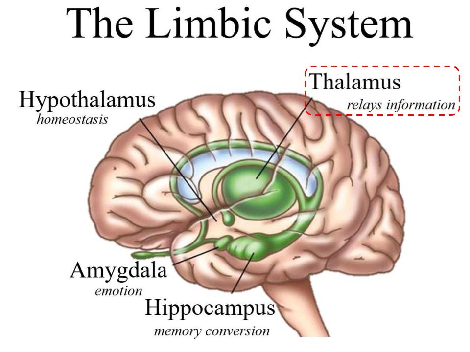
Hypothalamus
- sits anterior and inferior to the thalamus
command centre of autonomic nervous system and master regulator of the endocrine system
- regulates heart rate, blood pressure, digestion, thermoregulation
- links emotional and behavioural responses: fear, anger, pleasure, motivation to the limbic system
Limbic System
Structures that process and experience emotions
- Hippocampus: memory forming-centre
- Amygdala: processes fear, anxiety and emotional responses
- Septum: associated with pleasure
- Olfactory Bulb: processes smell with memory
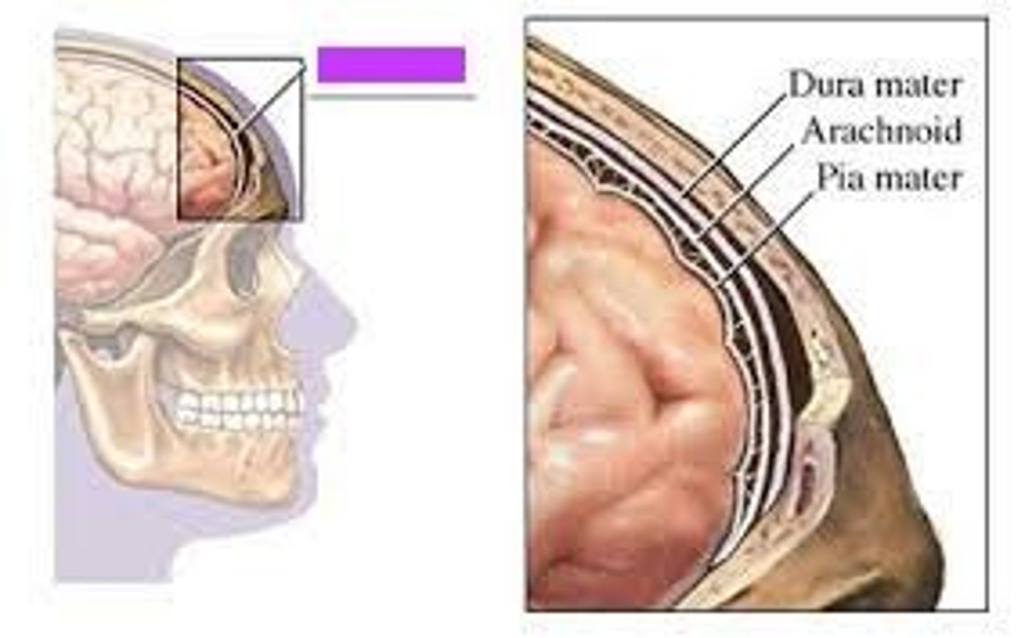
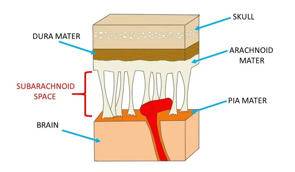
Meninges
- 3 layers of connective tissue that surround, protect and nourish the brain and spinal cord
- Dura mater: thick, fibrous and opaque. Innervated with sensory nerves
- Arachnoid Mater: it is partially composed of a delicate web of collagen and elastic fibres. Supports cerebral arteries and veins within the subarachnoid space. Helps contain and circulate CSF
- Pia Mater: thin layer of delicate areolar connective tissue that is tightly adhered to the surface of the brain and spinal cord
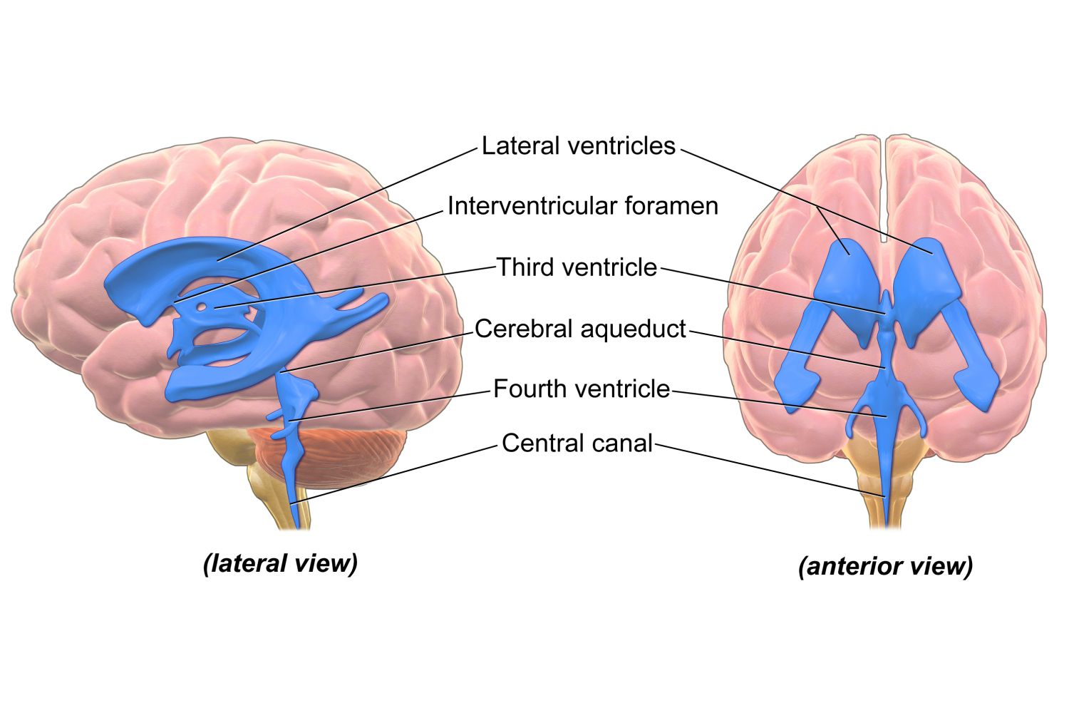
Ventricular System
- Produces CSF (via the choroid plexus in each ventricle)
- Transports CSF through the ventricular system and into the subarachnoid space
- Removes excess CSF via absorption into the venous system (through the arachnoid granulations into the superior sagittal sinus)
- largest, most rostral are the two lateral ventricles
- connected to the third ventricle via the interventricular foramen
- Cerebral aqueduct leads to the fourth ventricle
- Extends through the medulla oblongata to the spinal cord
Cerebrospinal Fluid (CSF) (Functions, extra info)
- Functions:
1. Provides buoyancy - reduces the effective weight of the brain by over 95%
2. Physically protects the CNS - shock absorber
3. Maintains chemical stability - washes the CNS - provides nutrients and removes waste
- brain produces around 500mL CSF/day but at any given time, only 100-160mL is present
- produced by ependymal cells in the choroid plexus within the ventricles
- CSF is reabsorbed by arachnoid granulations. Transport to superior sagittal sinus (dural venous sinus)
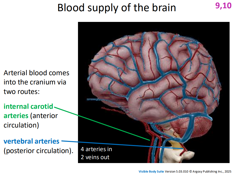
IMAGE FROM SLIDE 42
Blood supply of the brain
- Venous blood and CSF from the superior sagittal sinus leave the cranium via the internal jugular vein
- Internal carotid arteries (anterior circulation)
- vertebral arteries (posterior circulation)
Location of the 4th ventricle
between pons and cerebellum
Subarachnoid Space
a space in the meninges beneath the arachnoid membrane and above the pia mater that contains the cerebrospinal fluid
Key Takeaway about blood flow in the CNS
- there is one major pathway out, and two major pathways into the cranium for blood
DO THE LABELLING DIAGRAM FROM SLIDE 47
they said it can be part of the short answer section in exam