(3) Meninges, Ventricular System, & CSF
1/119
There's no tags or description
Looks like no tags are added yet.
Name | Mastery | Learn | Test | Matching | Spaced | Call with Kai |
|---|
No analytics yet
Send a link to your students to track their progress
120 Terms
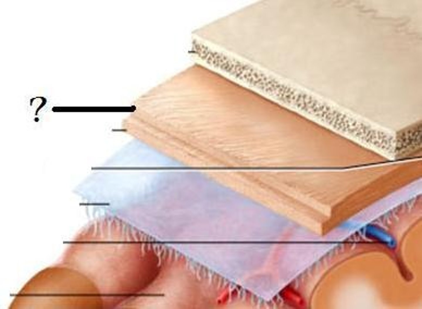
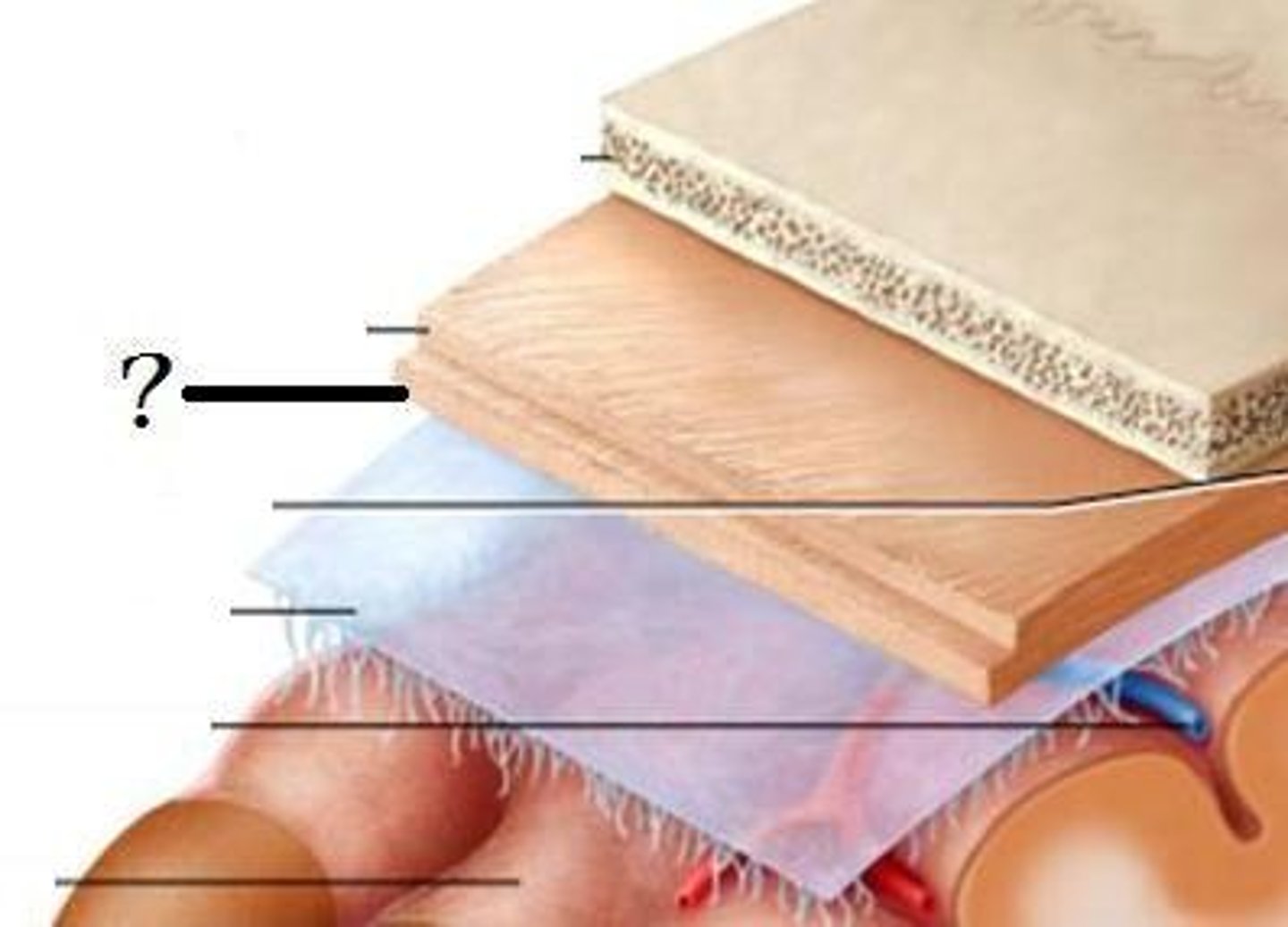
meninges
connective tissue membranes that envelop the brain and spinal cord
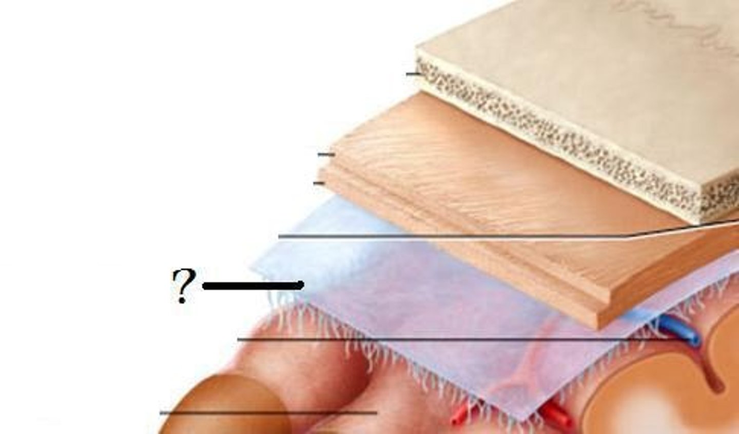
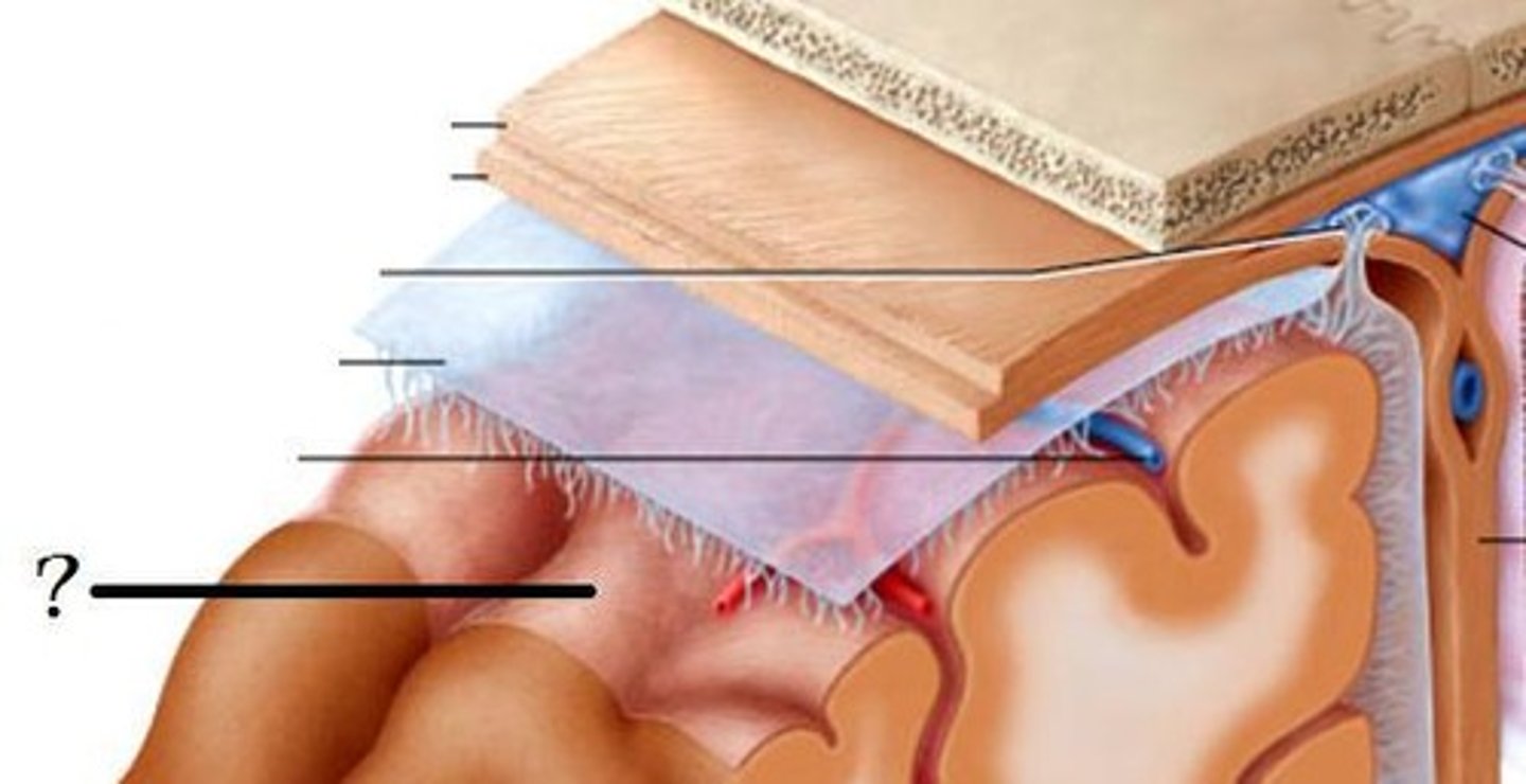
dura, arachnoid, pia
What are the 3 layers of meninges from outer to inner?
dura
Which meninx is the thickest?
pia
Which meninx is the thinnest?
dura mater
outermost and thickest meningeal layer
arachnoid mater
delicate middle meningeal layer
pia mater
very thin inner meningeal layer that is adherent to the surface of the central nervous system
leptomeninges
another name for the arachnoid and pia mater
pachymeninx
another name for the dura mater
arachnoid, pia
Which two meninges make up the leptomeninges?
arachnoid and pia mater
The subarachnoid space lies between which two layers of meninges?
true
(NOT a potential space)
True or false: The subarachnoid space between the arachnoid and pia mater is a real space.
CSF
(brain and spinal cord are basically suspended in this)
What fills the subarachnoid space?
periosteal
outer layer of the dura mater that also forms the periosteum on the inner side of the inner table of the skull bones that from the cranial vault; therefore, it is fused to the inside of compact bone of the parts of the skull that form the cranial cavity
meningeal
inner layer of the dura mater surrounding the brain that is thinner and has an inner surface lined with simple squamous epithelium; this forms tubular sheaths for cranial nerves as they exit the skull, transitioning into their epineurium
periosteal dura mater
meningeal dura mater
has periosteal and meningeal layers in the skull, just has meningeal layer in spinal cord
What is the difference between the dura mater in the skull and in the spinal cord?
sclera
The dural sheath of the optic nerve (consisting of the meningeal layer only) is continuous with what structure of the eye?
no, inner skull and dura mater are basically one and the same
In the spinal cord, there is a true epidural space between the bone of the vertebral column and the dura mater. Is this the same case with the skull and dura mater?
false
(normally we do not have one, not even a potential space, so if one is made by an epidural hematoma, this is abnormal and BAD)
True or false: We normally have an "epidural space" in the skull, just like in the spinal cord.
fluid is forced into the region between dura mater and surrounding bone, tearing the attachment
What causes an epidural space in the skull? Is it natural or created by something?
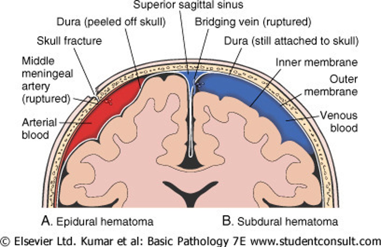
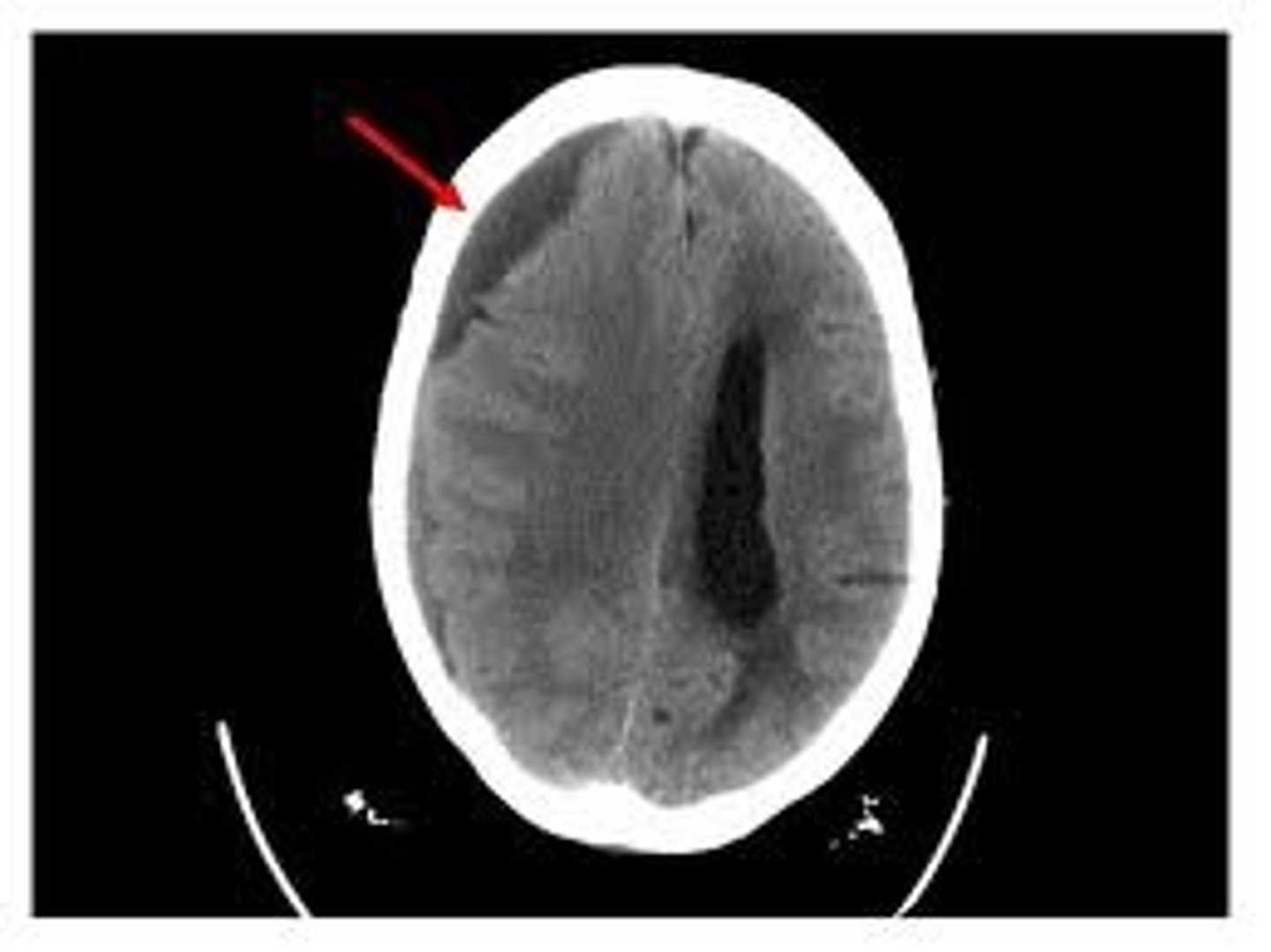
epidural (extradural) hematoma
refers to fluid being forced into the region between the dura mater and surrounding bone which are usually attached (pushes periosteum off of bone, not good), such as blood from damage to the meningeal arteries

meningeal arteries
An epidural hematoma typically results from blood gathering caused by damage to what vessels?
yes
The dura mater and inner surface of the skull are attached to each other, so there isn't really a potential space between the two. What about between the dura mater and arachnoid mater? Is there a potential space there?
potential
Is the subdural space between the dura mater and arachnoid mater a real or potential space?
subdural hematoma
What results when fluid (e.g., blood) gathers in the subdural space between the dura mater and arachnoid mater?
**usually results from damage to superficial cerebral veins and/or dural venous sinuses

superficial cerebral veins, dural venous sinuses
A subdural hematoma typically results from blood gathering caused by damage to what vessels?

epidural hematoma
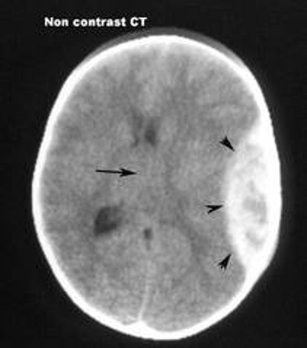
subdural hematoma
S2 (anchored to coccyx by coccygeal ligament)
Where does the dura mater end?
dural root sleeves
tubular sheaths of dura mater that transition into the epineurium of the spinal nerves
dural folds
septa that partially divide the cranial cavity into compartments, formed when the meningeal layer of the cranial dura mater is reflected inward
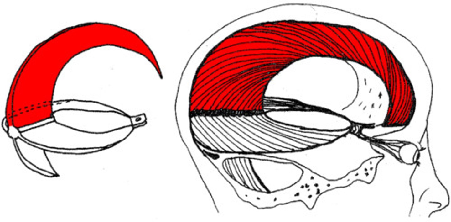
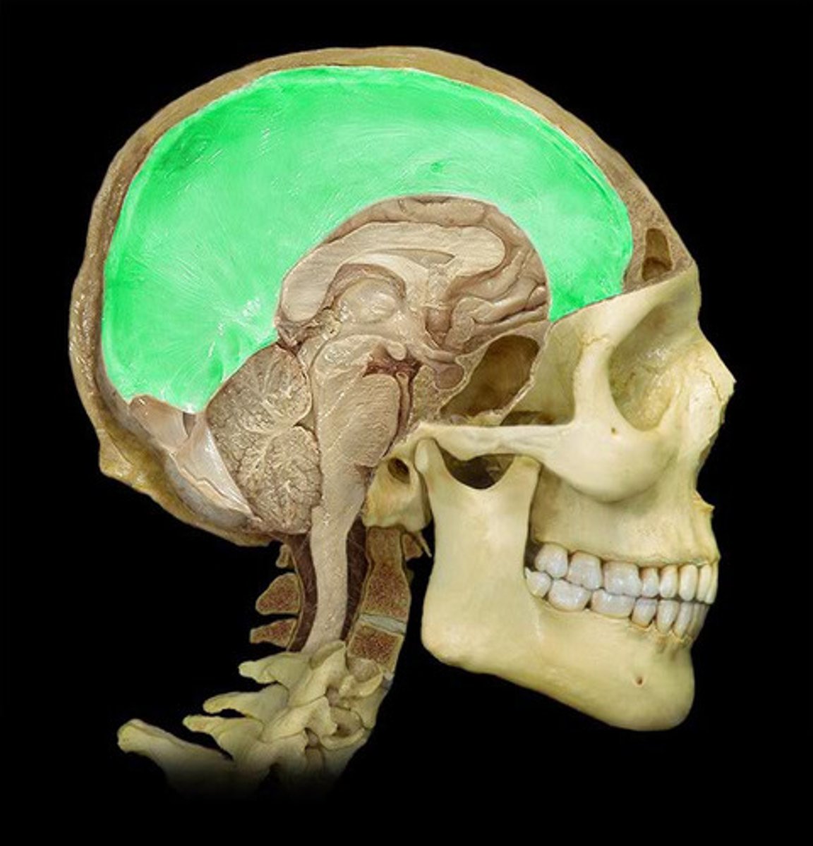
falx cerebri
largest of the dural folds that is a sickle-shaped vertical partition that lies in the longitudinal fissure and has the following attachment sites:
anteriorly - crista galli
superiorly - midline of cranial vault (under sagittal suture)
posteriorly - internal occipital protuberance
inferiorly - midline portion of tentorium cerebelli (only about the posterior 1/4, rest is a free edge)
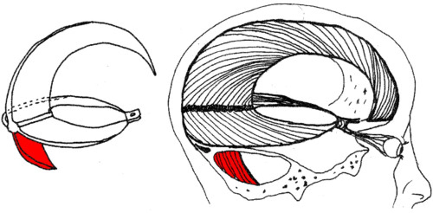
falx cerebelli
vertical dural fold in the midline that is smaller and lies between the cerebellar hemispheres; has the following attachment sites:
anteriorly - free edge
posteriorly - internal occipital crest
superiorly - falx cerebri & tentorium cerebelli
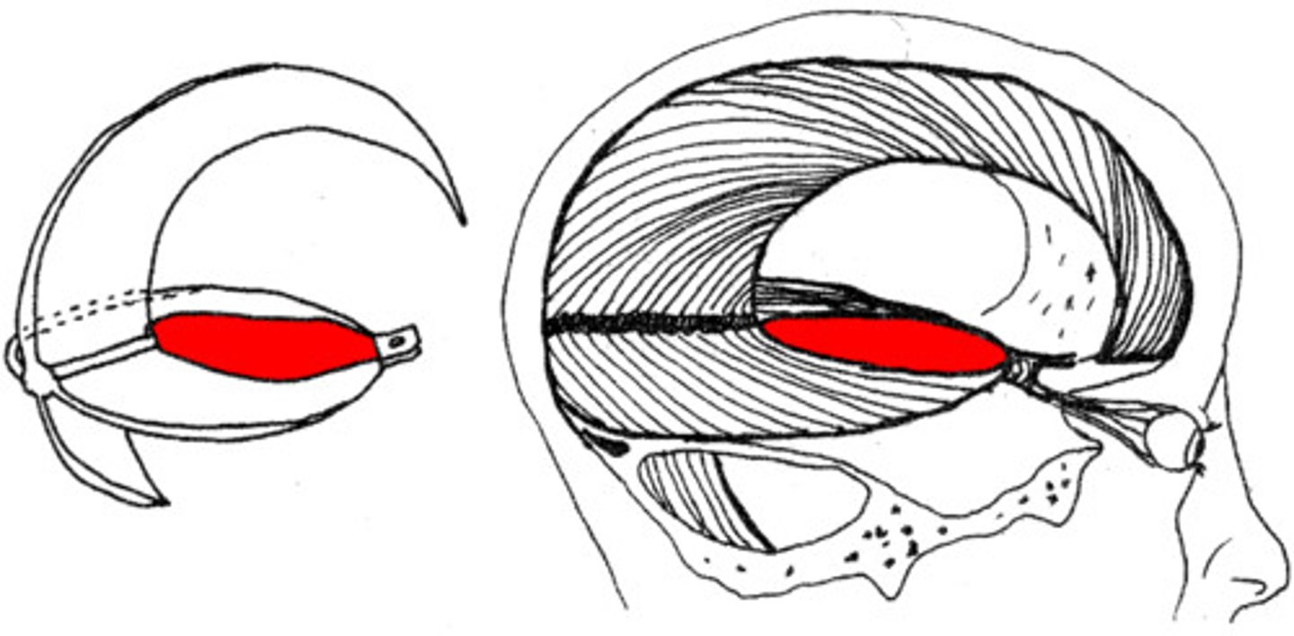
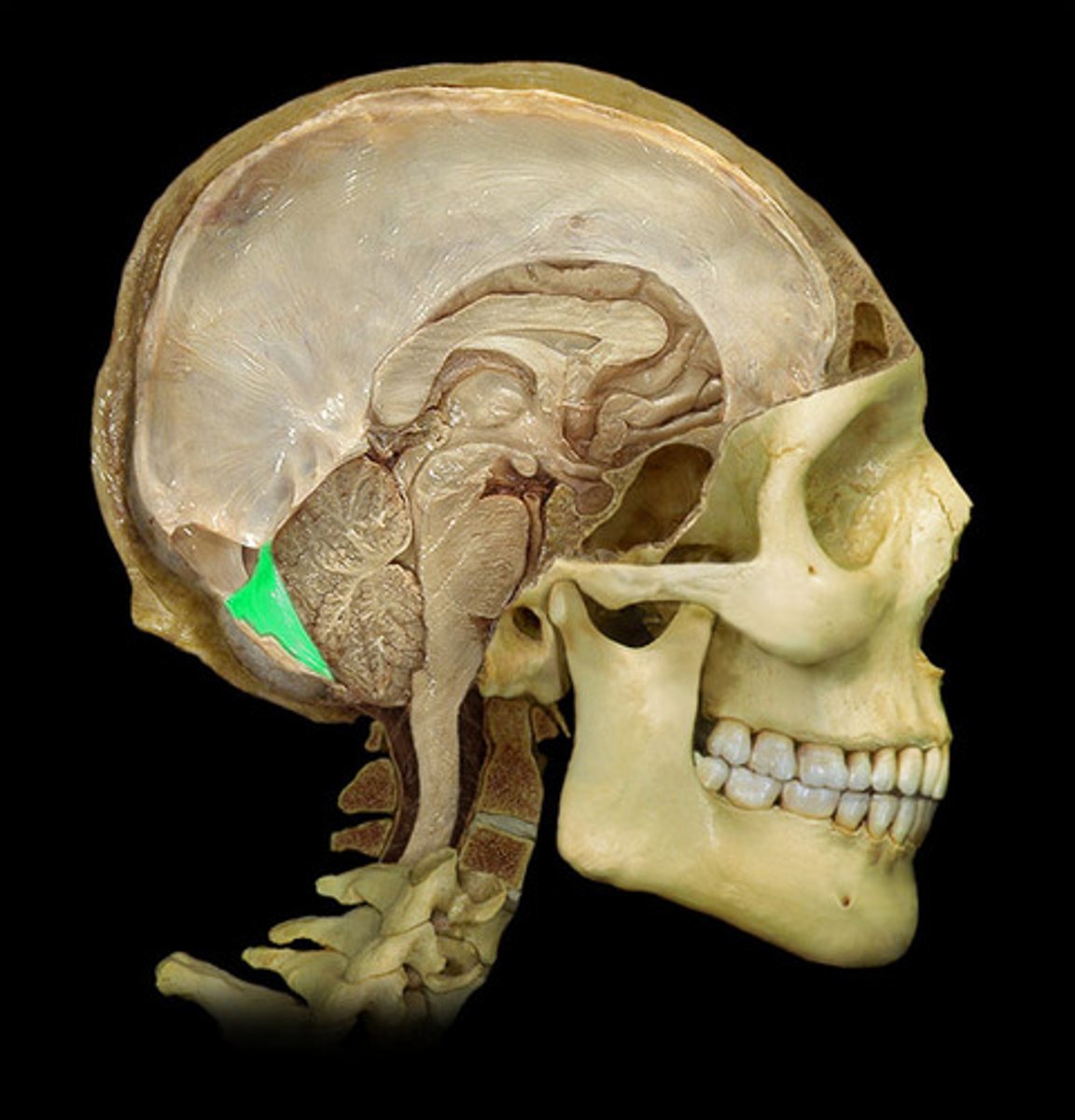
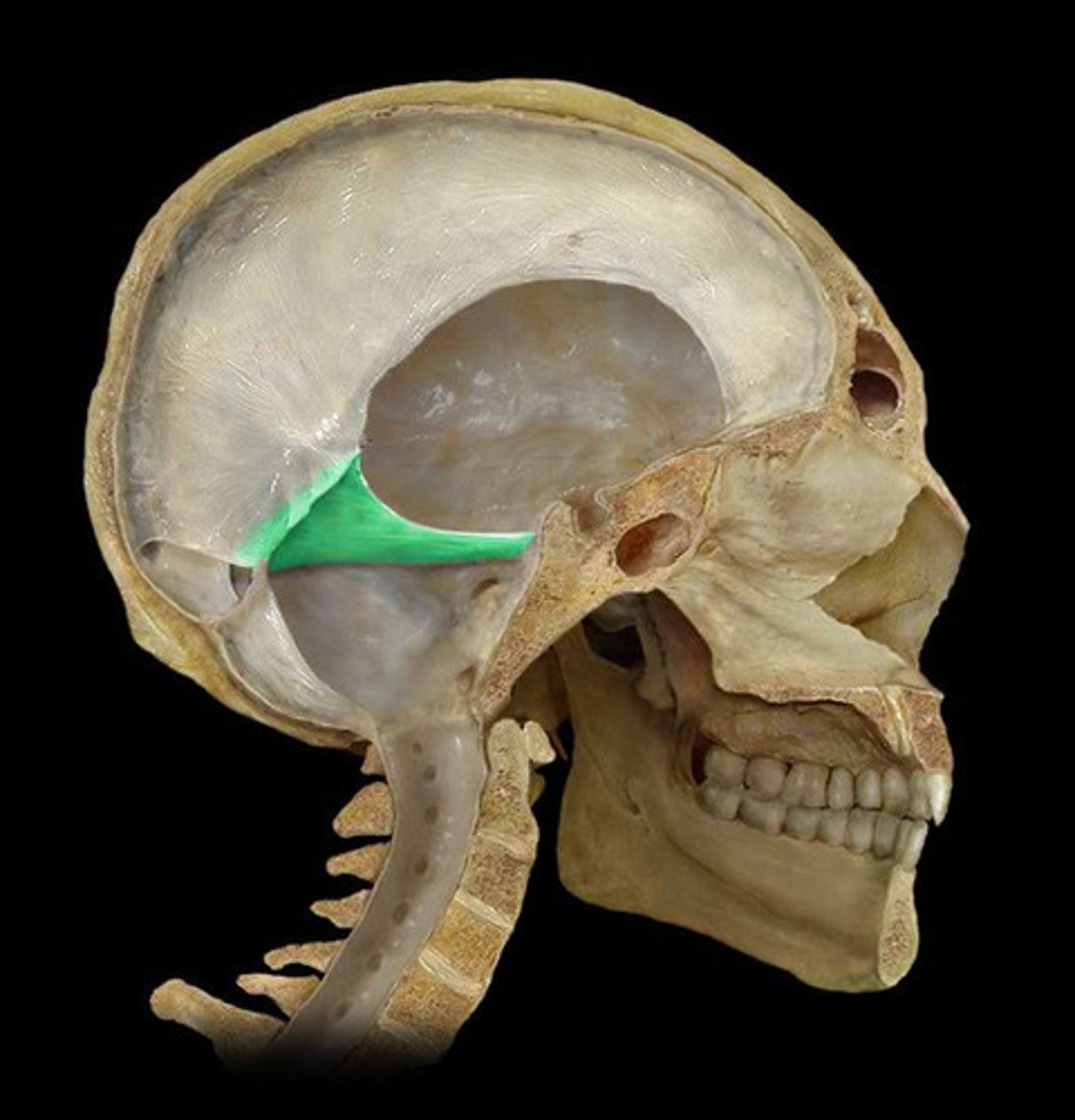
tentorium cerebelli
single dural fold that has a horizontal orientation, located between the occipital lobe and cerebellum; also has a namesake notch where the midbrain is found; attachments include:
-posterior clinoid processes
-petrous portions of temporal bones
-grooves for transverse sinuses
-internal occipital protuberance
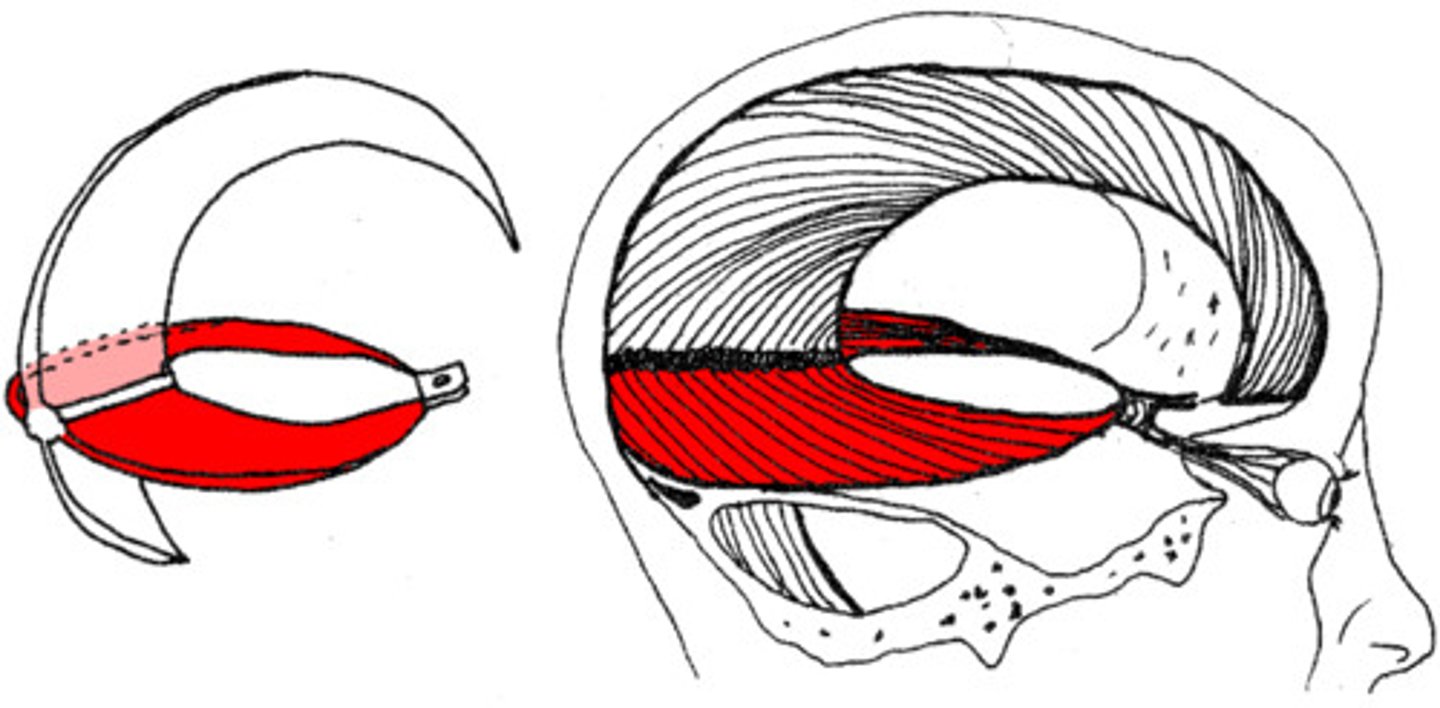
tentorium cerebelli
The occipital lobes extend posteriorly above which dural folds?
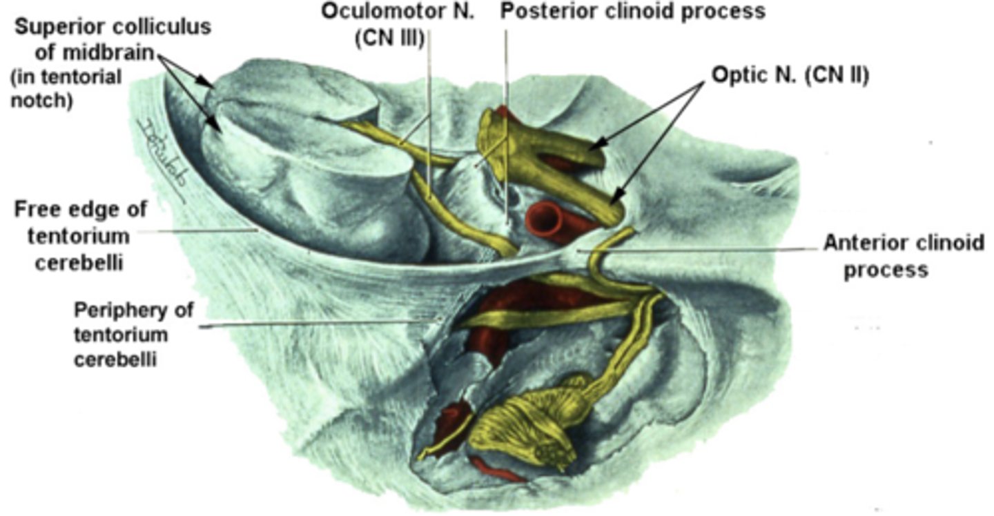
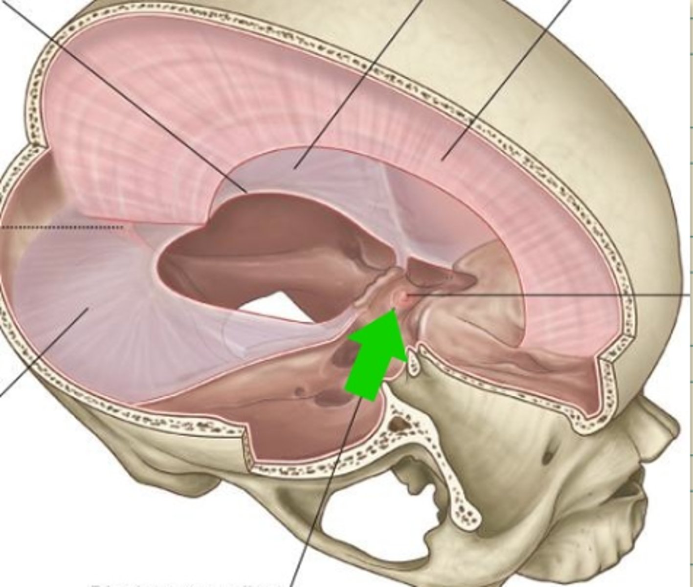
tentorial notch (incisure)
large, curved hiatus between the free margin of the tentorium cerebelli and dorsum sellae that contains the midbrain
CN III (oculomotor)
Which cranial nerve runs between the attachment sites of the tentorium cerebelli with the anterior and posterior clinoid processes?
**potential site of injury for this nerve
supratentorial
1 of 2 compartments that the tentorium cerebelli divides the cranial cavity into, contains the forebrain
**connected to the other compartment by the narrow gap between the midbrain and border to tentorial notch/incisure; this region can get obstructed, leading to ventricular dilation
infratentorial
1 of 2 compartments that the tentorium cerebelli divides the cranial cavity into, contains the hindbrain
**connected to the other compartment by the narrow gap between the midbrain and border to tentorial notch/incisure; this region can get obstructed, leading to ventricular dilation
CN III (oculomotor)
Space-occupying lesions in the supratentorial compartment may cause the medial portion of the temporal lobes to herniate through the tectorial incisure. The midbrain gets displaced laterally and can get injured by the firm edge of the tentorium cerebelli. In addition, what cranial nerve may be stretched, compressed, or both?
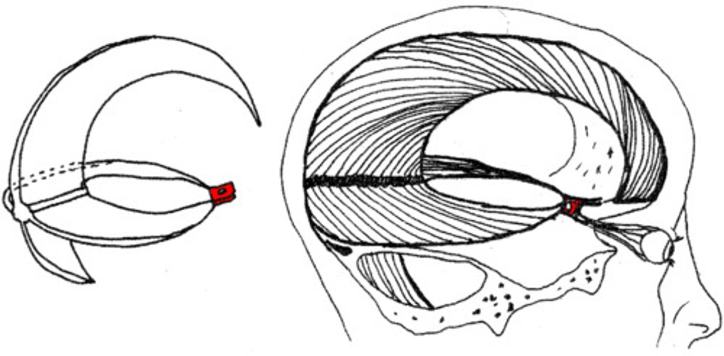
diaphragma sellae
smallest of the dural folds that is a round horizontal sheet that forms a roof over the sella turcica
has a hole in its center that allows the pituitary stalk to run through the this fold on its way from the hypothalamus to attach to the pituitary gland
falx cerebri
falx cerebelli
tentorium cerebelli
diaphragma sellae
meningeal arteries
What is the blood supply for the meninges and also the bones of the skull?
anterior (meningeal)
smallest of the meningeal arteries that arises from the branches of the ophthalmic artery
middle (meningeal)
largest of the meningeal arteries that arises from the maxillary artery and enters the cranial vault through the foramen spinosum
posterior (meningeal)
small meningeal artery that arises from the occipital or vertebral arteries
ophthalmic artery
What does the anterior meningeal artery arise from?
maxillary artery
What does the middle meningeal artery arise from?
occipital and/or vertebral arteries
What does the posterior meningeal artery arise from?
true
(since the brain is not sensitive to touch, the dura mater is to protect the brain)
True or false: The dura mater receives a rich nerve supply and is very sensitive to pain.
CN V1
Portions of dura mater in the anterior cranial fossa, tentorium cerebelli, lining of the superior aspect of the cranial cavity, and falx cerebri receive nerve supply from which nerve?
CN V2 and V3
Portions of dura mater in the middle cranial fossa receive nerve supply from which TWO sources?
CN X, C1-3
Portions of dura mater in the posterior cranial fossa receive nerve supply from which TWO sources?
forehead, temples, eyes
(since supplied by ophthalmic division of trigeminal nerve)
Problems in the anterior cranial fossae, superior aspect of the cranial cavity, falx cerebri, and tentorium cerebelli tend to result in referred pain to what areas?
cheeks, jaw, mouth
(since supplied by maxillary and mandibular divisions of the trigeminal nerve)
Problems in the middle cranial fossa tend to result in referred pain to what areas?
neck, behind the ear
(since supplied by vagus and cervical nerves)
Problems in the posterior cranial fossa tend to result in referred pain to what areas?
false
True or false: The arachnoid mater is vascular.
arachnoid villi (also called arachnoid granulations or pacchionian villi)
specializations of the arachnoid mater that recycle CSF back into the blood
subarachnoid cisterns
The supratentorial and infratentorial regions of the subarachnoid space are connected by the narrow gap between the midbrain and the border of the tentorial incisure. This region can get obstructed, leading to an abnormal ventricular dilation.
There are several places where the subarachnoid space is normally dilated. These expanded regions of the subarachnoid space serve as reservoirs of CSF and are known as what?
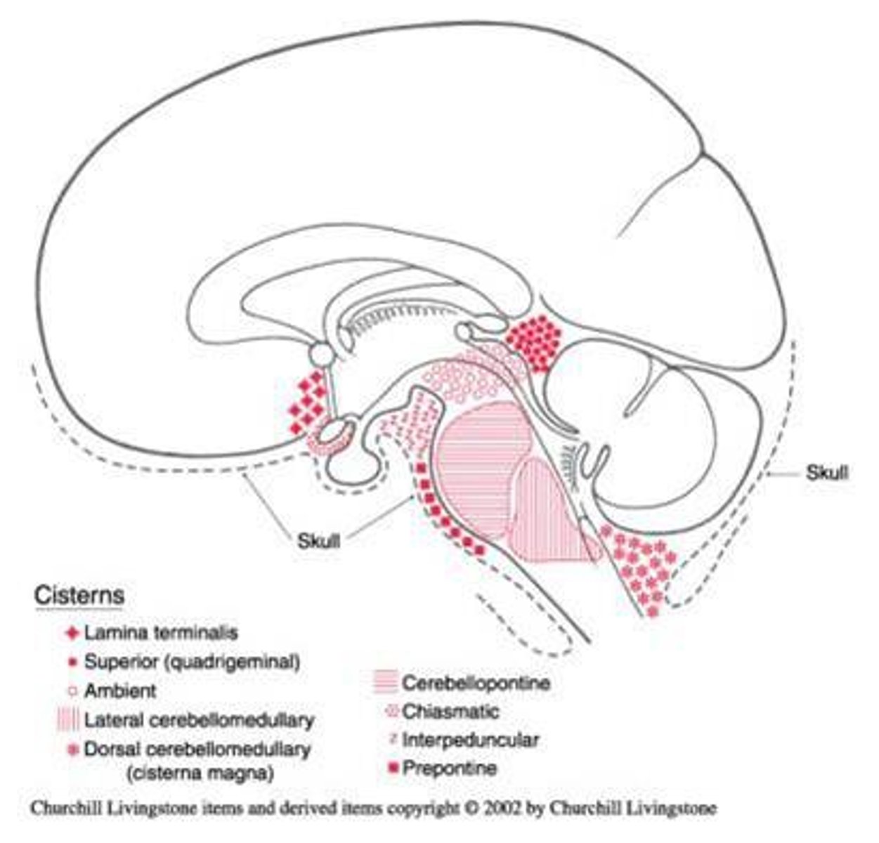
cerebellomedullary cistern, cisterna magna
subarachnoid cistern that is the largest located posterior to the medulla and inferior to the cerebellum (just above the foramen magnum); single midline structure
superior cistern, cisterna vena magna cerebri
subarachnoid cistern that is located deep in the space between the cerebral hemispheres and the cerebellum; posterior to the midbrain and superior to cerebellum; single midline structure, contains the great cerebral vein
mesencephalic cisterns, cisternae ambiens
subarachnoid cisterns that are not midline and located on the lateral sides of the midbrain and superior to the cerebellum; CN IV runs through each one and they connect the superior cistern and the interpeduncular cistern
interpeduncular cistern, cisterna basales
subarachnoid cistern that is located between the cerebral peduncles just anterior to the midbrain; single midline structure and contains CN III
chiasmatic cistern
subarachnoid cistern that is a small dilation of the subarachnoid space located just above the optic chiasm; single midline structure
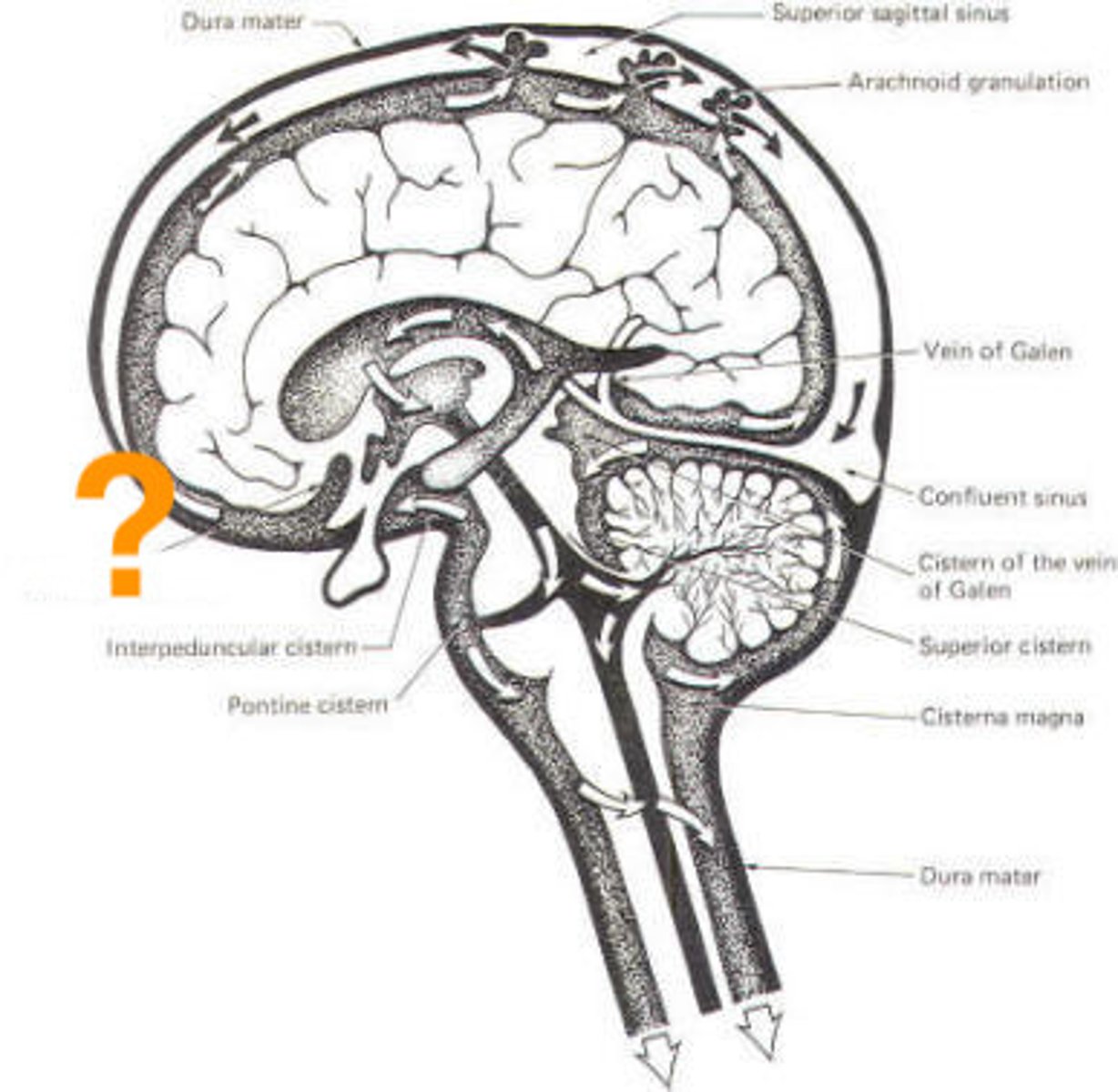
pontine cistern
subarachnoid cistern that is found just anterior to the pons and contains the basilar artery; single midline structure that contains CN VI
cisterns of the lateral cerebral fissure (of Sylvius)
subarachnoid cisterns that are not midline and located in the anterior aspect Sylvian fissures between the frontal and temporal lobes of the cerebral hemispheres
lumbar cistern
subarachnoid cistern that is the only one NOT in the head that is in the lower lumbar region of the spine and contains the caudal equina; this is where CSF samples are usually taken; single midline structure
false
(only a single layer of cells, so basically invisible)
True or false: The pia mater is just thick enough that it can be seen with the naked eye.
true
True or false: The pia mater is vascular.
arachnoid mater
pia mater
denticulate ligaments
folds of extra thick pia mater located on the lateral aspects of the spinal cord; span the subarachnoid space to attach to the deep surface of the arachnoid
**helps to anchor the spinal cord in the vertebral canal
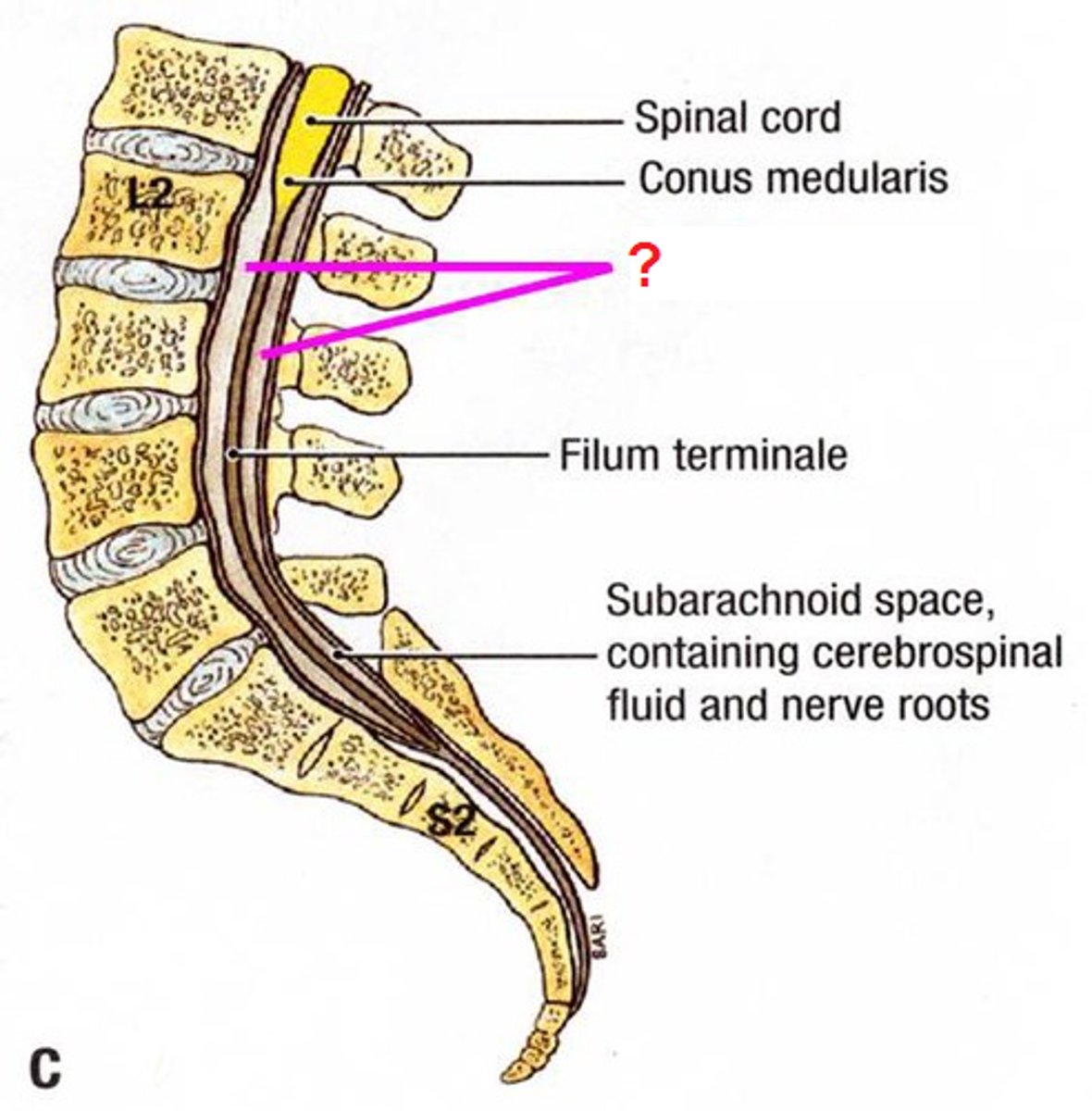
filum terminale (internum)
single midline continuation of pia mater that attaches the conus medullaris to the inferior aspect of the dural sac at S2
**anchors the end of the spinal cord in the vertebral canal
tela choroidea
pia mater specializations found in specific regions in each of the ventricles that support the choroid plexus, which makes CSF
ventricles
fluid filled (CSF) spaces inside the brain (remnants of neural tube cavity)
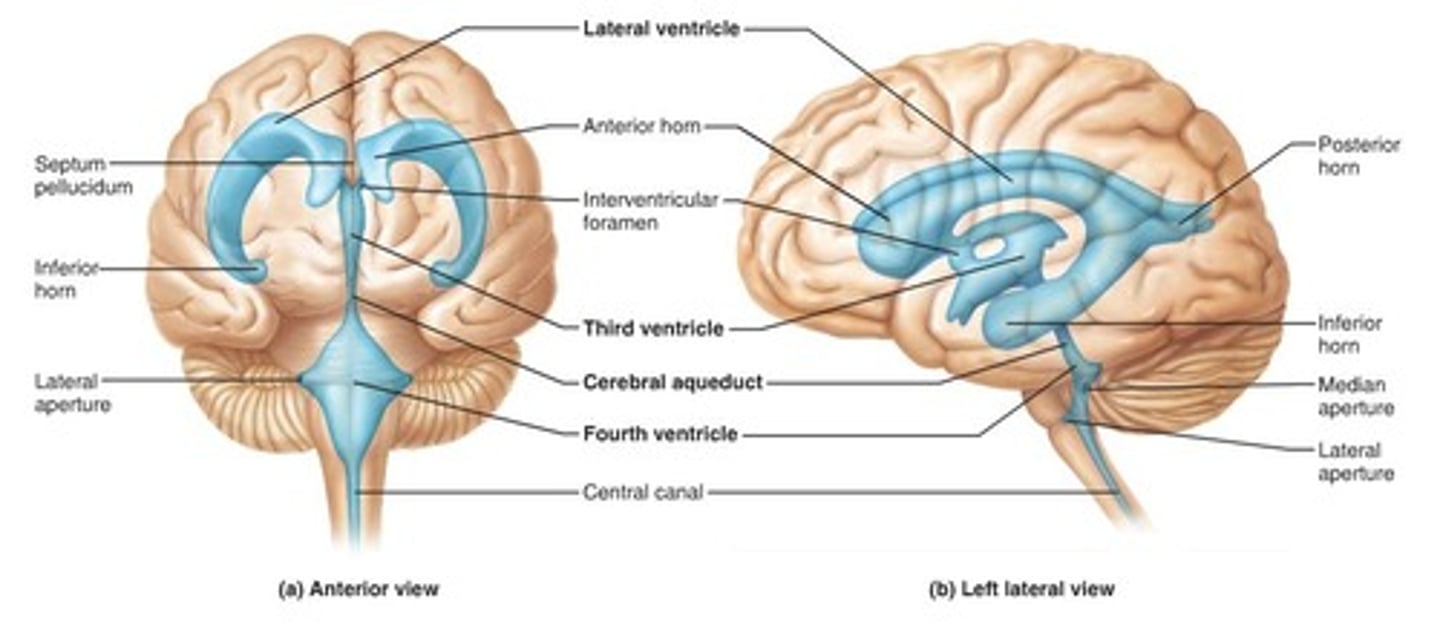
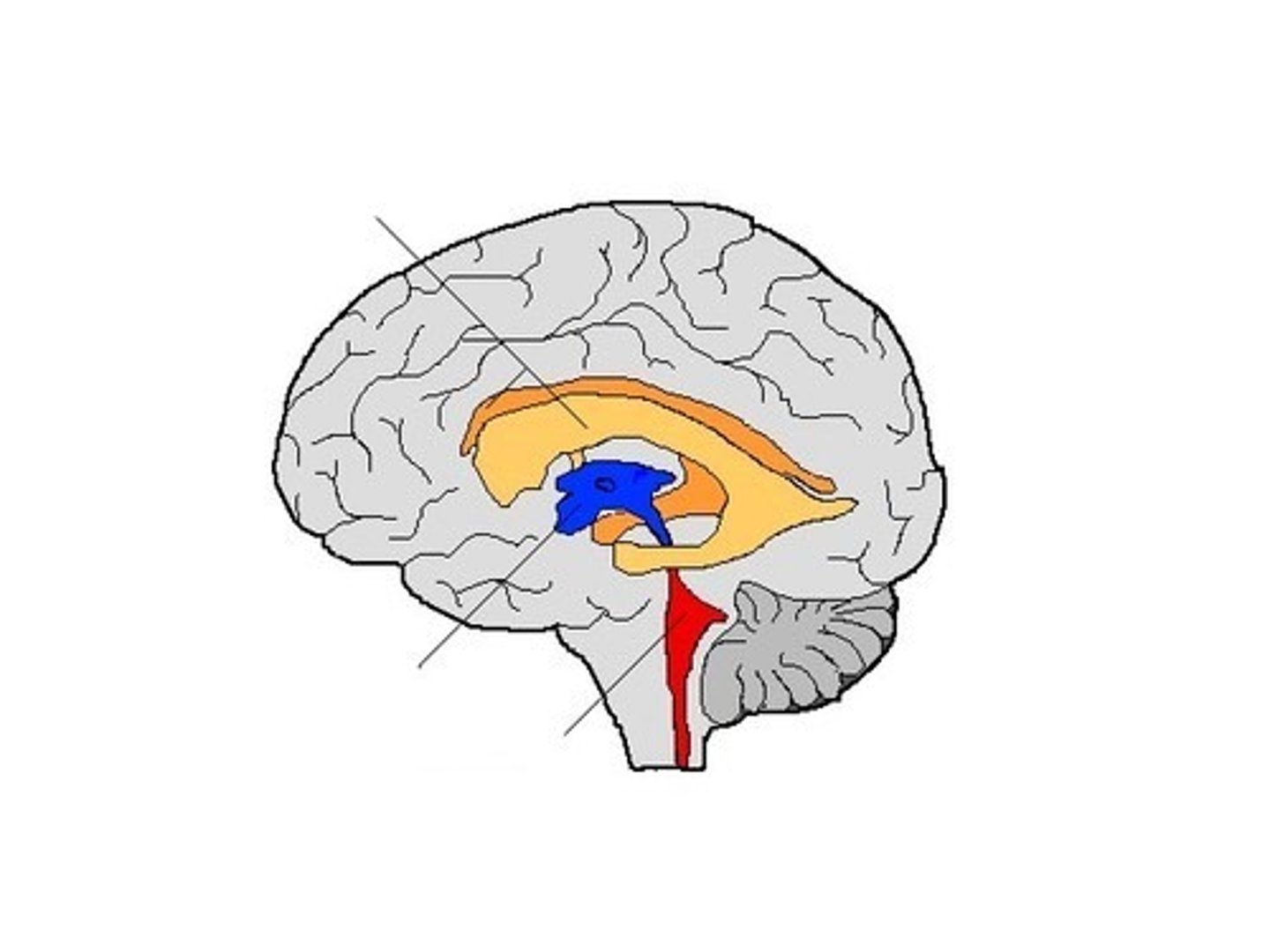
lateral
Which ventricles? (orange)

parietal
In which lobe(s) of the brain is the body of the lateral ventricles found?
frontal
In which lobe(s) of the brain are the anterior horns of the lateral ventricles found?
occipital
In which lobe(s) of the brain are the posterior horns of the lateral ventricles found?
temporal and limbic
In which lobe(s) of the brain are the inferior horns of the lateral ventricles found?
septum pellucidum
double glial membrane that separates the two anterior horns of the lateral ventricles from each other
thalamus
Which brain structure helps form the floor of the body of the lateral ventricles?
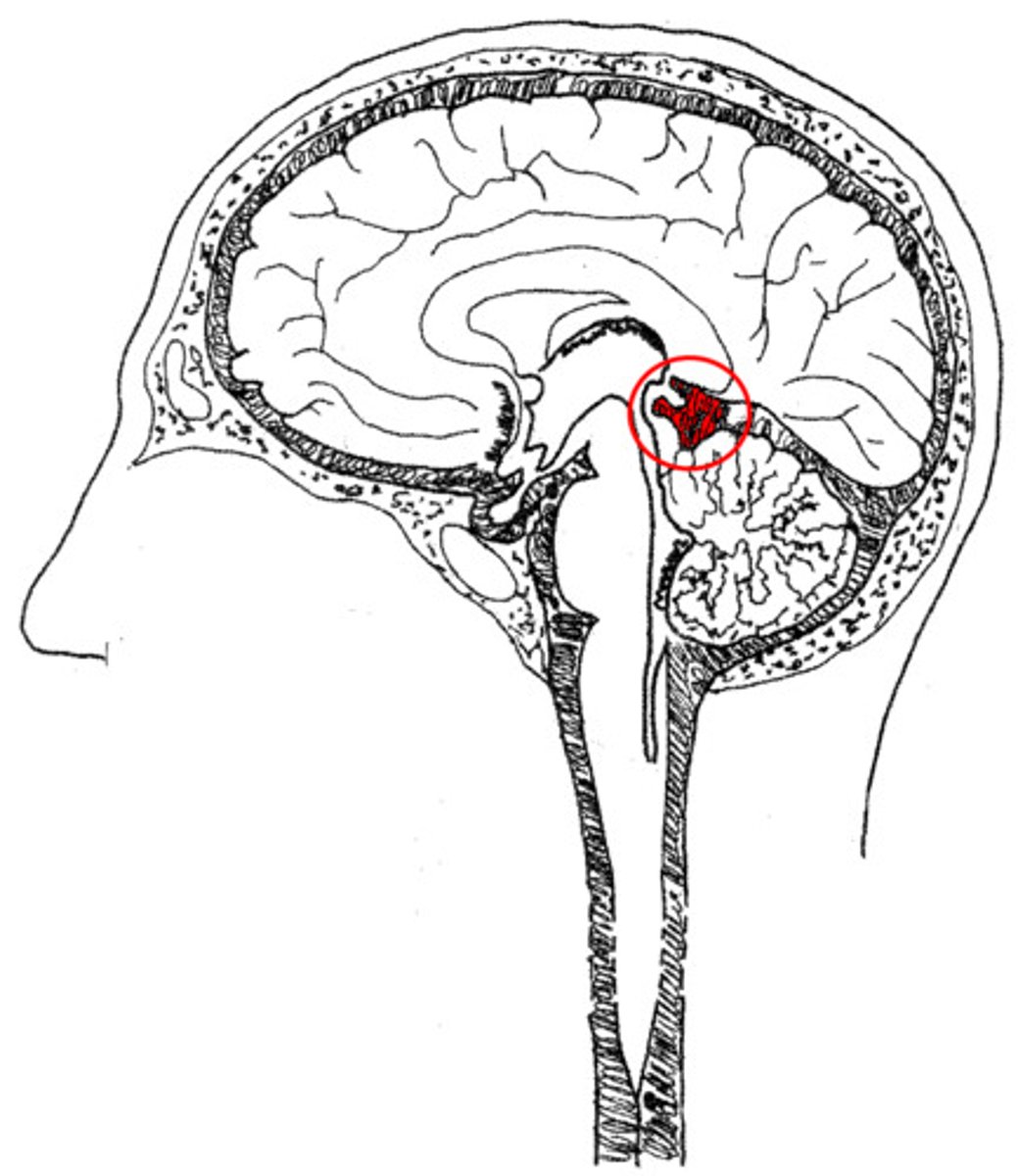
third
Which ventricle? (blue)
diencephalon
Where in the brain is the III ventricle found?
interthalamic adhesion
refers to the right and left halves of the thalamus meeting each other in about 50% of the population, which forms a solid bridge across the III ventricle
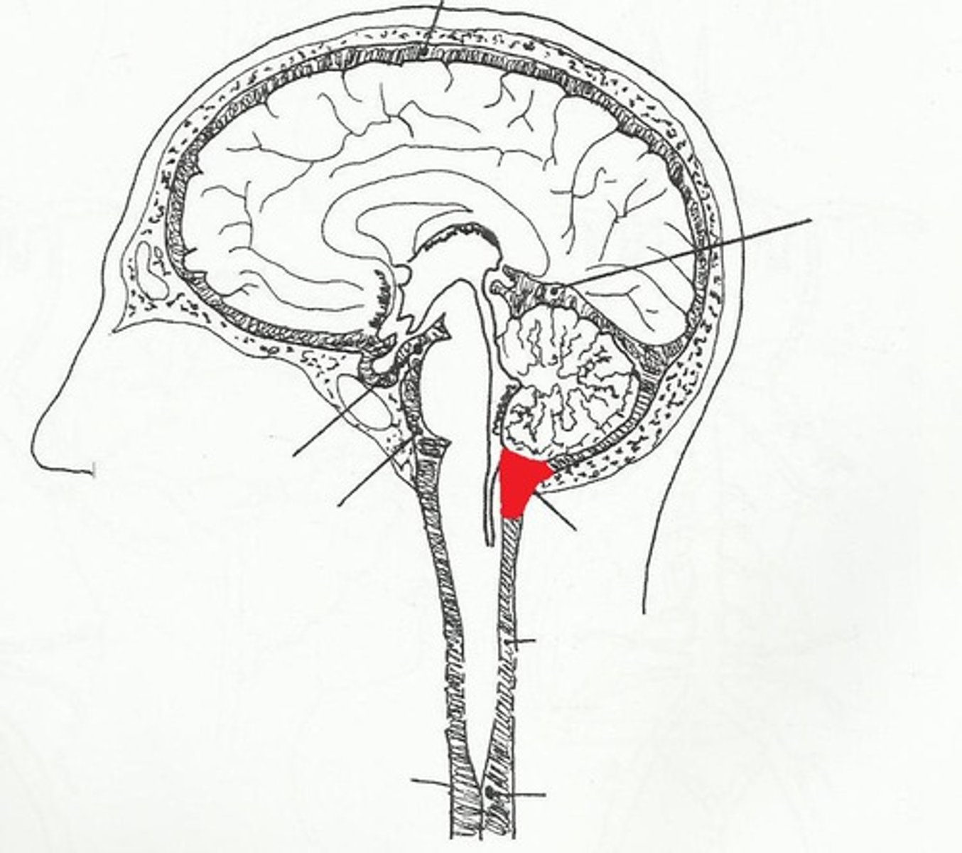
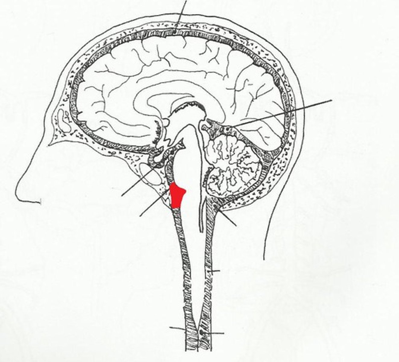
fourth
Which ventricle? (red)

pons and upper medulla
Where in the brain is the IV ventricle found?
superior and inferior medullary velum
2 parts of the roof (dorsal/posterior side) of the IV ventricle
**each one separates CSF from cerebellum
cerebral aqueduct, central canal, foramen of Magendie, 2 foramina of von Luschka
5 openings of the IV ventricle
interventricular foramen of Monro
connects each lateral ventricle to the III ventricle
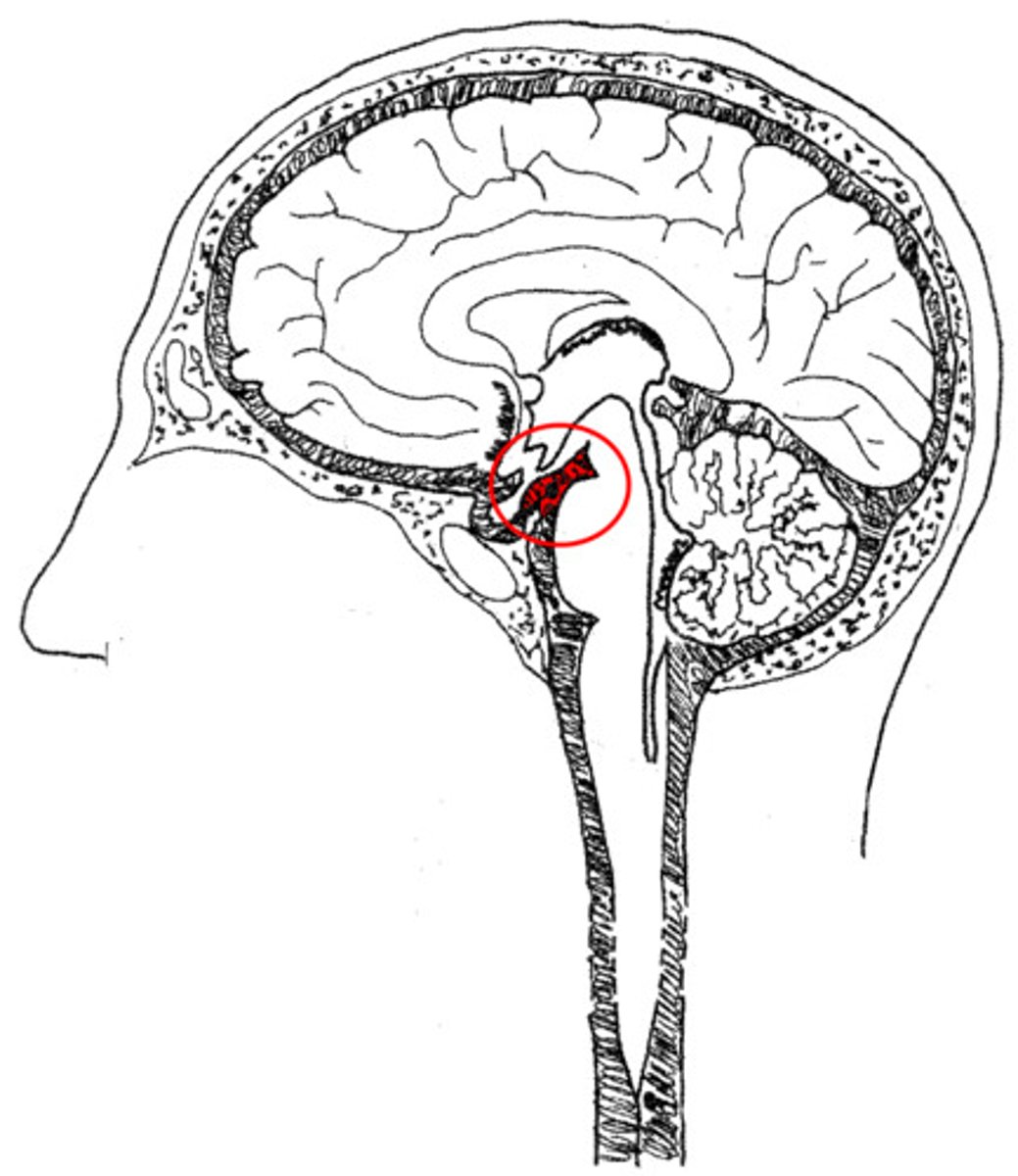
cerebral aqueduct of Sylvius (also caller iter)
connects the III and IV ventricles, narrow channel running through the midbrain
midbrain
Where in the brain is the cerebral aqueduct found?
median aperture of Magendie, 2 lateral apertures of von Luschka
three openings that connect the IV ventricle with the subarachnoid space
foramen of Magendie
What connects the IV ventricle to the cisterna magna?
foramina of von Luschka
What connects the IV ventricle to the pontine cistern?