PSYC 3334 Cognitive Chapter 2
 Call Kai
 Learn
Learn Practice Test
 Spaced Repetition
 Match
Match Flashcards
 Knowt Play
1/25

There's no tags or description
Looks like no tags are added yet.
Name | Mastery | Learn | Test | Matching | Spaced | Call with Kai | Chat |
|---|
No analytics yet
Send a link to your students to track their progress
26 Terms
2.1 Mind: the element of a person that enables them to be aware of the world and their experiences, to think, and to feel; the faculty of consciousness and thought (aka mind = cognition)
OLD- Dualism: the idea, credited to the seventeenth century philosopher Rene Descartes, that there is a distinction between the physical world, where the brain resides, and a nonphysical world where the mind and soul reside
The mind and the brain are separate
Believe the mind exists outside the brain and body, both receiving info from the brain and direction the body via the brain → distinguishes between the physical and nonphysical world
NEW- Materialism: the view that the mind is entirely a product of the brain (in modern psychology and neuroscience)
all cognition has a neural basis, and there is no mind without the brain
“Behavior and neural function are perfectly correlated… one is completely caused by the other” - neuroscientist Donald Hebb
the tight relation between mind and body makes it essential to understand how the brain supports cognition
Legal System with Cognitive
tests legal systems because they wrestle with how to hold people responsible for their actions
some assume that all decisions and actions are the results of a person’s voluntary choice and control - Free will
but people’s actions can also reflect uncontrollable brain states, genetic predispositions, and environmental influences
Neuroethicists (scholars who consider the implications of cognitive neuroscience on ethical issues like criminal responsibility) believe that judicial focus should change from who to blame? to What to do with the accused?
Cognitive Neuroscience: interdisciplinary study of the neural mechanisms of cognition and behavioral
Goal: to better understand how we perceive, think, learn, communicate, and control action, but with focus on the role of brain mechanisms
will be hard to study cog psyc w/o considering the underlying brain systems (lack powerful and insightful perspective)
Human brain is most complex physical and computational devices, enabling all perceptions, thoughts, and emotions
brain allows us to constantly learn, adapt, change the environment
contains almost 100 billion neurons & 100 trillion connections for neural signals
neurons are distinct with different functions → can be organized into meaningful systems across the brain
2.2 Functional specialization (brain): principle that different brain areas serve different perceptual and cognitive skills → can lead to very specific impairment if have brain damage from stroke or injury
Removal of Hippocampus = memory loss
Damage to Broca’s area = inability to speak
damage to Wernicke’s area = impaired ability to understand language
Prosopagnosia: a neurological condition characterized by difficulty recognizing faces (aka face blindness) → caused by a damaged area in fusiform gyrus
EX: Dr. Oliver Sacks (shy but smart guy… called shy bc he couldn’t recognize faces lol)
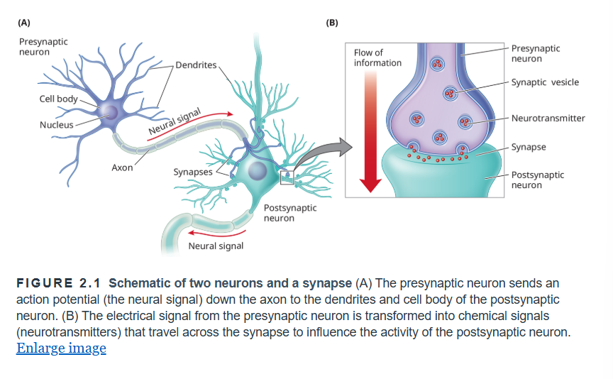
Neurons: specialized cells that receive and transmit signals in the nervous system including the brain (fundamental units of the NS)
little computer/employee that received input and makes a decision about whether to pass on a signal to other neurons in the network
Neuron’s three major components:
Dendrites - branchlike component of a neuron, receiving input from sensory receptors or other neurons
Soma (cell body) - the neuron’s core, which receives input and provides the metabolic machinery for the neuron
Axon - part of a neuron that carries nerve impulses away from the cell body, toward the receiving dendrites of other neurons
Neurons are separated by small gaps called synapses
most neurons communicate via chemical signals across the synapses between them
sending neuron is called presynaptic neuron
receiving neuron is called postsynaptic neuron
Signal transmission
To transmit signal, pre neuron releases chemical messengers called neurotransmitters (NT) from the ends of the axon (called axon terminals) into the synapse
neurotransmitters can either facilitate or inhibit whether the post neuron fires
At synapse, neurotransmitters bind to receptors located on the post neuron’s dendrites or cell body
this binding cause either positively or negatively charged ions to flow into the post neuron, leading to a buildup or reduction of electric charge within the cell body
if the charge exceeds a threshold, it triggers an electrical signal called action potential to flow down the length of the post neuron’s axon
Action potential occur in an all-or-nothing fashion (either fire or not fire)
if it does fire, when the AP reaches its axon terminal, it stimulates the release of neurotransmitters into the next synapse and so on
Neurons cont
neurons communicate across synapses has important function consequence: it enables learning and experience to modify the strength of transmissions between neurons
When two neurons communicate a lot, the pre neuron can begin to release more NT and sprout new axon terminals (forming additional synapses with post neuron)
Post neurons can also begin to grow new branches that reach out to receive incoming signals → density of receptors on post neuron increase
Neuroplasticity: the ability of neural connections to change with experience
learning and experience can make it more likely or less likely signals will transmit from one neuron to another → foundation of memory
Neuroplasticity can lead to remarkable changes in the brain
EX: violinist and guitarist use left hand → brain representing these left-hand fingers would be enlarged

Left and right hemispheres of brain
left hemisphere processes sensory and motor functions for the right side of the body
right hemi controls the left side of the body
language functions are stronger in left hemi
visuospatial tasks are more concentrated on right hemi
These tasks are not equally distributed bc…
based on processing efficiency
takes time for info to be shared across hemispheres
For complex skills like language use, a faster response is better
within-hemisphere processing improves efficiency in performing complex cognitive tasks
Corpus Callosum: a large bundle of neural fibers (axons) forming a massive interconnection between the two hemispheres of the brain
interesting difference in spilt brain pt where the pt’s corpus collosum has been severed
research shows that left hemisphere is better at language processing and analytic skills, and right hemi is more involved in tasks requiring spatial manipulation or attention
Brain anatomy
Cerebral cortex: a thin folded sheet of neurons constituting the outer layer of the brain
cortex has grooves (sulci) and bumps (gyri) to increase SA that can fit inside the skull → each hemi can further be divided into 4 lobes
Occipital lobe (back) = visual perception
Temporal lobe (low bottom) = complex perception, memory, and language
Frontal lobe (front) = thinking, planning, decision making, cog control
Parietal lobe (upper back) = controlling action, attention, number processing, spatial cognition and understanding intention
Cerebellum: responsible for control of fine motor movements and possibly the coordination of complex thought (near brain stem)
2.3 Neuroscience methods
Methods differ in
Spatial resolution: ability of a research method to pinpoint where neural activity occurs (ex: ranging from neurons (.001mm) to brain areas (5cm) of varying size)
Temporal resolution: ability to pinpoint neural activity occurs over millisec, seconds, min, or longer
Invasiveness: degree to which a research method impacts the individual’s brain who is being studied (EX: implanting or observation MRI)
Neuropsychology: study of the behavioral consequences of brain damage
damage is often from natural causes like stroke or illness, but can also be from surgery or trauma (unnatural cases)
provides early evidence supporting the idea that mind was dependent on the brain (materialism), rather than being separate (dualism)
For pt with acquired deficit like face blindness
neurologists will look for damage (plot on a map of the brain
with several pts’ data, can see if there’s an overlap across pt with common face blindness disorder
overlapping area of damage then becomes a candidate brain area that is important for the function that was impaired
Task of mapping the brain can be done bc
of functional specialization (principle that different brain area is specialized for different things)
Dissociation: a pattern where conditions affect one behavior or process more than another, indicating they involve different mechanisms
EX: pt that can understand spoken lang but can only speak “tan”
Broca’s area = area in the left frontal lobe, with functions linked to speech production
But sentences spoke by pts with this condition can also sound fluent even tho they don’t make sense
Double dissociation: dissociating (distinguishing) two different cognitive functions from each other, such as when they can be disrupted independently (Broca and Wernicke)
Double dissociation can be shown differently with damages in different brain areas
damage to Broca impairs speaking, but not comprehension
damage to Wernicke impairs comprehension, but not speaking
3 reasons that neuropsychology alone cannot fully advance the scientific study of the brain
brain lesions are imprecise and generally affect more than one region → chance that brain damage will be well defined or consistent across pt is low, making it difficult to associate patterns of brain damage with specific functions
bc brain damage usually impairs several functions, studies conducted on pts with brain damage are not easy to compare with studies of healthy controlled pt
clinicians must prioritize treating pt over testing them for scientific gains
Bc of all these reasons and limitations, need other methods to study brain function
Electrophysiology: direct measurements of electrical activity from neurons, either from single cells or from an aggregate of cells
measure of action potentials directly reveals whether a neuron is active or not
Goal: find experimental manipulations that change the firing rate of a neuron → demonstrating the neuron’s function
Single-cell Electrode Recordings
can measure AP by placing thin electrodes (needle like probes that measure electrical activity) into a network of neuron regions
electrode will pick up signals from multiple neurons, then computer algorithms can isolate individual signals
bc of neuron’s quality of all or nothing (0 or 1), neuron’s activity can be quantified by its firing rate
Baseline firing rate = the firing rate of neurons in the absence of an explicit task or stimulation
firing rate will change in response to stimulus (ex: face) or when neuron is involved in a task or action (ex: moving finger)
EX: to study function of visual neuron
place electrode in brain region hypothesized to be important for visual processing
measure neuron’s baseline firing rate while the participant is looking at nothing
research present various kinds of stimuli to see which of them make the neuron fire differently from baseline
Hubel & Wiesel discovered neurons in the visual cortex, area in the occipital lobe that receives visual info from the eyes
BUT not all visual neurons have the same function (they exhibit selectivity)
to establish neuron’s selectivity to face, researcher must compare how it responds to face-selective cells that respond vigorously to faces, but not to a range of other visual stimuli like bodies, fruits, and etc
a neuron that responds similarly to other visual stimuli (nonface body parts) is NOT FACE SELECTIVE
weaker response to scrambled face features = these neurons are sensitive not just to face parts but to also how they arrange (configured) relative to each other
EX: face cells in monkeys will respond strongly to human face as to a monkey face = these neurons categorize human faces together with monkey faces
Population coding: representation or coding by an ensemble (population, or group) of neurons → this is why we can distinguish between an image of San Fran and Miami Beach landscape
objects are represented as patterns across this population of four neurons → same sys can represent more objects (15) rather than just 4 objects in the single-cell coding
Pro and Con of single-cell electrode recording (electrophysiology)
Pro: spatial and temporal precision
by measuring in single neurons, offers high spatial resolution and temporally precise bc this method allows neuronal firing to be measured the instant it happens (at millisec resolution of AP_
Con: invasive procedure (inserting electrodes in brain) → these experiments can’t be done on humans just for scientific research…
there must be a clinical reason to help the person in order to insert
Electrocorticography or intracranial recordings: method of recording electrical signals from electrodes placed on surgically exposed cerebral cortex
only for conditions like severe epilepsy → electrode implantation can help localize the source of seizures as targets for intervention
when recording, clinicians are keen to preserve function so they don’t remove or disrupt brain tissues that underlies language or memory
Pro: high spatial and temporal resolution
Con: invasive method
when electrodes are placed on surface of brain, they aren’t arrange in a way that is designed to answer a theoretical research question… they are instead placed based on what would most benefit the pt and surgeons
recordings are from individuals who tend to be heavily medicated to control their epileptic seizures → since these pts have abnormal brain activity due to epilepsy, their brains are operated differently from control individuals (aka reduce the ability to generalize results)
Electroencephalography (EEG): a method using electrodes on the scalp to measure electrical activity in the brain (less invasive)
aggregate activity of active neurons transmit thru the head and can be detected on the scalp
EEG is widely used in clinical settings bc different states of consciousness and sleep produce different EEG patterns that can be reliably measured
in a lab, EEG can measure perceptual and cognitive functions in an individual who is performing a task (EEG produces signals that reveal perceptual and cognitive activity)
Event-related potential (ERP): time-locked EEG waveform that occurs in response to a stimulus, event or task, measured from average of many trials
EX: when recording from pt viewing several different kinds of objects, EEG reveals a different response for face stimuli than for other visual stimuli → this face-selective ERP is called the N170 bc it occurs about 170 milisec after stimulus onset and bc it’s electrical polarity is negative
Pro of EEG
high temporal precision
EEG signal is really rich and informative, where several different aspects of the signal reveals underlying perceptual and cognitive processes
inexpensive compared to fMRI → therefore very widespread clinical use
noninvasive and relatively convenient to use (no risks even to babies)
place of electrodes on scalp may require shampooing afterwards (bc conductive gel is applied between scalp and electrode to enhance signal)
Cons of EEG (opened path for fMRI)
poor spatial resolution
it sums activity across the entire brain and signals from different areas mingle with each other’s in ways that are difficult to pull apart
it usually is possible to distinguish whether if the activity came from front or back of brain… but more anatomical precision is required
Magnetic Resonance Imaging (MRI): a widely used technique for studying brain structure and cognition, involving use of a powerful magnetic field and brief radio pulses
structural MRI was initially developed to provide detail images of internal structures, organs, and brain
How it function (it does not reveal mental function)
since different brain structures and tissues have different magnetic properties, MRI scanner can detect these differences in 3D volume
by assigning different shades of gray, it can transform the differences in 2D images, taken as layered slices thru brain volume
in medical applications, physicians use the structural pics from MRI tech to identify abnormalities in pts
Brain imaging uses medical tech to study brain activity noninvasively
structural medical imaging provides detailed views of internal body structures and abnormalities ranging from fractures to tumors
Several imaging techniques: XRay, computerized tomography scans, and positron emission tomography
Functional magnetic resonance imaging (fMRI): a technique that uses MRI to infer brain activity from measurements of blood oxygen levels throughout the brain
takes advantage that magnetic properties of blood change depend on its oxygen content
bc blood oxygen level increase brain areas that are active, this blood-oxygen-level-dependent signal (signal used to generate images in fMRI, indicating blood oxy lvls throughout the brain that correlate with neural activity) can be used to infer brain activity
this signal can be superimposed on anatomical images of the brain to identify which regions are more active, like traffic info on google maps
if MRI gives you a map of the brain, fMRI tells you which area of the brain map is more active
Pro of fMRI
significantly better spatial resolution (detail) than other noninvasive methods like EEG
don’t require placing electrode on brain or scalp
Con of fMRI
measurements are indirect (they rely on changes in oxygenated blood flow to infer neural activity) → accuracy is limited in two ways: spatial resolution is coarse and temporal resolution is slow
spatial precision calculated in voxels (1×1×1 or 3×3×3) → fMRI = looking at a city vs electrophysiology = person (more specific)
participants can’t have any metals in their bodies bc of scanner’s high magnetic field
confined tube for scanning (kinda uncomfortable)
there is limitation in what a person can conclude from correlation data, so caution is needed
Functional brain mapping: the tasks of linking cognitive and perceptual functions to specific brain regions
Pro: allowing surgeons to understand or predict what may happen when a brain area is damaged or removed
EX: Visual processing of face and scenes
participants are scanned while viewing sequences of faces or scenes
bc both tasks involve visual stimulation, broad areas of the visual cortex become active in response to both
Subtraction method allows for comparison of patterns of brain activity for two categories of stimuli
when subtract activity drive by scenes from faces (F - S = fusiform face area), what remains are the brain regions that are important for face processing (found out it was fusiform gyrus, that spans the lower portions of occipital and temporal lobes)
when subtract face from scene (S - F = parahippocampal place area), reveals activity that is higher for scenes
Subtraction method: fMRI analysis technique that compares patterns of brain activity elicited by different stimuli or tasks
also used in EEG, removes activity common to both face and scene processing, isolating activity that is specific to one category versus the other
Brain imaging data are extremely useful in testing hypotheses about how the mind works
localization or mapping brain functions are the FIRST steps in understanding a complex behavior like face perception
Brain mapping establishes regions of interest (brain area that is focus of study)
the region of interest approach can be limited…
most perceptual or cognitive functions don’t have dedicated, specialized brain areas
EX: the brain doesn’t have separate area that remember shoes and another remembering bottles
fMRI Analysis Methods (Pattern and functional connectivity)
Pattern Analysis
similar to population coding
spatial pattens of neural activity within region looks different (measured in voxel)
computers learn (machine learning) the relationship between each show or watch and the corresponding pattern of fMRI activity it elicits → calculate if the fMRI pattern is more similar to shoe or watch pattern
Functional Connectivity
look at how different brain region interact
typically studied by examining their correlations in activity
if two regions operate independent of each other = 0 correlation in activity (can also have positive or negative correlation, line graph thingy)
Connectome: estimate functional connectivity for all pairs of regions across the brain (unique to each individual)
Clinical examples of connectome allow to measure from brain scans whether someone is suffering depression, anxiety, autism, dysfunction and etc.
Be careful how you interpret brain data
MUST AVOID reverse inferencing
EX: if observed that fusiform face area was active, you might infer that individual was viewing human faces… BUT can’t be certain bc it could be that the individual was thinking about faces or might viewed animal faces instead
avoid correlation and causation problem (especially in neuroscience)
EX: LED light on tv will correlate if your screen is powered on, but that don’t mean the signal caused your device to turn on or that a malfunction of the power light will prevent your screen from turning on
EX: Face area is active when viewing faces doesn’t logically dictate that the face area is necessary for face perception
if you want to prove necessity, you need to show that blocking face area activity will disrupt face perception (use brain stimulation methods)
Reverse inference: the illogical inference of a mental process (cognitive or emotional) from the activation of a particular brain region
when you observe that A causes B, you cannot infer that B causes A
EX: spraying water (A) on someone causes them to become wet (B), but just bc you see someone wet (B), it doesn’t mean that you can infer 100% that someone sprayed water (A) on them
Doesn’t mean to abandon use of fMRI for trying to decode the mind
simply highlights the need for caution in interpreting such data
statistical methods can help make reverse inferences more accurate (especially when you catalog many studies that map different functions to different brain areas)
Analysis methods based on pattern of activity (not averaged act) can further enhance precision and confidence in reverse inferencing
Brain Stimulation methods (the stimulation or disruption of brain activity to study causal effects on perceptual and cognitive function
Transcranial magnetic stimulation (TMS)
a method to temporarily disrupt brain activity using focal magnetic pulses targeted over different areas of the scalp
safe method for temporarily stimulating or interrupting brain activity
TMS-induced “virtual lesions” can help establish whether a brain region plays a causal role (compared to fMRI’s correlations)
How it works
a coil is placed at strategic location around an individual’s head
when electrical current runs thru coil, it causes a magnetic field to pass thru the skull, influencing neural act in the region under the coil
EX: TMS over the visual cortex can generate phosphenes (the perceptual sensation of visual speckles) or TMS over motor cortex can cause muscles to twitch
Pro of TMS
establish causality AND high temporal precision
EX: timing of TMS pulse to visual cortex determines whether it can “blind” a person to briefly presented visual stimulus
provide therapeutic benefits for some clinical disorders (like pain management, recovery after stroke, and treating drug-resistant depression)
Con of TMS
low spatial precision and reach
foci of TMS disruption are not well defined, and best precision seems limited to just a few mm (just small enough to stimulate individual fingers from brain area that represent fingers)
can only reach to around 3cm of scalp, not deeper structures
can cause discomfort of raise a small risk of seizures → have to follow rigorous guidelines to minimize these negative effects
Brain Stimulation methods cont
Transcranial direct current stimulation (tDCS)
a brain stimulation that involves applying a weak electrical current across the skull to modify brain activity, thereby increasing or decreasing the likelihood that underlying neurons will fire (no causing)
safe method and noninvasive bc electroudes are on scalp and lvl of electrical currents are minimal
How it works
Similarly to working on modes of a battery, tDCS uses two electrodes placed on scalp that create an electrical current flowing from one electrode to another
orientation of the current flow and placement of electrode affect how tDCS will modulate brain activity
Pro of tDCS
may have therapeutic benefits, but need more research (ex: changing the likelihood that neurons will fire can promote learning)
Deep brain stimulation
electrodes implanted in the specific brain area, allowing for direct measurement of neuronal activity and precise delivery of stimulation for therapeutic effect
most effective treatments by FDA to help control tremors in Parkinson’s disease
also being studied to alleviate symptoms of major depression, small cognitive improvement in memory, attention, and executive function
Compared to other methods used in humans, deep brain stimulation offer unprecedented temporal and spatial measurement and control of brain circuits
therapeutic potential is promising (even if how it works is not well understood…)
But since it requires surgical implantation (invasive), it’s mainly used for pts with serious medical conditions that are resistant to other types of treatments
Genes: heritable codes in almost every cell that dictate how an organism develops and functions
Genotype: entire set of genes that an organism carries
Phenotype: all of the observable traits resulting from the interactions of its genotype and its environment
include susceptibility to physical or mental illness
Behavioral Genetics
field that attempts to link behavior (phenotypes) and genes (genotypes)
EX: study of identical twins who were separated at birth → strong correlation in intelligence
Epigenetics
study of external factors that affect how genes operate
EX: maternal behaviors changes how genes were expressed in pups
EX: rats that received more maternal behaviors responded better to stress as an adult rat
Manipulation of genes
knock-out: research technique that involves rendering a gene absent or unexpressed
effects can be linked to different behaviors ranging from memory impairment to generation of false memories
CRISPR: precise gene editing (targeted gene disruption, replacement, or modification)
Computational neuroscience
field that uses math and computer modeling to understand brain function and behavior
brain data is complex → use machine learning to make sense of signals
AI algorithms enhance brain decoding → understanding the brain improves AI
Deep learning (in machine learning): a form of AI that uses deep neural nets like brain-like algorithms to attempt to interpret natural images

2.1 Summary
Cognitive neuroscience is the interdisplinary study of how the brain supports our ability to perceive, think, learn, communicate, and control behavior. The field assumes materialism, which views the mind as a product of the brain, as opposed to dualism, which considers the mind separate from the brain.
One example of the tight relationship between brain and behavior is when patients with brain damage (to specific regions involved in face perception) show impairments in face recognition
2.2 summary
The fundamental unit of the brain is the neuron, a cell in the NS that receives input from other neurons and decides whether to signal output to other neurons. The brain is a rich network of around 100 billion neurons, which are connected via synapses, across which neurons communicate with each other using chemicals called neurotransmitters.
These heavily interconnected neurons form networks within a brain area, which are also networked with other brain areas. They are organized into different systems across the brain, and many of these systems have specialized functions. At the largest scale, the brain’s anatomy is organized into left and right hemispheres, which communicate across a large bundle of neural fibers called the corpus callosum. Each hemisphere can be further divided into the occipital, temporal, parietal, and frontal lobes
2.3 summary
Safely protected by the skull, the brain is difficult to study. Neuroscientists have had to rely on a variety of clever techniques, each with strengths and limitations, especially with regard to spatial precision (whole brain to single neurons), temporal precisions (years to milliseconds), and invasiveness (brain lesions and surgery to brain imaging and behavioral measurements).
Neuropsychology studies the effects of brain damage. Electrophysiology measures action potential, that is, electrical activity from neurons, either from single cells or from an aggregate of cells. fMRI uses MRI to infer neural activity from different brain areas. Brain stimulation techniques allow for the manipulation or disruption of brain activity. Computational neuroscience uses math and computer simulations to understand brain function and behavior, which improves AI’s capabilities to benefit human lives.
Why do cognitive neuroscientists often combine evidence from lesions, recordings, imaging, stimulation, genetics, and computational models?
A) Different methods generally measure the same thing, so their results should be interchangeable.
B) Each method has different strengths and limitations, so converging results support stronger conclusions. (CORRECT)
C) A conclusion is valid only if every available neuroscience method is used in the same study.
D) Combining methods eliminates the need for a theory of cognition.
Which comparison of neuroscience methods is most accurate?
A) EEG and fMRI both directly measure neurotransmitter release, while TMS measures blood oxygenation.
B) EEG provides the best spatial localization, fMRI records individual action potentials, and TMS only measures existing activity.
C) EEG, fMRI, and TMS provide the same kind of evidence and mainly differ in cost.
D) EEG provides precise timing, fMRI provides better spatial localization but measures an indirect signal, and TMS can test causal contributions by perturbing processing. (CORRECT)
What does neural plasticity help explain?
A) Experience and learning can alter the strength and organization of neural connections. (CORRECT)
B) Each memory is stored by creating an entirely new neuron.
C) The adult brain’s connections remain fixed unless the brain is injured.
D) Learning occurs without producing any changes in the nervous system.