University of Waterloo PSYCH 207 - Chapter 2
 Call Kai
 Learn
Learn Practice Test
 Spaced Repetition
 Match
Match Flashcards
 Knowt Play
1/49

There's no tags or description
Looks like no tags are added yet.
Name | Mastery | Learn | Test | Matching | Spaced | Call with Kai | Chat |
|---|
No analytics yet
Send a link to your students to track their progress
50 Terms
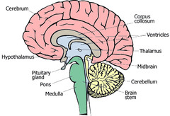
The Hindbrain
- Originally developed as one of three bulges in the embryo's neural tube
- The most primitive
The three major structures of the hind brain
1. Medulla oblongata (or 'medulla') transmits information from the spinal cord to the brain and regulated life support functions such as respiration, blood pressure, coughing, sneezing, vomiting, heart rate
2. The Pons facilitates the 'cross over' of information between the left side of the body and the right side of the brain (and vice versa)
3. The Cerebellum contains neurons that coordinate muscular activity (one of the most primitive structures)
The Midbrain
- KEY: Involved in relaying information between brain regions
- Includes
- Reticular formation (keeps people awake and alert, responds to shocking stimulus)
- tectum
- tegmentum
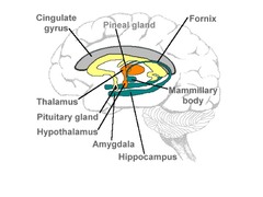
Thalamus
Switching station for sensory information; also involved in memory
- Relays information to the cerebral cortex
Hypothalamus
Regulates basic biological functions, including hunger, thirst, temperature, sexual arousal
- Also involved in arousal
- Controls the pituitary gland by releasing hormones
Hippocampus
Involved in learning, memory (long term) and emotions
Amygdala
Involved in memory and emotion (the strength of emotional memories & learning), particularly fear and aggression
Basal Ganglia
Involved in the production of motor behaviour
Spinal Cord
Transmits signals between the brain and rest of the body
The Cerebellum
A large structure of the hindbrain that controls fine motor skills.
What are the two approaches to studying brain lesions?
What function is supported by a given brain region?
- Examine a group with similar lesions
What brain regions support a given cognitive function?
- Examine a group with similar impairments, examine the brain regions common to this deficit
Perceptions is not complete determined by the [ ]
stimulus display
- requires the perceivers active participation
- key: conscious experience is an active reconstructive process
Visual Searching
Bottom up processing -> when there is a lot of overlapping of features (requires more fine grain discrimination), it takes longer to process
It is easier to identify things when:
They are in their associated context (context effects) (key: top-down processes)
Cerebrum
largest part of the brain
- Key: Contains the cerebral cortex
Cerebral cortex
Carries information between the cortex and thalamus, or between different parts of the cortex
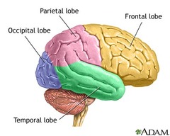
cerebral cortex lobes
frontal, parietal, occipital, temporal
The right and left sides of the brain's temporal lobes are connected by . . .
the anterior commisure
What divides the lobes?
frontal and parietal: central sulcus
temporal (within): lateral sulcus
Where is the somatosensory cortex located? What does it do?
parietal lobe (post-central gyrus)
- Processing sensory information of the body/skin
occipital lobe
A region of the cerebral cortex that processes visual information
temporal lobe
A region of the cerebral cortex responsible for hearing and language (KEY: and also faces)
- Key: because of their location in the brain, damage to these lobes can cause memory disruption
Three regions of the frontal lobe
Motor cortex - fine motor movement
Premotor cortex - planning movements
Prefrontal cortex - executive functioning
Faculty Psychology
The theory that different mental abilities, such as reading or computation, are independent and autonomous functions, carried out in different parts of the brain.
What is Phrenology? What are its problems?
Psychological strengths and weaknesses could be correlated to the size of different brain areas
Problems: the size thing, and that each area is completely independent
Aphasia
impairment of language, usually caused by left frontal lobe damage either to Broca's area (impairing speaking)
Wernicke's aphasia (fluent aphasia). What is it? Where is it?
condition characterized by poor language comprehension and impaired ability to remember the name of objects
KEY: Left temporal lobe
primary somatosensory cortex. What is it? Where is it?
Located in the Parietal lobe
Each part of it receives information from a specific part of the body
KEY: Size of area devoted to the leg is much smaller than area devoted to finger tips
Penfield. Who was he? What did he do?
Canadian neurosurgeon who used electrodes and electrical stimulation techniques to "map" out different parts of the brain during surgery
ablation. What study showed this
the removal of a body part or the destruction of its function
Rats doing poorly in a maze correlated to amount of brain removed, not from a specific region
Lateralization
specialization of the two cerebral hemispheres for particular operations (especially language)
left hemisphere
- Most people show specialization for language here
- Larger in size
- Better at processing SERIALLY
right hemisphere
Larger in the parietal and temporal areas
- Better processing of visual and auditory information and better spatial processing (puzzles, navigation)
- Better at processing WHOLES
CAT scan (computerized axial tomography)
a method of creating static images of the brain through X rays
- Detects density -> can create a picture of what's up
- Slides that are captures at different depths
- Earliest imaging method
EEG (electroencephalogram)
An amplified recording of the waves of electrical activity that sweep across the brain's surface. These waves are measured by electrodes placed on the scalp.
- Used to detect different states of consciousness
- Continuous measure
MEG
Like EEG, but more precise, uses magnets
ERP (event-related potential)
Measures an area of the brain's response to a specific event
PET (positron emission tomography) scan
Involves injecting a radioactive compound to measure blood flow of different regions of the brain
MRI (magnetic resonance imaging)
Similar to CAT in that it shows a picture of neuroanatomy, but doesn't require radiation and shows clearer pictures
- Uses radio waves
- Makes cross sections
- Can't do this if you have metal in your body
fMRI (functional magnetic resonance imaging)
Relies of the fact that blood has magnetic properties (blood becomes less magnetic the further it is from the heart)
subtraction technique
The technique used in brain imaging in which baseline activity is subtracted from the activity generated by a specific task. The result is the activity due only to the task that is being studied.
Medulla
Controls vital functions such as breathing and heart rate
What are the four different brain planes
Should include:
- Horizontal Plane as "axial horizontal plane"
- "Midsagittal Plane"

Diagram of the brain
Limbic System Diagram
Regions of the brain diagram
Electrical Activity
event-related potentials (ERPs) derivative from EEG
Blood flow increases . . .
as a function of neural activity
- KEY: fMRI signal depends on the difference in the magnetic properties of oxygenated and de-oxygenated hemoglobin