musculoskeletal system
 Call Kai
 Learn
Learn Practice Test
 Spaced Repetition
 Match
Match Flashcards
 Knowt Play
1/108

There's no tags or description
Looks like no tags are added yet.
Name | Mastery | Learn | Test | Matching | Spaced | Call with Kai | Chat |
|---|
No analytics yet
Send a link to your students to track their progress
109 Terms
functions of the skeletal system
support- framework for the body
movement- provides locomotion with attachment of muscles, ligaments, & tendons
protection- protects vital organs (heart, liver, brain, lungs)
hematopoiesis- blood formation (WBCs, RBCs and platelets)
storage- calcium, phosphorus
“locomotor system”
bones; joints/cartilage; ligaments; muscles; tendons
bones
206 bones total (more than half in wrist, hands, feet, and ankles)
babies born with 300 bones most fused by age 25
make of calcium, phosphorus, sodium, minerals, protein, collagen
hyoid bone is only bone not connected to any other bone
4 main types of bone: long, short, flat, irregular
long bones
support/strength
femur, tibia, radius
short bones
facilitates movement
irregular/cube shape
carpals, tarsals
flat bones
muscle attachment
protections
skull, scapulae, ribs, sternum
irregular bones
attachment within joints
pelvis, vertebrae, middle ear
diaphysis~ bone structure
main shaft portion of bone, compact bone
epiphysis~ bone structure
end of bones, spongy bone
epiphyseal plates~ bone structure
growth plates
medullary cavity~ bone structure
contains yellow marrow, in the middle of diaphysis
endosteum~ bone structure
membrane that lines medullary cavity
periosteum~ bone structure
dense white fibrous membrane covering entire surface of bone
articular cartilage
smooth covering over ends, cushions joints
red marrow
found in the ends of long bones. bodies of vertebrae and flat bones. manufactures red blood cells, white blood cells, and platelets (hematopoiesis)
yellow marrow
found in the medullary cavity of long bones mostly fat; no longer produces blood cells
axial skeleton (midline)~ divisions of skeleton
ossicles, skull, hyoid bone, ribcage, vertebral column
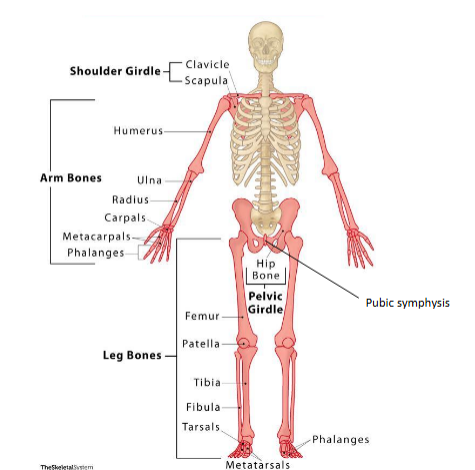
appendicular (hangs from) skeleton~ divisions of skeleton
shoulder girdle; clavicle, scapula
arm bones; humerus, ulna, radius, carpals, metacarpals, phalanges
pelvic bone; pubic symphysis, hip bone
leg bones; femur, patella, tibia, fibula, tarsals, metatarsals, phalanges
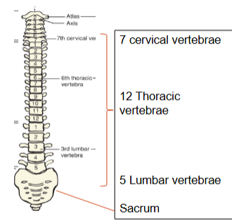
spine~ major bones
7 cervical vertebrae > 12 thoracic vertebrae > 5 lumbar vertebrae + sacrum
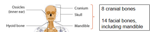
skull~ major bones
8 cranial bones, 14 facial bones, including mandible
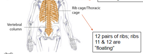
rib cage~ major bones
12 parts of ribs; ribs 11 & 12 are floating
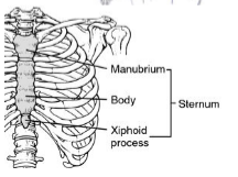
sternum~ major bones
manubrium
body
xiphoid process
joints
classified by
functional classification
plus, synovial joints by type of motion
synarthrosis joint
immovable joint
ex: skull sutures, epiphyseal plates, joint between first rib and manubrium of sternum
ampiarthrosis joint
slightly moveable joint
ex: vertebral joints, joint of the symphysis pubis
diarthrosis “synovial” joint
freely movable joint
ex: joints of the extremities, shoulder joints, hip joints
ball and socket~ types of joints and movement
movement in all directions (hip and shoulder)
hinge~ types of joints and movement
bending and straightening in one direction (knee and elbow)
pivot~ types of joints and movement
limited rotation (neck)
saddle~ types of joints and movement
movement back and forth and side to side; no rotation (base of thumb)
condyloid~ types of joints and movement
2 oval shaped bones that fit together. similar to ball and socket but can’t rotate (jaw, finger joints)
gliding~ types of joints and movement
joints glide over each other (carpals and tarsals)
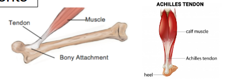
tendon
attaches muscle to bone
injury= strain
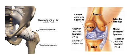
ligaments
attach bone to bone
injury= sprain
cartilage
soft & flexible tissue
reduces friction in joints
slick surface for rotation
distributes weight bearing
absorbs shock
bursae
synovial fluid filled sac
produces synovial fluid
reduces friction
between bone/muscle or bone/tendon
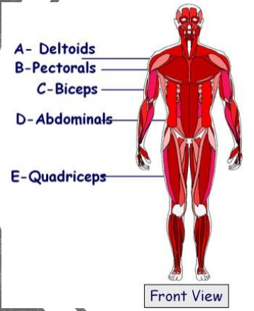
major muscle groups; front view
A- Deltoids~ shoulder
B- Pectorals~ chest
C- Biceps~ arms
D- Abdominals~ abdomen
E- Quadriceps~ front thigh
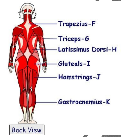
major muscle groups; back view
F- Trapezius
G- Triceps
H- Latissimus Dorsi
I- Gluteals
J- Hamstrings
K- Gastrocnemius
muscles of respiration
intercostals in between ribs
diaphragm dome shaped contracts and flattens with breathing
skeletal muscle~ types of muscle
close to skeletal components
striated
voluntary
locomotory actions~ movement
smooth muscles (visceral muscles)~ types of muscle
inner walls of hollow organs like alimentary canal
unstriated
involuntary~ don’t have to think about digestion
transportation of food and gametes
cardiac muscles~ types of muscle
heart
striated
involuntary
pumps heart
contractability~ muscle characteristics
the ability to become shorter/thicker
irritability/excitability~ muscle characteristics
ability to respond to stimulus
extensibility~ muscle characteristics
ability to lengthen or stretch
elasticity~ muscle characteristics
ability to return to original shape
tone~ muscle terms
state of slight tension
fatigue~ muscle terms
when overexerted, loses some power
spasm~ muscle terms
involuntary contraction/sustained contraction
atrophy~ muscle terms
degeneration or loss of muscle fiber due to lack of use/loss of nerve supply
voluntary movement~ functions of muscle
walking, sitting, standing, maintain upright position
balance
involuntary movement~ functions of muscle
heart to pump blood, gastrointestinal tract movement, respiration, reflexes
protection~ functions of muscle
protect body in emergency
cover, surround, support internal organs
misc.~ functions of muscle
produce heat
provides shape to body
maintains body temp
shivering, chills, goose flesh
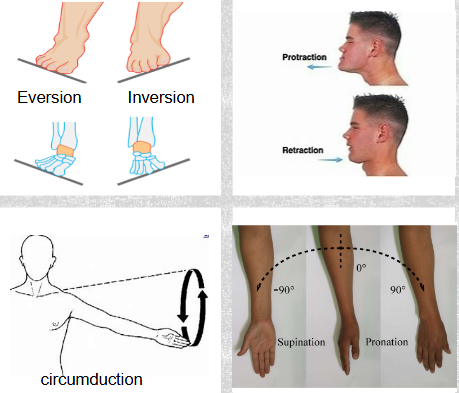
musculoskeletal movements
ankle bones: inversion~ inward; eversion~ outward
jaw: protraction~ forward; retraction~ backward/resting
shoulder: circumduction~ circular movement
arm: supination~ upward; pronation~ flat down
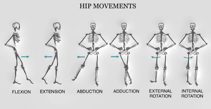
hip movements
flexion- forward/upward
extension- behind your body
abduction- out/away
adduction- toward/across midline
external rotation- outward
internal rotation- inward
effects of aging on the musculoskeletal system
bone mass/calcium loss
vertebral column shortens
decrease joint flexibility
muscle cells lost (decrease strength/endurance)
muscle cell replaced by fat
elasticity muscle fibers lost
ESR (Erythrocyte Sedimentation Rate)
test for inflammation related to possible infection or inflammatory disorders
(rheumatoid arthritis, polymyalgia rheumatica, osteomyelitis, systemic lupus erythematosus, bone tumors)
CBC (Complete Blood Count)
WBCs, RBCs, platelets, etc.; indication of overall condition of health and musculoskeletal system
RF (Rheumatoid Factor)
measures the amount of rheumatoid factor in blood; indicator for rheumatoid arthritis
uric acid
elevated level indicative of gout
CK (Creatinine Kinase)
an enzyme found in skeletal muscle, cardiac muscle, and brain. Elevated CK indicates muscle cell injury
CRP (C-Reactive Protein)
a protein made by the liver that increases in the blood in response to inflammation, as it binds to damaged cells to mark them for removal
calcium and phosphorus
essential mineral components of bone structure and metabolism
x-ray~ imaging studies
can diagnose fractures, dislocations, arthritis, osteoporosis, and bone tumors
non-invasive
CT (computerized tomography)~ imaging studies
provides 3-dimentional radiographic view diagnosing bone fracture, ligament/tendon injuries, soft tissue damage, tumors, lumbar disc herniation
does not show nerve roots clearly
MRI (Magnetic Resonance Imaging)~ imaging studies
magnetic field enables scanning machine to produce detailed 3D images
does not use x-rays
shows detail of soft tissues; disc, nerve roots, tumors
ultrasound
non invasive
use of sound waves/echoes
can show soft tissue masses, osteomyelitis, infection, bone mineral density, sports injuries/fracture
used to guide needle placement for aspiration or injection
myelogram~ diagnostic test
injects dye or air into vertebral column; used for spinal cord/vertebral column diagnostics- lesions on the spine
arthrogram~ diagnostic test
x ray or MRI study of joint; dye is injected into the joint, then a series of x rays or MRI taken; joint diagnostics
bone scan~ diagnostic test
nuclear imaging technique (IV) injection of radioisotope, which accumulates in areas of increased bone activity. A gamma camera is used for visualization.
Diagnose bone tumors, osteomyelitis, osteoporosis, inflammation, bone/joint infections, stress fx
arthroscopy~ diagnostic test
surgeon uses small endoscope, called an arthroscope; has a lens and a light source that transmits a picture to a video monitor. flexible scope used in viewing joints for diagnostic and treatment purposes.~ consider allergy to dye
example: removal of bone spur
arthrocentesis~ diagnostic test
uses needle to aspirate synovial fluid or pus from joint cavity. used to diagnose infection/inflammatory conditions and bleeding.
EMG (Electromyogram)~ diagnostic test
test of electrical conductivity; measures muscle response or electrical activity in response to a nerve’s stimulation of the muscle
the test is used to help detect neuromuscular abnormalities
biopsy~ diagnostic test
bone or soft tissue sample is taken; sent to lab
used for detection of cancer
degenerative disorders
muscular dystrophy
carpal tunnel syndrome
muscular dystrophy~ degenerative disorders
chronic, degenerative disease of skeletal muscles that often inherited. Varying degrees of progressive weakening & muscle wasting
cause: not known; possibly related to inadequate enzyme production
there are multiple types that affect different muscle groups
Duchenne type is caused by mutations on the X chromosome, leading to dysfunction of dystrophin, a protein crucial for maintaining muscle cell integrity. Females are carriers and usually don’t have symptoms. Male children inherit the disease. The condition progresses to loss of ability to walk and heart failure; death usually late teens, early 20s, but new treatment might change that
tx: support to continue activities as normally as possible
exercise programs and splints/braces; can permit ambulation
prevent upper respiratory tract infection
Repetitive strain injury: carpal tunnel syndrome~ degenerative disorders
compression neuropathy of median nerve in wrist
causes: repetitive movements, arthritis, trauma, gout, tumor
s/s: progressive forearm, wrist pain, numbness, tingling, weak grip, positive Tinel sign
treatment: wrist splinting, rest, NSAIDs corticosteroid injections, surgery
systemic disorders w/ musculoskeletal manifestation
gout
lupus
scleroderma
rickets & osteomalacia
Gout~ systemic disorders w/ musculoskeletal manifestation
purine rich foods are broken down into uric acid. if the body produces too much uric acid or cannot excrete it, uric acid crystals form in the joints leading to gout attacks. Usually affects big toe, instep, ankle, or knee but may be in any joint
causes: genetic defects in purine metabolism enzymes or uric acid transporters; eating too much food in high in purines, high fructose diet, dehydration, drinking alcohol (especially beer), surgery, injury, diet, stress
s/s: joint swelling, redness, severe pain
Dx: uric acid lab test, joint aspiration for crystals
tx: diet, avoid alcohol and high purine foods: liver, anchovies, red meat, sardines, salmon, turkey, some shellfish
medications (allopurinol, colchicine, corticosteroids, NSAIDs)
weight loss, drink plenty of fluids
Lupus~ systemic disorders w/ musculoskeletal manifestation
2 types:
Discoid Lupus Erythematosus is a chronic disease with skin manifestations of disc like patches with raised, reddish edges and depressed centers
Systemic Lupus Erythematosus: SLE (“lupus”) is an autoimmune systemic disorder that affects many body systems
chronic multisystem autoimmune disorder; effects nearly every organ
more common in women
inflammation, tissue injury, organ damage
s/s: butterfly rash on face, joint pain muscle aches, fatigue, fever, headache, appetite loss
no cure, NSAIDs, Corticosteroids, immunosuppressive drugs, avoid sun, PT
Scleroderma (“Hard skin”)~ systemic disorders w/ musculoskeletal manifestation
autoimmune disorder that involves chronic hardening and shrinking of connective tissues (CREST)
localized: skin, muscle, bone
generalized: skin, muscles, joints, internal organs
treatment: symptom management; similar to tx of arthritis conditions
CREST~ symptoms of scleroderma
Calcinosis- calcium deposits in the skin
Raynaud’s phenomenon- spasm of blood vessels in response to cold or stress
Esophageal dysfunction- acid reflux and decrease in motility of esophagus
Sclerodactyly- thickening and tightening of the skin on the fingers and hands
Telangiectasias- dilation of capillaries causing red marks on surface of skin
Rickets & Osteomalacia~ systemic disorders w/ musculoskeletal manifestation
Rickets: caused by vitamin D deficiency in childhood, causing bones to be weak and become bowed when the child starts walking; rare in the U.S. but more common in areas with little sunlight and poor nutrition (Alaska)
Osteomalacia: Vitamin D deficiency in adults (bones softening)
tx: vitamin D, sunshine, mikl
spinal column disorders
chronic back pain; kyphosis; lordosis; scoliosis; spondylolisthesis; spinal stenosis; herniated disc
chronic back pain~ spinal column disorders
nearly 80% of all people have some type of back pain at some point in their lives
8% of Americans have chronic back pain
one of the most common reasons people go to the ER
Causes:
muscle deconditioning leading to weak back muscles
herniated or ruptured discs, spinal abnormalities
trauma, aging process, poor body mechanics
prevention: maintain healthy wt, strengthen core muscles, keep proper posture and body alignment
tx options for chronic back pain
medication: NSAIDs, muscle relaxants, antidepressants, opioids for severe pain
epidural steroid injections: reduce inflammation around spinal nerves
nerve blocks: to block pain signals
radiofrequency ablation (RFA): to disrupt pain signals
surgical procedures: used when herniated discs or nerve compression is the cause of the pain
physical therapy: to retain posture, strengthen core muscles and teach body mechanics
cognitive behavioral therapy: to change the negative thought patterns that worsen and use relaxation techniques and mental imaging to give a sense of control over the pain
complimentary treatments: acupuncture, chiropractic care, biofeedback, yoga
dietary counseling: to lose body weight
kyphosis, lordosis, scoliosis~ spinal column disorders
abnormal curvature of spine
kyphosis- abnormal bowing of the spine at the thoracic area (hunchback)
scoliosis- lateral curvature of the back
lordosis- abnormal inward curvature of the lumbar region (swayback)
treatment: analgesics, NSAIDs, Vitamin D, Physical therapy, weight loss/diet, brace, surgery
spondylolisthesis~ spinal column disorders
one of the vertebrae in the spine slips out of alignment and presses down on the vertebra below it
causes: degeneration or fracture from injury or osteoporosis. can be congenital
s/s: low back pain, sciatica, stiffness, difficult walking or standing, numbness weakness or tingling in feet
tx: NSAIDs, corticosteroids, PT, back brace, surgery
spinal stenosis~ spinal column disorders
narrowing of the spinal canal in the spinal column, causing compression on the spinal cord or roots
causes: bone spurs caused by osteoarthritis, Paget’s disease (excessive bone growth), herniated disks, thick ligaments, tumors, spinal injury
s/s: numbness, tingling or weakness in hand, leg, foot, or arm, problems with walking and balance, neck pain, problems with bowel or bladder continence (L4-5 or L5-S1)
tx: NSAIDs, corticosteroids, PT, surgery
herniated disc~ spinal column disorders
causes: gradual wear and tear; trauma i.e., lifting/twisting
s/s: arm or leg pain; numbness or tingling; weakness
dx: CT, MRI, CT Myelogram; MRI gold standard for dx back pain. Shows nerve roots
tx: PT, lumbosacral brace, antispasmodic, analgesic, heat/ice, surgery, excercises
inflammatory disorders
bursitis, rheumatoid arthritis; osteoarthritis; polymyalgia rheumatica
bursitis~ inflammatory disorders
inflammation of the bursa
causes: mechanical irritation, infection, trauma, gout
s/s: swelling, pain, tenderness, decreased mobility
tx: ice for first 72 hours, then heat, rest, NSAIDs, corticosteroids, drainage of bursa
rheumatoid arthritis~ inflammatory disorders
chronic, systemic, autoimmune inflammatory disorder primarily affecting symmetrical joints; also has systemic effects
inflammation of synovial membranes leading to pain, swelling, stiffness. Can lead to crippling ulnar drift in hands and hard nodules on joints
morning stiffness > 30 min
systemic effects: increased risk of heart disease, pericarditis, interstitial lung disease, pleuritis, muscle weakness, fatigue, osteoporosis, carpal tunnel, peripheral neuropathy, Sjogren’s syndrome (dry eyes, mouth, skin), anemia, Felty’s syndrome (splenomegaly, neutropenia, rheumatoid arthritis), infections
cause: autoimmune; virus can trigger
dx: elevated ESR, positive for rheumatoid factor
tx: heat/ice, PT, NSAIDs, ASA, rest, ROM, steroids, good nutrition
osteoarthritis~ inflammatory disorders
wear and tear on joint cartilage causes cartilage to wear away, exposing bone; bone ends rub together causing osteophytes to form. Overgrowth of bone causes stiffness, crepitus, restricted ROM
affects asymmetrical joints
morning stiffness <30 minutes
osteophytes in joints of fingers called Heberdon’s nodes
not systemic
particles of cartilage break off and float around
causes: obesity, injury, overuse, genetics
tx: NSAIDs, ASA, steroids, removal spurs, heat/ice
osteoarthritis vs rheumatoid arthritis
osteoarthritis: degenerative disease; morning stiffness <30 minutes; Heberden’s nodes; cartilage loss; asymmetrical
rheumatoid arthritis: autoimmune disease; morning stiffness >30 minutes; extraarticular involvement; inflamed synovium; symmetrical
polymyalgia rheumatica (PMR)~ inflammatory disorders
systemic inflammatory disorder affecting muscles. More common in women
s/s: muscle pain and stiffness, especially in the shoulders, hips, and neck; difficult raising the arms above the shoulders; systemic s/s include fever, fatigue, weight loss
cause: unknown, but likely autoimmune with possible environmental triggers (infections, vaccinations, and some medications, such as statins)
dx: based on s/s, elevated ESR, elevated CRP (C reactive protein, which is a protein produced by the liver in response to inflammation)
tx: low dose corticosteroids, physical therapy, analgesics, exercise and stress management
prognosis: most improve within a few months, but some experience relapses or have long term effects
traumatic injuries
sprain/strain; dislocation; fracture; amputation
strains~ traumatic injuries
muscles or tendons
strains affect muscles or tendons- thick bands that attach muscles to bones
they occur in reaction to a quick tear, twist, or pull of the muscle
they usually result from overstretching or over contraction
typical symptom are pain, weakness, and muscle spasms
sprains~ traumatic injuries
ligaments
sprains occur in ligaments- thick bands of cartilage that attach one bone to another
they occur in response to a stretch or tear of ligament
they usually result from traumas like a fall or an external force that displaces the surrounding joint from its normal alignment
typical symptoms are bruising, swelling, instability, and painful movement