BIO Bites: 10.5 Muscular System
1/23
Earn XP
Description and Tags
Created 6.5.25
Name | Mastery | Learn | Test | Matching | Spaced | Call with Kai |
|---|
No analytics yet
Send a link to your students to track their progress
24 Terms
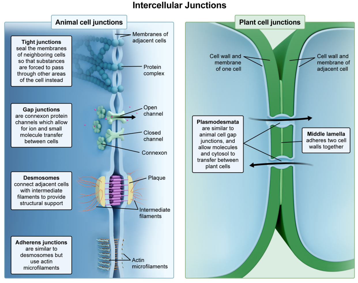
what are intercalated discs made of?
A. desmosomes and gap junctions
B. desmosomes and tight junctions
C. Hemidesmosomes and adherens junctions
D. desmosomes and tight junctions
A. desmosomes and gap junctions…which connect cardiac muscle cells, allowing synchronized contraction and electrical communication.
Which of the following best describes the sarcolemma?
A. connective tissue that surrounds the entire muscle
B. protein filament responsible for muscle contraction
C. structure responsible for calcium storage in muscle cells
D. cell membrane of a muscle fiber
D. cell membrane of a muscle fiber…which is involved in excitation-contraction coupling and maintaining the ionic environment.

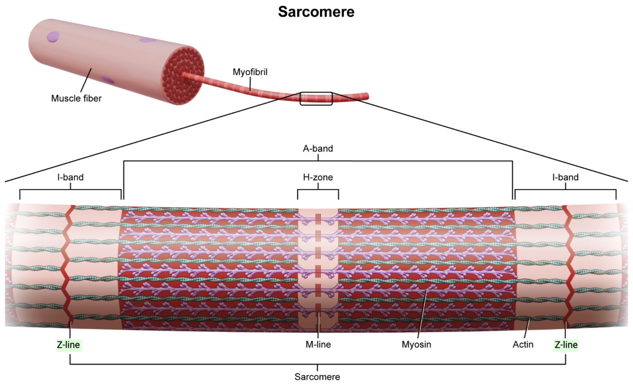
Which structure contains repeating units of sarcomeres?
A. sarcolemma
B. myofibrils
C. muscle fibers
D. muscle fascicles
B. myofibrils…contain repeating units of sarcomeres, which are the contractile units of muscle
What are the components of sarcomeres that enable muscle contraction?
A. muscle fascicles
B. myofilaments
C. myofibrils
D. myocytes
B. myofilaments…which include thick filaments (myosin) and thin filaments (actin) that slide over one another during contraction.

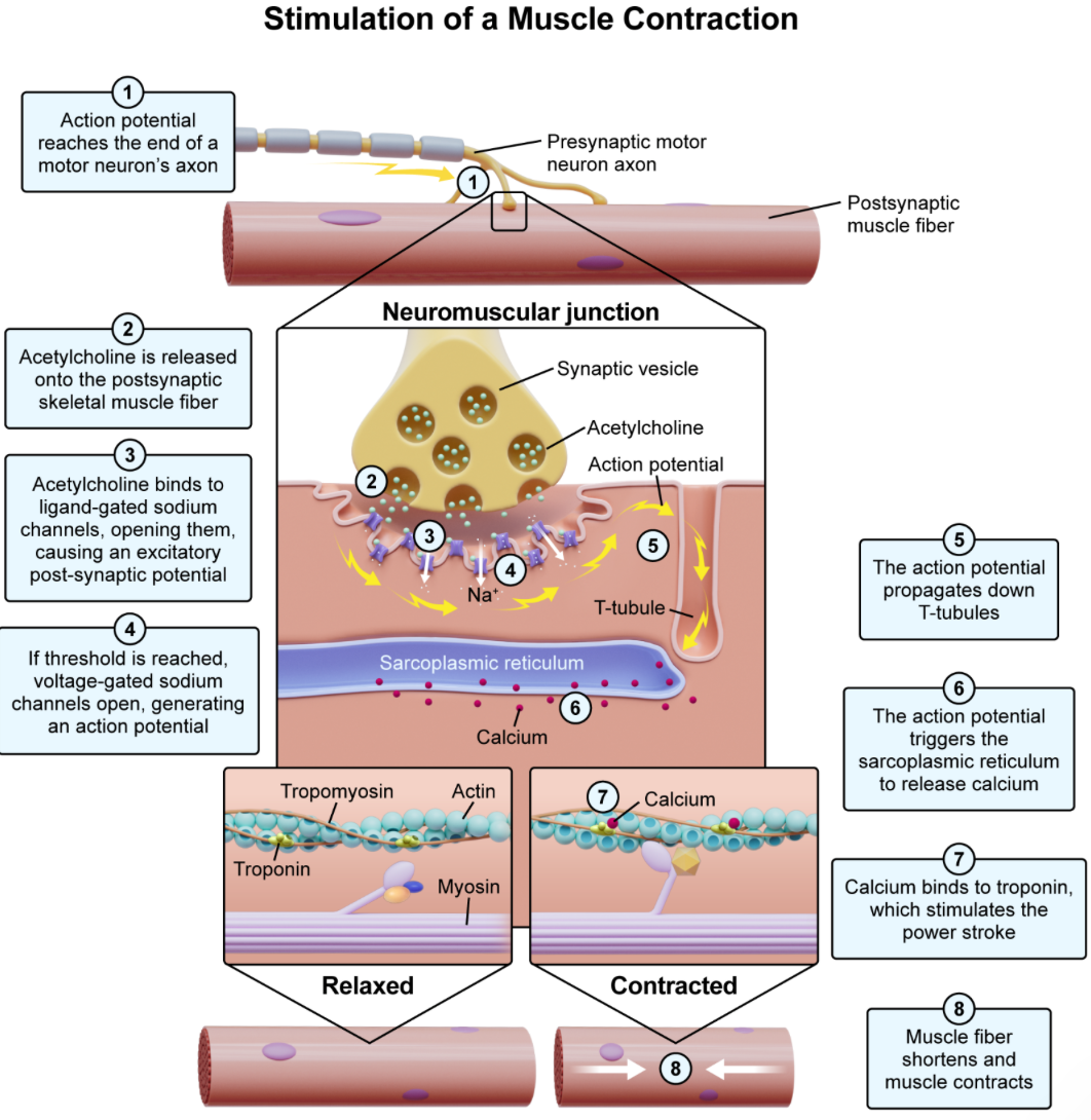
which channels are activated when acetylcholine is released into the neuromuscular junction?
A. ligand-gated sodium channels on the motor neuron
B. voltage-gated potassium channels on the sarcolemma
C. ligand-gated sodium channels on the muscle fiber
D. voltage-gated calcium channels on the sarcoplasmic reticulum
C. ligand-gated sodium channels on the muscle fiber…which allow sodium ions to enter the muscle cell, leading to depolarization and triggering muscle contraction.

What happens after acetylcholine binds to ligand-gated sodium channels?
A. the depolarization opens nearby voltage-gated sodium channels
B. an action potential is transferred from the muscle to the motor unit
C. calcium ions are released from the sarcoplasmic reticulum
D. the action potential is propagated by T-tubules
A. the depolarization opens nearby voltage-gated sodium channels… which leads to further depolarization and the initiation of a graded potential, which then opens nearby voltage-aged sodium channels.

Which structure propagates an action potential through the muscle fiber?
A. acetylcholine
B. T-tubules
C. Sarcoplasmic reticulum
D. tropomyosin
B. T-tubules…which are extensions of the cell membrane that penetrate into the muscle fiber, allowing the action potential to quickly spread throughout the muscle.
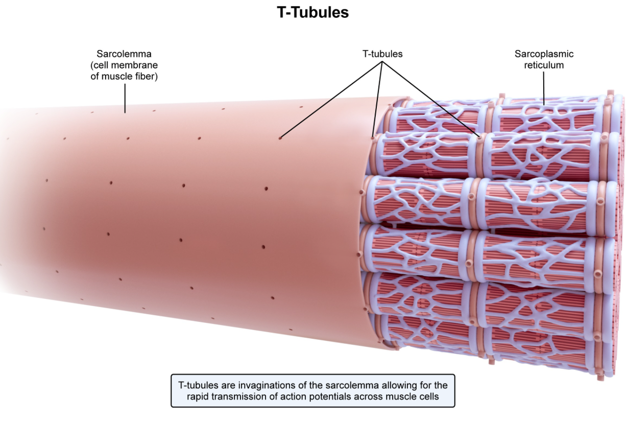
During muscle contraction, where are calcium ions released from?
A. calcium vesicles
B. myosin
C. sarcoplasmic reticulum
D. T-tubules
C. The sarcoplasmic reticulum…a specialized type of ER in a muscle cell that stores calcium ions. These calcium ions are released from the sarcoplasmic reticulum via voltage-gated calcium channels.
What can be found along the membrane of a sarcoplasmic reticulum?
A. ligand-gated Na channels
B. voltage-gated Na channels
C. ligand-gated Ca channels
D. voltage-gated Ca channels
D. voltage-gated Ca channels…The sarcoplasmic reticulum is a special type of ER that stores Ca ions. When depolarization travels doesn't the T-tubules, it causes voltage-gated Ca channels to open. This releases Ca ions into the sarcoplasm, which initiates muscle contraction.

What is the function of tropomyosin?
A. covers actin’s binding site for troponin
B. covers actin’s binding site for myosin
C. covers myosin’s binding site for actin
D. holds troponin in place
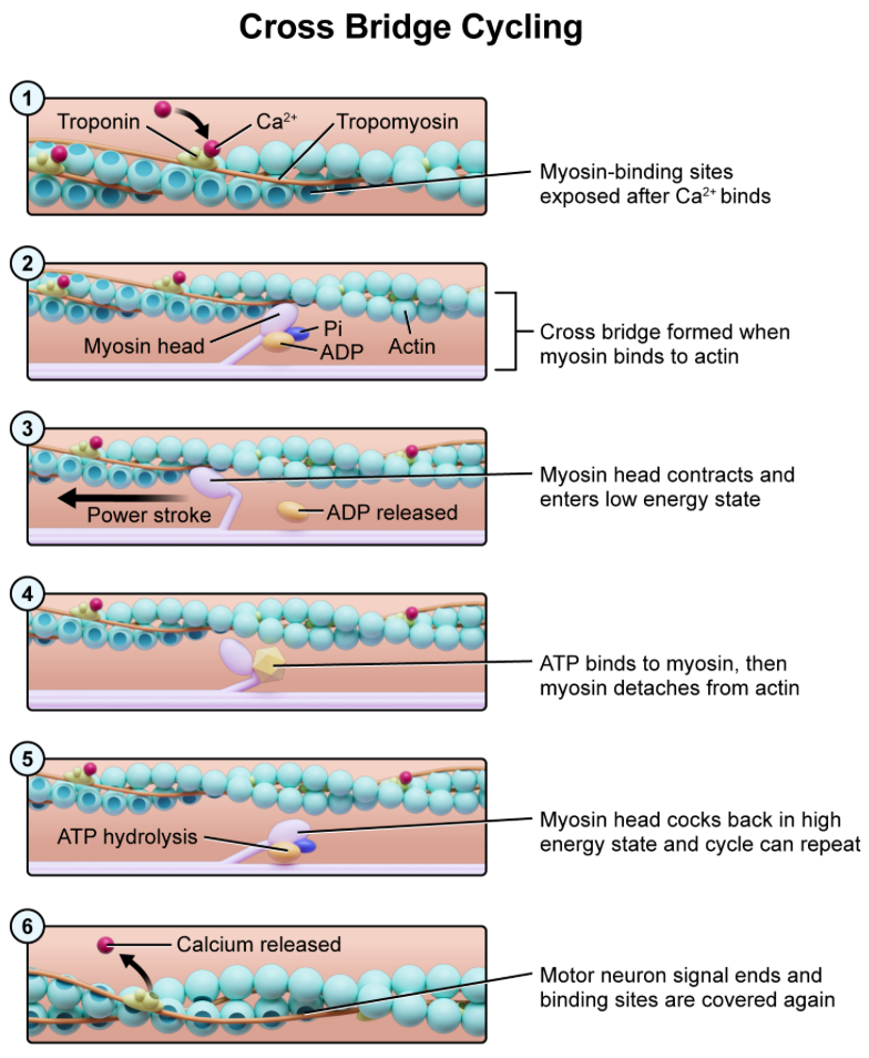
B. covers action’s binding site for myosin…Tropomyosin is a regulatory protein found in muscle fibers. In the relaxed state, it blocks the binding sites on actin for myosin, preventing cross-bridge formation. Tropomyosin is stabilized in this position by troponin.

What stimulates the removal of tropomyosin from actin binding sites?
A. an electrical gradient
B. ATP hydrolysis
C. Influx of sodium
D. release of calcium
D. release of calcium…an action potential triggers the release of calcium ions from the sarcoplasmic reticulum into the sarcoplasm. These calcium bind to troponin, causing the removal of tropomyosin from the binding sites on actin. This allows actin and myosin to interact, leading to muscle contraction.

At which point of a muscle contraction is a cross bridge formed?
A. during the power stroke
B. when ATP binds to myosin
C. once the myosin enters its low-energy state
D. when myosin cocks back into its high-energy state
D. when myosin cocks back into its high-energy stage…when the myosin head cocks back into its high-energy state, it binds to actin, forming a complex called the cross-bridge

what happens during a power stroke?
A. the actin head pulls myosin toward the center of the sarcomere
B. the actin head pulls myosin toward the edge of the sarcomere
C. the myosin head pulls actin toward the center of the sarcomere
D. the myosin head pulls actin toward the edge of the sarcomere
C. the myosin head pulls actin toward the center of the sarcomere…following the formation of a cross-bridge, the myosin head releases ADP and inorganic phosphate (Pi), triggering a power stroke. During the power stroke, the myosin head pivots from its high-energy conformation, pulling the actin filament toward the center of the sarcomere

What causes myosin to detach from actin during muscle contraction?
A. binding of ATP
B. Hydrolysis of ATP
C. Release of ADP + Pi
D. Ca2+ binds troponin
A. binding of ATP…after a power stroke, the myosin head is in a low-energy state and tightly bound to actin. The binding of ATP to the myosin head induces a conformational change that leads to its detachment from actin

What represents the midpoint of each sarcomere?
A. M line
B. H zone
C. A band
D. Z line
A. M line

What represents the end of the sarcomere?
A. M line
B. H zone
C. I band
D. Z line
D. Z line

Which of the following is a correct statement?
A. each muscle fiber requires a unique motor neuron
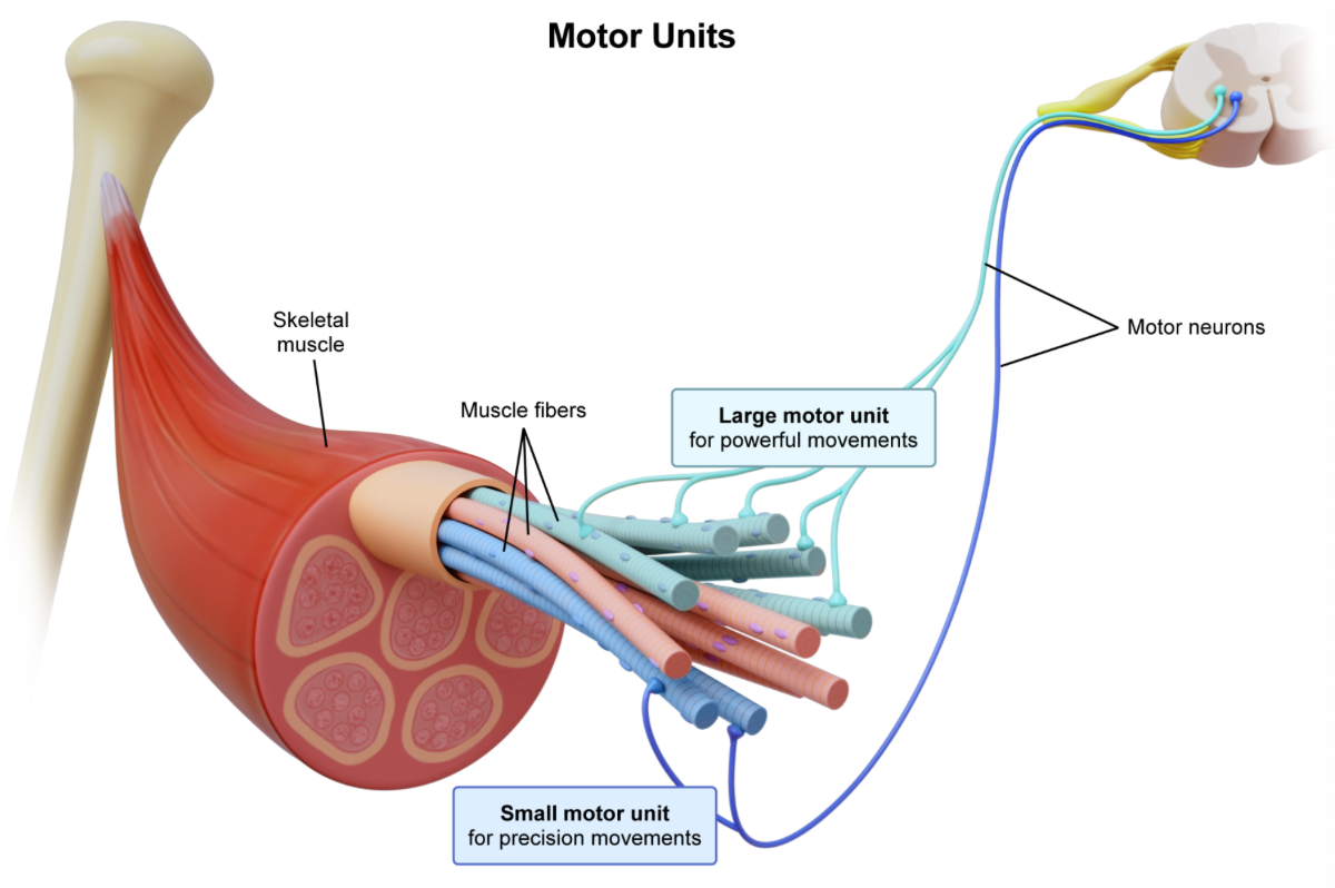
B. a motor neuron coordinates the activity of various motor units
C. a motor unit is all the motor neurons controlling a muscle fiber
D. a motor unit is all the muscle fibers innervated by one motor neuron
D. a motor unit is all the muscle fibers innervated by one motor neuron…small motor units include only a few muscle fibers and are used in precision movements, while large motor units include many muscle fibers and are used in powerful movements.
What does the size principle of motor unit recruitment refer to?
A. motor units are recruited from smallest to largest
B. motor units are recruited from largest to smallest
C. various motor units are recruited with no preference of size
D. small motor units are recruited for precise movements, and large ones for powerful movements
A. motor units are recruited from smallest to largest…helps reduce fatigue by allowing motor units within the same muscle to be activated a different times, giving them time to rest between contractions
What type of muscle cells are connected by intercalated discs?
A. smooth
B. cardiac
C. skeletal
B. cardiac
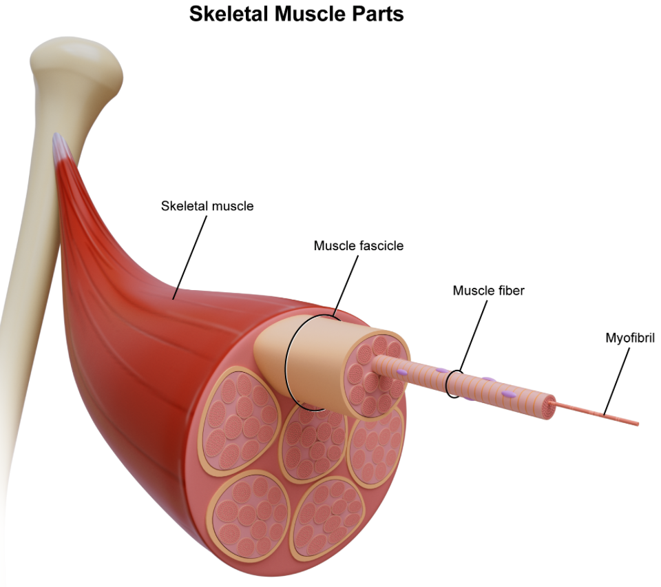
Which of the following correctly arranges the components of muscle tissue from smallest to largest?
A. myofibril, muscle fiber, muscle fascicle, skeletal muscle
B. myofibril, muscle fascicle, skeletal muscle, muscle fiber
C. muscle fascicle, myofibril, skeletal muscle, muscle fiber
D. muscle fiber, myofibril, muscle fascicle, skeletal muscle
A. myofibril, muscle fiber, muscle fascicle, skeletal muscle
What is myosin bound to when it is in its high-energy state?
A. ATP
B. ADP + Pi
C. Calcium
D. Troponin
B. ADP + Pi
What represents the area of the sarcomere where only actin is present?
A. A band
B. H zone
C. I band
D. Z line
C. I band

What represents the area of the sarcomere where only myosin is present?
A. A band
B. H zone
C. I band
D. M line
B. H zone

Which of the following areas of the sarcomere does not shorten during muscular contraction?
A. A band
B. H zone
C. I band
A. A band